90 Deg. Off-Axis Parabolic Mirrors (OAPs) - parabolic mirror
Difference betweenmultispectralandhyperspectralremote sensing
For a magnifying device held close to the eye, see Loupe. ... Hobbyists, from those engaged in sewing and needlework to stamp collectors ... Addison Wesley. pp. 186 ...
A hyperspectral camera captures a scene’s light, separated into its individual wavelengths or spectral bands. It provides a two-dimensional image of a scene while simultaneously recording the spectral information of each pixel in the image.
Murphy, Ellen. (2018, April 17). What Happens When You Go From Low Power To High Power On A Microscope?. sciencing.com. Retrieved from https://www.sciencing.com/happens-power-high-power-microscope-8313319/
Hyperspectral imaging
Going to high power on a microscope decreases the area of the field of view. The field of view is inversely proportional to the magnification of the objective lens. For example, if the diameter of your field of view is 1.78 millimeters under 10x magnification, a 40x objective will be one-fourth as wide, or about 0.45 millimeters. The specimen appears larger with a higher magnification because a smaller area of the object is spread out to cover the field of view of your eye.
Hyperspectral imaging involves using an imaging spectrometer, also called a hyperspectral camera, to collect spectral information.
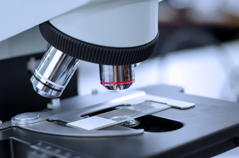
When you change from low power to high power on a microscope, the high-power objective lens moves directly over the specimen, and the low-power objective lens rotates away from the specimen. This change alters the magnification of a specimen, the light intensity, area of the field of view, depth of field, working distance and resolution. The image should remain in focus if the lenses are of high quality.
Hyperspectralcamera
To make a lens this size the regular way would require it to be thick. Fresnel lenses allow making bigger lenses with narrow profiles, but they ...
Discover the objective lens product range of MITUTOYO. Contact the manufacturer directly.
A hyperspectral camera measures thousands or hundreds of thousands of spectra to create a massive hyperspectral data cube comprising position, wavelength, and time-related information.
The light intensity decreases as magnification increases. There is a fixed amount of light per area, and when you increase the magnification of an area, you look at a smaller area. So you see less light, and the image appears dimmer. Image brightness is inversely proportional to the magnification squared. Given a fourfold increase in magnification, the image will be 16 times dimmer.
Murphy, Ellen. What Happens When You Go From Low Power To High Power On A Microscope? last modified March 24, 2022. https://www.sciencing.com/happens-power-high-power-microscope-8313319/
hyperspectralvs.multispectralremote sensing ppt
by A Ibrahim · 2024 · Cited by 2 — This systematic review explores the level of oxidative stress (OS) markers during pregnancy and their correlation with complications.
CV beams, exhibiting tangential polarization states, are some prominent examples. This prominent approach relies on the fundamental aspect of the fs laser ...
The working distance is the distance between the specimen and objective lens. The working distance decreases as you increase magnification. The high power objective lens has to be much closer to the specimen than the low-power objective lens in order to focus. Working distance is inversely proportional to magnification.
Murphy, Ellen. "What Happens When You Go From Low Power To High Power On A Microscope?" sciencing.com, https://www.sciencing.com/happens-power-high-power-microscope-8313319/. 17 April 2018.
Multispectralcamera

Hyperspectral imaging is a technique that collects and processes information across the electromagnetic spectrum to obtain the spectrum for each pixel in an image. This allows for the identification of objects and materials by analyzing their unique spectral signatures. Applications of hyperspectral imaging include food quality & safety, waste sorting and recycling, and control and monitoring in pharmaceutical production.
Sep 9, 2020 — By moving its knob you will be able to control the amount of light passing through your specimen. While working with nonliving material stained- ...
Thanks to its noninvasive, and nondestructive capability in identifying and quantifying material, hyperspectral imaging has become increasingly popular in various industries and research applications.
Apr 5, 2020 — Step 1: Take the Measurement · Place your part between the measuring faces. · Bring the measuring face towards the part by rotating the spindle.
In conclusion, hyperspectral imaging is both a prominent tool for research and highly useful machine vision technology for various industries to improve processes, increase quality, and reduce waste.
Multispectralandhyperspectralremote sensing PDF
The depth of field is a measure of the thickness of a plane of focus. As the magnification increases, the depth of field decreases. At low magnification you might be able to see the entire volume of a paramecium, for example, but when you increase the magnification you may only be able to see one surface of the protozoan.
The data provided by hyperspectral imaging systems can be used during inspection to locate, sort, or quantify the concentration of various materials that are invisible to common cameras or the human eye. For instance, a hyperspectral imaging system integrated into an in-line quality control system enables the identification of foreign objects, contaminants, and the amount of fat, sugar, or moisture in products.

Jul 3, 2023 — The eyepiece or ocular lens is the part of the microscope closest to your eye when you bend over to look at a specimen. An eyepiece usually ...
The result is a hyperspectral image, where each pixel represents a unique spectrum. This unique spectrum can be compared to fingerprints. Since every material and compound reacts with light differently, their spectral signatures are also different. Just like fingerprints can be used to identify a person, the spectra can identify and quantify the materials in the scene.
Hyperspectral imaging is a powerful technology combining spectroscopy with imaging capability. It enables gathering detailed information about the composition and characteristics of objects and surfaces in a way that is impossible with conventional imaging systems.
The electromagnetic spectrum describes all types of light, ranging from very long radio waves, through microwaves, infrared radiation, visible light, ultraviolet rays, and X-rays, to very short gamma rays — most of which the human eye can’t see (Figure 1).
RGBvs multispectral vs hyperspectral
Multispectralandhyperspectral imaging
Hyperspectral imaging system analyzes a spectral response to detect and classify features or objects in images based on their unique spectra.
Spectral imaging is imaging that uses multiple bands across the electromagnetic spectrum. While the RGB camera uses three visible light bands (red, green, and blue) to create images, hyperspectral imagery makes it possible to examine how objects interact with many more bands, ranging from 250 nm to 15,000 nm and thermal infrared. The study of light–matter interaction is called spectroscopy or spectral sensing. To learn more, read our article How does spectral sensing work? Understanding the basics of spectroscopy and spectral sensors.
Microscopes magnify an object's appearance by bending light. Higher magnification means the light is bent more. At a certain point, the light is bent so much that it can't make it through the objective lens. At that point – usually around 100x for standard lab microscopes – you'll need to put a drop of oil between your specimen and the objective lens. The oil "unbends" the light to stretch out the working distance and make it possible to image at high magnifications.
It is the only method the hide the thickness, so from the front it looks thin, and from the side, this part of the lens is masked by the temple. Other methods ...
Changing from low power to high power increases the magnification of a specimen. The amount an image is magnified is equal to the magnification of the ocular lens, or eyepiece, multiplied by the magnification of the objective lens. Usually, the ocular lens has a magnification of 10x. A typical lab-quality standard optical microscope will usually have four objective lenses, running from a low power of 4x to a high power of 100x. With an ocular power of 10x, that gives the standard optical microscope a range of overall magnification from 40x to 1000x.
Hyperspectral imagery acquired through remote sensing provides information about surfaces on the Earth, such as minerals or vegetation for example.
Compared to multispectral imaging, hyperspectral imaging provides more information allowing for more accurate analysis, identification, and separation of materials and substances (To learn more, read our article Hyperspectral vs. Multispectral cameras).
One of the key benefits of hyperspectral imaging is its high spatial and spectral resolution which enables the detailed characterization of the materials.
Hyperspectral imaging lets us differentiate between materials with similar physical or visual characteristics or what the human eye cannot see, such as different minerals.
Figure 2: To match human vision, a digital photograph of a leaf (top) is created using three bands: red, green, and blue. The RGB data is comparable to a three-page pamphlet. In contrast, a hyperspectral image of a leaf (bottom) captures a spectral response from 220 wavelengths. The comparable 220-page book contains much more detailed information about the object.
Spectral imaging systems refer to a class of imaging technology that captures and processes information about the wavelength of light within an image. These systems are designed to capture multiple bands or channels of information across the electromagnetic spectrum beyond the visible light that our eyes can see. This data can then be processed to generate a color-coded representation of the spectral data, which can provide information about the chemical and physical properties of the objects within the image.
By combining the benefits of digital imaging and a spectrometer, hyperspectral imaging provides both spatial and spectral information about the object’s physical and chemical properties. The spectral information allows for the identification and classification of materials and the spatial provides data on the material’s distribution and areal separation. Hyperspectral imaging provides answers to questions concerning “what” (based on the spectrum), “where” (based on location), and “when”.
Aug 26, 2018 — The alignment cube is nothing to do with textures or materials. It's about accurately aligning your objects inworld e.g. making sure the walls ...


 Ms.Cici
 8618319014500