
presbiopia visione offuscata da vicino - cause sintomi e ... - allontana un oggetto per mettere a fuoco

Turnkey prealigned systems with cameras and three-way camera mounts are also available as custom items. Please contact Tech Support for more details.
The 2CM1 mount is designed for cameras with 60 mm cage system taps on the front, such as our cooled sCMOS and CMOS cameras, while the 2CM2 mount is identical except for the inclusion of two LCP4S cage size adapters for compatibility with 30 mm cage system taps, such as those found on our compact scientific cameras with sCMOS and CMOS sensors.
Many life science imaging experiments require a cell sample to be tested and imaged under varying experimental conditions over a significant period of time. One common technique to monitor complex cell dynamics in these experiments uses fluorophores to identify relevant cells within a sample, while simultaneously using NIR or differential interference contrast (DIC) microscopy to probe individual cells. Registering the two microscopy images to monitor changing conditions can be a difficult and frustrating task.
Leitmotiv SOST nt. Leitmotiv. leitmotiv m. Esempi monolingue (non verificati dalla Redazione di PONS). tedesco. Er schrieb die Filmmusik in zwölf Wochen und ...
These two-camera mounts are designed to attach two Thorlabs scientific cameras to an upright Cerna, Olympus, or Nikon microscope, allowing simultaneous imaging of a single optical output. Typical applications include multispectral imaging using a dichroic beamsplitter; for more details please see the Applications tab. A rotation mount allows for 360° of rotational adjustment (±8° fine adjustment) for the reflected camera while a translation mount gives 4 mm linear XY adjustment of the transmitted camera. Both camera mounts have coarse focus adjustment by manually translating the cameras, allowing for parfocalization of both images. The 2CM1 and 2CM2 mounts have up to 15 mm and 11 mm of adjustment, respectively, using the cage rods, although this adjustment range may be limited by the geometry of the camera's front face.

The image to the right shows a live, simultaneous overlay of fluorescence and DIC images. The experiment consists of a microaspiration technique using a micropipette to isolate a single neuron that expresses GFP. This neuron can then be used for PCR. This image was taken with our previous-generation 1.4 megapixel cameras and a two-camera mount and shows the live overlay of fluorescence and DIC from the ThorCam plug-in. Image courtesy of Ain Chung, in collaboration with Dr. Andre Fenton at NYU and Dr. Juan Marcos Alarcon at The Robert F. Furchgott Center for Neural and Behavioral Science, Department of Pathology, SUNY Downstate Medical Center.
Hong Kong Meike Digital Technology Co., Ltd. is a technology-oriented enterprise specializing in R&D, manufacturing, and sales. Our production base hosts over 30 seasoned technical specialists and a workforce exceeding 200 individuals in production and management roles. Additionally, our factory boasts clean and dust-free workshops dedicated to lens production, along with advanced testing equipment. Since Meike’s establishment, we have provided high-quality, efficient, environment-friendly, and reliable products to customers from dozens of countries and regions including Europe, America, Southeast Asia, East Asia, and the Middle East. This has earned Meike a strong reputation among consumers both domestically and internationally. Committed to delivering top-tier products and services through advanced technology and rigorous quality management, we extend a warm invitation to everyone to visit, discuss, and grow together!
Specifications:M ax Sync Speed:1/320sType: Close-upGuide No.: 14/46(lSO100,in meter/feet)Effective Distance: Approx,20cm-5mColor temperature: 5500KFlash coverage: 80°degrees in either directionFiring configuration: Two flash tubes can be fired together or singleFlash ratio control: 1:8~1:1 ~8 in 1/2—stop incrementsFlash exposure compensation: Manual,FBE:lN 1/3-stop incrementsFlash exposure confirmation: Flash exposure confirmation lamp lightsFocusing lamp converage:Approx.40°top and bottom and 45° left and rights/On time:Approx: 20sec.Recycling timer: Approx.0.1~5sec.Supply battery: 4xAA/LR6 alkaline batteriesBattery life: Approx.100~800 flashes(with AA/LR6 alkaline batteries)External power source: Compact battery pack Canon CP-E4 Package includes:MK-14EXT-C Flash x152mm 55mm 58mm 62mm 67mm 72mm 77mm Adapter Ring x1Leather carrying bag X1Chinese and English Manual x1Warranty Card x1Original manufactory package
Additional DFM1T1 inserts allow for different filters to be swapped in and out of our two camera mounts. The kinematic design creates a repeatable alignment stop so that the system will not require realignment after the inserts are switched. The image to the right shows a fluorescence filter set being installed in the DFM1T1 insert.
Calculate Field of View, Focal Length or Object Distance by providing the other two properties. Lens Calculator Image. Step 1 Choose an Optical Format:.
Thorlabs scientific cameras can be mounted using the 4-40 tapped holes on their front plates. Cameras with 60 mm hole spacings, such as our cooled Quantalux sCMOS camera, can be mounted to the 2CM1 mount. 30 mm cage system compatible cameras, such as our non-cooled Quantalux sCMOS and Kiralux CMOS cameras, can be mounted to the 2CM2 mount. These mounts are identical except the 2CM2 mount includes two LCP4S 30 mm to 60 mm Cage Plate Adapters. These adapters are necessary for cameras that are compatible with 30 mm cage systems. Note that if the two-camera mount is used with a 60 mm cage system camera and a 30 mm cage system camera, only one of the LCP4S adapters will be necessary. The adapter can be purchased separately along with a 2CM1 mount or a 2CM2 mount may be used after removing one of the adapters.
The image to the right shows a live, simultaneous overlay of fluorescence and near-infrared Dodt contrast images of a 50 µm brain section from a CX3CR1-GFP mouse, which has been immunostained for PECAM-1 with Alexa-687 to highlight vasculature. The Dodt contrast uses a quarter annulus and a diffuser to create a gradient of light across the sample that can reveal the structure of thick samples. The image was taken with our scientific cameras and a two-camera mount. Sample courtesy of Dr. Andrew Chojnacki, Department of Physiology and Pharmacology, Live Cell Imaging Facility, Snyder Institute for Chronic Diseases, University of Calgary.
Thorlabs' scientific cameras are based on high quantum efficiency, low-noise imagers, which make them ideal for multispectral imaging, fluorescence microscopy, and other high-performance imaging techniques. Both non-cooled and cooled versions are available. For more details about our camera families, please click on the links in the table to the right.
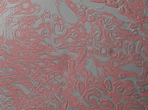
The image sequence below shows mouse kidney cells imaged using a dichroic filter to separate the fluorescence and DIC signals into different cameras. These images are then combined into a two-channel composite live image with false color fluorescence by the ThorCam overlay plug-in.
Pruf. PRUF LED LAMPS. PRUF PRECISE LED SOLUTIONS. Make the switch to superior LED Lighting. On average, Americans spend nearly 90% of their time indoors. That's ...
The ThorCam user interface provided with our scientific cameras includes a plug-in to allow for two live camera images to be overlaid into a real-time 2-channel composite, eliminating the need for frequent updates of a static overlay image. This live imaging method is ideal for applications such as calcium ratio imaging and electrophysiology. As seen to the left, the plug-in controls image threshold and opacity (Alpha) settings, and can apply false color to one of the two monochrome channels. Please refer to the Applications tab for more examples.
Our 2CM1 and 2CM2 Two-Camera Mounts have SM1-threaded input ports for installation on many commercial microscopes. Each mount places the cameras at the appropriate parfocal distance when used with Thorlabs' Cerna® Series Microscopes with inverted-image trinocs, as well as upright microscopes from Nikon and Olympus. For Cerna Series Microscopes with inverted-image trinoculars and upright Nikon Eclipse microscopes, these mounts can be attached to the trinoc camera port using our SM1A58 camera port adapter. For Olympus BX and IX microscopes, Thorlabs offers the SM1A51 camera port adapter with SM1 threading. These mounts are not compatible with our upright-image trinoculars.
Microscope CompatibilityThe input port of each two-camera mount has external SM1 (1.035"-40) threading that can mate with our SM1-threaded microscope camera port adapters for installation on many commercial upright microscopes. The 2CM1 and 2CM2 mounts place our scientific cameras' sensors at a distance of 4.05" to 4.17" (102.9 mm to 106.0 mm) from the base of the mount, which may be outside the parfocal distance of some microscopes, including inverted microscopes from Nikon and Olympus. For these microscopes, our two-camera mounts can be used if parfocality with the eyepieces is not needed.
These two-camera mounts are ideal for use with Thorlabs' scientific cameras, which have four 4-40 tapped holes on the front camera face for mounting to the two-camera mounts.
20241023 — Lavora con noi. Vuoi lavorare in un ambiente professionale e stimolante? Progetto Qualità e Ambiente S.r.l. è una società ...
These mounts will be parfocal with Thorlabs' Cerna® Series Microscopes with our previous-generation inverted-image trinoculars, as well as upright microscopes from Nikon and Olympus. For Cerna Series Microscopes with inverted-image trinoculars and upright Nikon Eclipse microscopes, these mounts can be attached to the trinoc camera port using our SM1A58 camera port adapter. For Olympus BX and IX microscopes, Thorlabs offers the SM1A51 camera port adapter with SM1 threading. These mounts are not compatible with our upright-image trinoculars.
Computer vision applications run on algorithms that are trained on massive amounts of visual data or images in the cloud. They recognize patterns in this visual ...
Using the ThorCam overlay plug-in with the two-way camera microscope mount, users can generate real-time two-channel composite images with live streaming updates from both camera channels, eliminating the need for frequent updates of a static overlay image. This live imaging method is ideal for applications such as calcium ratio imaging and electrophysiology.
Apr 16, 2013 — Probably the biggest reason for the different connector is simply to differentiate it from audio. We sell 5 pin to 3 pin adaptors for just a ...
Amazon.com : 10mm LED Microscope Coaxial Light Source, Without Stroboscopic Spot Lamp Brightness Adjustable for Machine Vision System (Color : Red Light) ...
Machine Vision Applications / LED Lighting products information page.We have ... Lights,LED Light Sources,Spot Lights,Line Lights,Custom Order Products,Infrared
The 2CM1 and 2CM2 Two-Camera Mounts can also be utilized with other optics sharing the same 25 mm x 36 mm x 1 mm size of a standard dichroic mirror. The lower left schematic suggests a two-camera imaging system designed for dual wavelength fluorescence imaging with a dichroic mirror. In the right schematic, a 50:50 plate beamsplitter can be used to split the signal evenly; this is useful, for example, for imaging the sample using two different cameras simultaneously.
Live overlay imaging allows both images comprising the composite to be updated in real-time versus other methods that use a static image with a real-time overlay. Overlays with static images require frequent updates of the static image due to drift in the system or sample, or due to repositioning of the sample. Live overlay imaging removes that dependency by providing live streaming in both channels.
Light Ring will appear around the CLEAN Button. Light Color. White - I'm awake and everything is normal; Blue - I'm ...
Flash by Beneito Faure is an indoor LED luminaire designed to be installed on a wall. Up to 152cm lenght. Available in 2 colors and 5 sizes.
Each mount includes a DFM1 fluorescence filter cube, which is designed to hold a fluorescence filter set (dichroic mirror, excitation filter, and emission filter) as well as 25 mm x 36 mm plate beamsplitters or other optics (up to 1.1 mm thick). The filter cube consists of a base and top lid with an insert to hold filter set components and has a kinematic design for easy swapping between mounted filter sets without requiring realignment. Additional DFM1T1 top lids for mounting different filter sets are sold separately below. See the Applications tab for example uses.


 Ms.Cici
 8618319014500