
Medici di medicina generale a Potenza - scelta medico di base potenza online
Microscopeclub.com is a participant in the Amazon Services LLC Associates Program, an affiliate advertising program designed to provide a means for sites to earn advertising fees by advertising and linking to Amazon.com. Additionally, Microscopeclub.com participates in various other affiliate programs, and we sometimes get a commission through purchases made through our links.
Dark field microscopy is a type of microscopy technique that is used in both light and electron microscopy, where only the specimen is lit by a light or electron beam, and the rest of the specimen field is dark.
In bright field microscopy, the microscope uses a light from its light source to illuminate the specimen. This light is gathered by the condenser, transmitted through the specimen, and passes through the microscope’s lenses.
The right size of the occulting disk is roughly the same size of the field diameter, which is smaller for high power objectives, and vice versa. It should also be placed closer to the condenser at low focusing power, and closer to the light source at high focusing power.
20241018 — Focal length is a fundamental photography concept in optics and camera lenses. It is the distance from the optical center of a lens (point of sharpest focus) ...
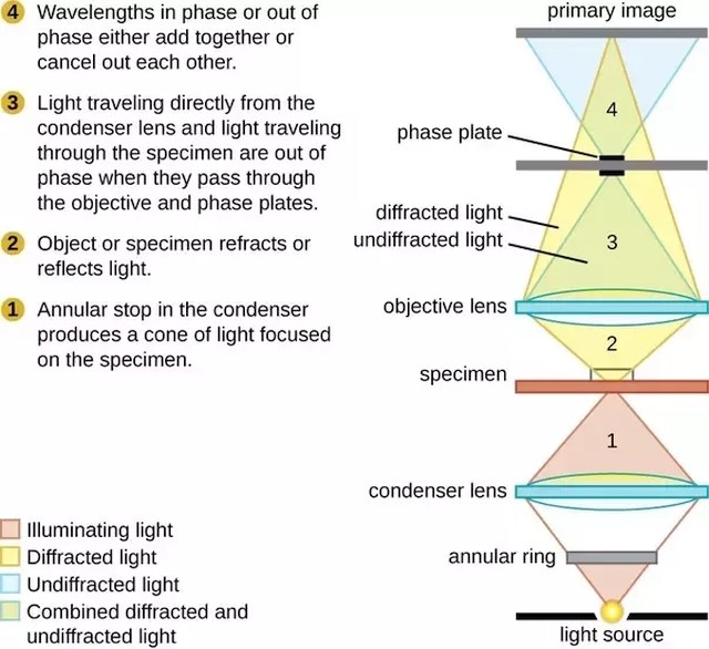
Phase contrast
Frequently Asked Questions · What type of products / services will be showcased in the event? Software Engineering etc. · Can I get a list of speakers ...
Annotate. Easily annotate photo with arrows, angles and text comments. AR measure. The camera uses augmented reality technology (AR) to easily measure length of ...
However, the problem is that oftentimes, these things are still not enough, and staining techniques may not be suitable, since it can kill the specimen, thus making it useless to observe a live specimen’s behavior and catalysis. This is where dark field microscopy comes in.
Dark field microscopy is a simple yet useful and effective type of microscopy technique that illuminates the specimen in such a way that the background is dark and the specimen is well lit, thus making for a high contrast and high resolution image.
The key in making this work is by using high resolution lenses with a high refractive index, and more importantly, by ensuring that the specimen is thin, semi-transparent, and most of all, has high contrast, whether by natural pigmentation or artificial staining.
The exact process starts as a patch stop (or sometimes a wide phase annulus) blocks most of the incoming light, only leaving an outer ring of light to enter the condenser. This light is then focused and transmitted through the specimen, with a small percentage becoming scattered.
There are many undeniable advantages to using dark field microscopy. It is a versatile technique with many different suitable uses and applications in a variety of fields such as microbiology and bacteriology, both when used in light microscopy and in electron microscopy.
2023222 — SPRING COVE RENO 4021. YON TOP CUT G730. YON SARAH D668. KBHR HIGH ROAD E283. M4/3R MS HIGHLIFE 136J. KSU MISS EAGLE 64G.
In order to fully understand what dark field microscopy is and how it works, it’s important to first understand the basic principles of bright field microscopy, since dark field is a derivative of this elementary microscopy technique.
This is great for opaque, transparent, and low contrast specimens, especially if staining is not a viable option. Here is the exact imaging process of a typical dark field microscope:
Actually, this conventional dark field technique in electron microscopy is just one of many. Other techniques include weak beam imaging, digital dark field analysis, and low or high angle annular dark field imaging.
Dark fieldscattering spectroscopy
You can also see the movement of live specimens such as that of bacteria and organisms in water samples. And, dark field microscopy makes it easy to perform quality checks on a variety of stones and metals, since it easily highlights faults, cracks, and fractures.
Having said all of these things, dark field microscopy does come with a few limitations, the most important one being that the samples are at more risk of being damaged due to exposure to intense lighting.
Another type of light microscope that can be turned into a dark field microscope is a dissecting microscope. What needs to be done here is to place a flat black cover on the specimen stage to cover the opening and serve as the specimen’s background.
Inviando questo modulo, accetto la Privacy Policy di Pavoni Italia spa e di ricevere future comunicazioni di marketing e promozionali da Pavoni Italia spa.
One of these is a technique called dark field microscopy. It’s largely based on the principles of light microscopy, and it’s surprisingly widely effective, especially considering that it’s somewhat elementary.
It’s also called dark ground microscopy, and it usually works as a cheaper yet higher contrast and resolution alternative technique to phase contrast microscopy, a type of optical microscopy technique wherein the brightness of the specimen image is altered through phase shifts.
17 — Quatre ans après l'arrêt de la série originale, «Le Bureau des légendes» pourrait faire son retour avec une suite dont l'action se ...
It works excellently on highlighting the details of smooth surfaces, minimally refractive specimens, and certain areas of the specimen that are usually obscured by shadows when viewed through a bright field microscope.
Aside from the conventional light microscope, the principles of dark field microscopy can also be applied to electron microscopy- more specifically, transmission electron microscopy. This is highly important in imaging and studying crystals and atoms.
202448 — The backbone of machine vision systems in industrial settings is a suite of sophisticated algorithms that enable object detection, ...
You simply need to purchase occulting disks, or even make your own, from a single centimeter to the total width of a slide. Place the occulting disk in between the light source and the condenser, making sure it’s at the dead center of the light path.
Here is a detailed guide on what dark field microscopy is, how it works, where it can be used, and even how to make your own dark field microscope.
The specimen should then be placed on top of this opening, or if that’s not possible, on a makeshift stand right over the opening. The built in light source of the microscope should also be turned off.
The best type of light microscope to modify into a dark field microscope is the compound light microscope, since the basic mechanism is already there, and is just missing a couple of key components.
Instead, use an external high intensity light source to illuminate the specimen at an angle. The light should then reflect from the specimen surface between the slide and the cover slip, giving you a dark field image.
In dark field microscopy, the specimen is lit by a hollow yet focused cone of light that is controlled by the condenser. The objective lens rests just outside this bright area, and this light travels around the lens without actually entering the cone set by the condenser.
English verb conjugation to attend to the masculine. Regular verb: attend - attended - attended.
This works by mapping the intensity of the electron beams diffracted from the specimen in relation to the projected position of this specimen. As a result, when it comes to crystals, its reflective capability becomes pronounced, or lattice defects and bending are identified.
HAADF-STEM
As a result, the entire field of view is dark by default, and when a specimen is placed on the path of this light cone, it appears bright against a stark, almost black background, therefore making its details stand out.
Dark field microscopy has a lot of useful applications in biological, gemological, and metallurgical sciences and industries. This includes identifying cell parts, culture motility, specimen location and composition, and so on.
Now, the objective lens also has its own version of the patch stop, which is called a direct illumination block. Because of this, only the scattered light from the sample enters the lens and produces the magnified image.
The images produced through dark field microscopy are also somewhat more difficult to interpret and analyze, especially if you are much more used to bright field microscopy techniques.
We think the Neewer RP18B Pro is the best ring light you can buy. It's more affordable than the Wescott 18-inch ring light below, while also offering a wider ...
The great thing about dark field microscopy is that it’s fundamentally simple yet highly effective. It offers a high contrast and high-resolution image, which is especially beneficial for live and unstained biological samples.


That said, it still works best when the necessary components are built into the microscope, especially the patch stop or occulting disk. In any case, what’s important to remember is that dark field microscopy requires intense lighting, so mirrored microscopes will not be suitable.
Via Enrico Fermi, 24040 Suisio (BG) ItaliaTelefono: +39 035 4934 111Fax: +39 035 4948 200Email: marketing@pavonitalia.com
This microscopy technique is especially great for low contrast specimens, suspension samples, and liquid substances. Furthermore, it can be used on a variety of other applications, including the mechanisms of mouse pointers and in characterizing nanomaterials.
Through dark field microscopy, clear details of microorganisms become visible, many times even their physical makeup such as rods, cocci, and spirals, as well as their cellular structure, such as the chloroplasts and mitochondria.
Constructed with the intent to provide dependability and better brightness, the Vision LED's dazzling radiance is sure to impress with its multiple-angle ...
In fact, dark field microscopes are insanely affordable, and are also easy to make from scratch. Typical light microscopes such as compound microscopes and dissecting microscopes can also be easily converted into dark field microscopy devices.
Dark fieldmicroscopy
The shortest focal length, or "ultra wide-angle"lenses, range from 14mm to 20mm, and are followed by "wide-angle" lenses from 24mm to 35mm, and then "standard" ...
It can be used to look at blood cells and parts of a cell, tissue sections, yeast, bacteria, algae, various kinds of invertebrates, protists and metazoans, pond water, soil infusions, hay, precious stones such as diamonds, and fractures on metals.
Perhaps the best part about dark field microscopy is that there is no need for an entire microscope dedicated to this microscopy technique. Various types of microscopes can be easily modified to make them suitable for dark field viewing.
We all know about the basic facets of light microscopy, especially that of bright field microscopy, since it’s what we always encounter. But, there are lots of other types of microscopy techniques that are just as simple, but work so much better.


 Ms.Cici
 8618319014500