
What is the Difference Between 365nm and 395nm UV ... - 365 nm light
Types of illumination PDF
The specimens used are prepared initially by staining to introduce color for easy contracting characterization. The colored specimens will have a refractive index that will differentiate it from the surrounding, presenting a combination of absorption and refractive contrast.
Illumination lighting meaning
This microscope is used to view fixed and live specimens, that have been stained with basic stains which gives a contrast between the image and the image background. It is specially designed with magnifying glasses known as lenses that modify the specimen to produce an image seen through the eyepiece.
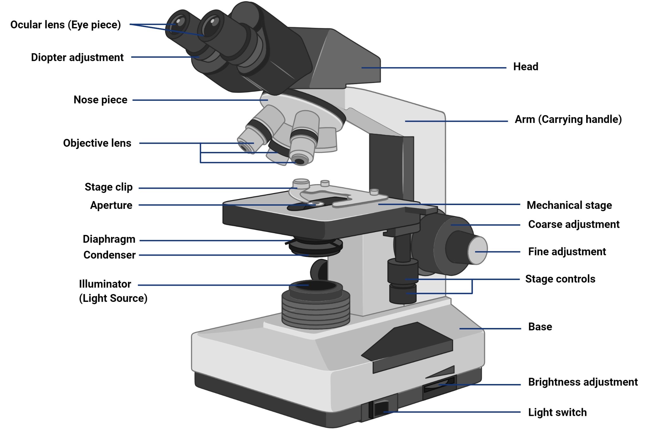
Brightfield Microscope is an optical microscope that uses light rays to produce a dark image against a bright background. It is the standard microscope that is used in Biology, Cellular Biology, and Microbiological Laboratory studies.
Types of illumination in slit lamp
The functioning of the microscope is based on its ability to produce a high-resolution image from an adequately provided light source, focused on the image, producing a high-quality image.
Brightfield Microscope is used in several fields, from basic biology to understanding cell structures in cell Biology, Microbiology, Bacteriology to visualizing parasitic organisms in Parasitology. Most of the specimens to be viewed are stained using special staining to enable visualization. Some of the staining techniques used include Negative staining and Gram staining.
For a specimen to be the focus and produce an image under the Brightfield Microscope, the specimen must pass through a uniform beam of the illuminating light. Through differential absorption and differential refraction, the microscope will produce a contrasting image.
Sorry, we just need to make sure you're not a robot. For best results, please make sure your browser is accepting cookies.


 Ms.Cici
 8618319014500