
Outdoor Spotlights - LED Spotlights - Spot lights - mountable spotlight
Fiber Optic Lights for Crafts
A polarising microscope is an optical microscope composed of a detector, lenses and polarising filters. Its process includes illumination of the sample with polarised light and is useful for better visualisation and understanding of birefringent materials (materials that have two different refractive indices). This microscope is operated through the use of a polarized filter can be turned and fixed in the light path beneath the specimen, usually below the stage. This particular device is known for its anti-reflective properties which is deemed essential for deep analysis of an isotropic particles that requires high integrity of light transmission.
The light microscope, or optical microscope, is a microscope that uses visible light and a system of lenses to magnify images. These days there are many complex designs of them which have been developed with the aim of improving resolution and sample contrast.
Fibre Optic Lights for ceiling
This is a pretty neat way to glow up. Light in the fiber optic cable zooms through the core by constantly bouncing off of the edge of the molded plastic, a principle called "total internal reflection" (which also kinda sounds like a very expensive meditation island retreat). When the light pipe doesn't have total reflection, you can get a nifty lighting effect from the light illuminating the plastic tubing.

OpticalFiberLight
Fluorescence microscopy is done with an optical microscope that uses a mercury arch lamp as a source of UV light. The microscope will also comprise excitation filter, dichromatic mirror and an emission filter. Fluorescence, used to observe the specimen, begins where a molecule absorbs light of high frequency and emits light of lower frequency. Fluorescence microscopy uses reflected light. In a fluorescence microscope the light source travels in a different trajectory than in the basic light microscope. An advantage of fluourescence microscopy is that it can be used to detect and visualise multiple fluorescent molecules e.g. cells glowing as they are doing their work. iOLight sell a microscope for mobile digital fluorescence microscopy, which is also great for field microscopy uses.
Fiber OpticLightCable
If you buy less than a full reel, you'll get a single strip, but it will be a cut piece from a reel which may or may not have a connector on it. If the piece comes from the end of the reel, the connector may be on the output end of the strip!
This type of microscope was developed in response to drawbacks with fluorescence microscopes (principally that they use high intensity UV light which means the samples are continuously exposed to it, causing photo bleaching and blurring in some samples). Two major modifications were made to address this downside: use of laser light instead of mercury arch lamp and images taken using a digital camera with a pin hole. The pin hole functions to allow light of only one focal plane to be focused on the digital camera. A laser beam focused and scanned over the sample produces 3D and 2D images therewith.
DIC creates contrast in a specimen by creating a high-resolution image of a thin optical section. With differential interference contrast microscopy, two closely spaced parallel rays are generated and made to interfere after passing through an unstained sample. The background is made dark and the interference pattern is particularly sharp at boundaries. Specimens will appear really bright in contrast to the dark background.
Dark field vs bright field microscopy: Bright field microscopy uses the most basic and the common type of optical microscope. Bright field microscopes usually have many components and the light sources used are either a halogen lamp or LED. This type of microscope tends to have low contrast owning to the biological samples transmitting most of the light. Staining if often required to combat this problem, which comes with the disadvantage that live imaging is difficult due to staining killing the cells. Dark field microscopy is generally preferred therefore over light field. With a dark field microscope a special aperture is used to focus incident light meaning the background stays dark. The light does not pass directly through the sample being studied. Instead light is reflected off the specimen, making it appear to be emitting light. Brightfield microscopy shows clear magnification while the dark field image shows minute details.
If you disable this cookie, we will not be able to save your preferences. This means that every time you visit this website you will need to enable or disable cookies again.
Fiber optic lighting for homes
Adafruit NeoPixel Digital RGB LED strips come to us in 4 or 5 meter reels with a 2 or 3-pin JST SM connector on each end and separated power/ground wires as shown in the picture below. If you order a full 4 or 5 meters, you get the full reel with both connectors installed.

Fiber OpticLightPrice
Phase contrast microscopes were invented to combat the problem of live cell study with a bright field microscope. Phase contrast microscopy is an optical microscopy technique in which phase shift is converted into change in amplitude/intensity of light. The phase shifts when light travels through dense medium and its velocity decreases, concurrently there is a shift in the phase. When the two waves meet at a certain point it will result in a destructive interference, decreasing amplitude and thereby density. Phase contrast microscopy is useful for looking at specimens that are both colourless and transparent.
Fluorescence microscopy is done with an optical microscope that uses a mercury arch lamp as a source of UV light. The microscope will also comprise excitation filter, dichromatic mirror and an emission filter. Fluorescence, used to observe the specimen, begins where a molecule absorbs light of high frequency and emits light of lower frequency. Fluorescence microscopy uses reflected light. In a fluorescence microscope the light source travels in a different trajectory than in the basic light microscope. An advantage of fluourescence microscopy is that it can be used to detect and visualise multiple fluorescent molecules e.g. cells glowing as they are doing their work. iOLight sell a microscope for mobile digital fluorescence microscopy, which is also great for field microscopy uses.
This website uses cookies so that we can provide you with the best user experience possible. Cookie information is stored in your browser and performs functions such as recognising you when you return to our website and helping our team to understand which sections of the website you find most interesting and useful.
All you need is a way to get a ton of photons into your light pipe, and that's what this is! It's Fiber Optic Light Source in a cool white color. It's a high powered LED cluster, you need to power it with 9-12V and 100mA of current, the metal body acts as a heat sink. Then, installation is a breeze. Just loosen the clear knob on the LED emitter and slide one of our fiber optic tubes.
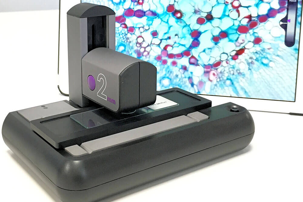
ioLight has invented a portable microscope, with a resolution of better than 1μm, which produces beautiful pictures of animal and plant cells and displays them directly onto your tablet or mobile phone.


 Ms.Cici
 8618319014500