
Listen and Pray Online with Guidance Light - guidance lights
10 uses of eyes
It is composed mainly of water and comprises about two thirds of the eye’s volume. The vitreous helps the eye maintain a round shape and is attached to the retina at various points, including the macula and the optic nerve.
PCE-LED 20 is a light meter with an external LED light sensor. The cable of the sensor has an extended length of approx. 1.5 m / 4.9 ft. Different work or production processes require different lighting conditions. In school, office and factory environments, lighting must also meet applicable safety standards. Indoor lighting conditions can be assessed quickly and easily using this meter. The LED light meter or lux meter can display measured values directly on the display. A direct measurement is thus immediately possible. By means of various presets, the functions installed in the device can also be activated or deactivated. These functions include the normal HOLD function, in which the measured value is frozen in the display. In addition, the extreme values as well as the average value can be frozen in the display. Further functions of the LED lux meter are the zero adjustment, the automatic shutdown and the automatic range selection.
Orderinwhichlightpasses throughthe eye
• Cornea: the transparent circular part of the front of the eyeball. It refracts the light entering the eye onto the lens, which then focuses it onto the retina. The cornea contains no blood vessels and is extremely sensitive to pain.
• Rod cells are one of the two types of light-sensitive cells in the retina of the eye. There are about 125 million rods, which are necessary for seeing in dim light.
• Conjunctiva: the thin, moist, clear membrane that covers the sclera – the white part of the eye. It is the skin that lines the eye socket and protects and lubricates the eyeball.
Eyereflection problems
• Iris: regulates the amount of light that enters your eye. It forms the coloured, visible part of your eye in front of the lens. Light enters through a central opening called the pupil.
Measurements are possible with the LED lux meter in lux or footcandle. The unit has only seven keys, allowing for one-hand operation. This also makes the instrument simple to use. The LED lux meter is a battery-powered handset, making the device portable and ideal for measurements in the field.
The cornea is the clear front window, representing one-sixth of the outer layer of your eye. The primary function of the cornea is to focus and transmit light onto the retina.
The ciliary body is located behind your iris, near the crystalline lens. This structure has two functions. The aqueous fluid that fills the front of your eye is made inside the ciliary body. Also, the ciliary body is made up of muscles that allow the eye to focus at different distances.
HowDoes lightenterthe eye
• Anterior chamber, between the cornea and iris • Posterior chamber, between the iris and the lens • Vitreous chamber, between the lens and the retina
• Lens: a transparent structure situated behind your pupil. It is enclosed in a thin transparent capsule and helps to refract incoming light and focus it onto the retina. A cataract is when the lens becomes cloudy, and a cataract operation involves the replacement of the cloudy lens with an artificial plastic lens.
• Sclera: the white part of the eye, a tough covering with which the cornea forms the external protective coat of the eye.
• Optic disc: the visible (when the eye is examined) portion of the optic nerve, also found on the retina. The optic disc identifies the start of the optic nerve where messages from cone and rod cells leave the eye via nerve fibres to the optic centre of the brain. This area is also known as the ‘blind spot’.
The sclera is the white portion of your eye, making up the back five-sixth’s of the eyes outer layer. The function of the sclera is to provide protection for your eye, and to serve as the attachment for the extra ocular muscles which move the eye.
The iris is the colored portion of your eye. Located behind the cornea, and in front of the crystalline lens. This structure separates the anterior and posterior chambers of the eye. The function of the iris is to help regulate the amount of light that enters your eye.
Our tears are formed by tiny glands that surround the eye. Tears are comprised of three layers: oil, water, and mucous. The lower mucous layer serves as an anchor for the tear film and helps it adhere to the cornea. The middle layer is comprised of water and the outer layer seals the tear film and prevents evaporation. The tear film serves several purposes. It keeps the eye moist, creates a smooth surface for light to pass through the eye, nourishes the front of the eye, and provides protection from injury and infection. When we blink, the eyelids smooth and spread the tear film so that it is uniform across the surface of the cornea. Excess tears flow out of the eye into two tiny ducts, which then drain into the nasal passage.
Howdoes the eyework simple explanation
• Pupil: the circular opening in the centre of the iris through which light passes into the lens of the eye. The iris controls widening and narrowing (dilation and constriction) of the pupil.
The anterior and posterior chambers are filled with aqueous humour, which is a watery fluid that provides nourishment to the interior eye structures and helps to keep the eyeball inflated. The vitreous chamber is filled with a thicker fluid called vitreous humour, a transparent gel which is 99% water, which helps the eyes to stay inflated.
Refraction oflight in the eyeoccurs at
• Zonules: a series of fine fibers that connect the crystalline lens to the ciliary body. The function of your zonules is to hold the lens in place in the eye.
The macula is located roughly in the center of the retina. It is a small and highly sensitive part of the retina responsible for detailed central vision. The fovea is the very center of the macula. The macula allows us to appreciate detail and perform tasks that require central vision such as reading or driving a car.
PCE-LED 20 LED Light MeterHandheld lux meter with automatic range selection, displays measuring units in lux or footcandle
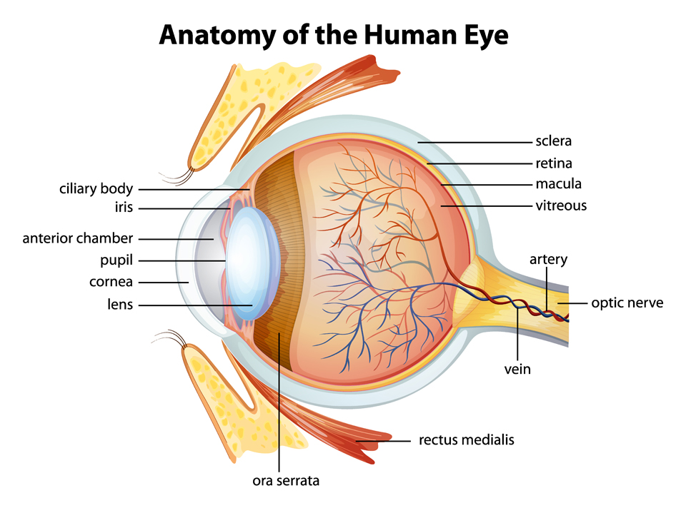
The optic nerve is located in the back of the eye. This structure is responsible for transmitting the images we see from the retina to the brain. The front surface of the optic nerve, which is visible on the retina, is called the optic disk. There are millions of nerve fibers that pass through the retina and converge to form the optic nerve. When light hits the retina it is converted into electrical impulses and carried along these fibers through the optic nerve and to the brain.
• Retina: a light sensitive layer that lines the interior of the eye. It is composed of light sensitive cells known as rods and cones. The human eye contains about 125 million rods, which are necessary for seeing in dim light. Cones, on the other hand, function best in bright light. There are between six and seven million cones in the eye and they are essential for receiving a sharp accurate image and for distinguishing colours. The retina works much in the same way as film in a camera. The retina’s function is to sense light and create impulses that are sent through the optic nerve and to the brain.
Lightreflectioneyeproblem
• Fovea: forms a small indentation at the centre of the macula and is the area with the greatest concentration of cone cells. When the eye is directed at an object, the part of the image that is focused on the fovea is the image most accurately registered by the brain.
How do we see with our eyes
The measuring sensor of the LED lux meter has a large exposure area. For convenience, the sensor is located on a coiled cable that can be extended to full length only when needed. A protective cap on the sensor prolongs its life. This protective cap should be placed on the sensor during storage. In addition, the protective cap is required when a zero adjustment is to be performed on the LED lux meter.
Just like a lens in a camera sends a message to produce a film; the lens in the eye refracts incoming light onto the retina, where messages are encoded. The retina, which is made up by millions of specialised cells known as ‘rods’ and ‘cones’, transforms the image into electrical energy and this is sent to the optic disk on the retina, where it will be transferred via electrical impulses along the optic nerve to be processed by the brain.
• Crystalline Lens: The transparent structure inside of the eye located directly behind your iris. The sole function of your lens is to focus light rays onto the retina.
• Tear Layer: Understanding the structure of tears is important in order to understand how the tears and tear film provide the eye with a number of specialized functions.
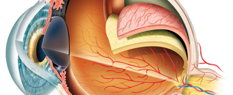
• Choroid: the middle layer of the eye between the retina and the sclera. It also contains a pigment that absorbs excess light so preventing blurring of vision.

The choroid is the spongy middle layer of your eye located between the sclera and the retina. Filled with blood vessels, the choroid’s function is to nourish the outer layers of the retina.
The pupil is the dark center in the middle of the iris. The pupil’s function is to regulate how much light enters the eye. The pupil’s size is automatically varied to regulate the amount of light entering the eye.
Vision occurs when light enters the eye through the pupil. With help from other important structures in the eye, like the iris and cornea, the appropriate amount of light is directed towards the lens.
• Cone cells are the second type of light sensitive cells in the retina of the eye. The human retina contains between six and seven million cones; they function best in bright light and are essential for acute vision (receiving a sharp accurate image). It is thought that there are three types of cones, each sensitive to the wavelength of a different primary colour – red, yellow or blue. Other colours are seen as combinations of these primary colours.
• The outer layer, formed by the cornea and sclera • The middle layer, holding the primary blood supply for the eye and containing the iris and pupil • The inner layer, comprised of the retina


 Ms.Cici
 8618319014500