
Linear LED Light Bars - lights for a bar
Even though the magnification of 1000x is achieved using 100x objective of 1.4 NA and 10x eyepiece, it does not provide image as sharp as that obtained with magnification of 400x using 40x objective of 0.65 NA and 10x eyepiece.
Convergent beam oflightdiagram
Electron microscope works on a principle similar to that of compound microscope only that electron beams replace light and electromagnets replace the optical lenses. A high voltage electron gun is used to produce a beam of electrons. Since air can scatter electrons, the chamber of EM is a vacuum. As electrons have poor penetrative power the specimen must be very thin.
Numerical Aperture:The numerical aperture of the lens is an important consideration in optics as it dictates the angle at which the light enters it. The light-gathering ability of a microscope objective is quantitatively expressed in terms of the numerical aperture. Higher values of numerical aperture allow increasingly oblique rays to enter the objective front lens, producing a more highly resolved image.
Electron beams are focused by means of a pair of condenser lenses on the specimen. The resulting image is produced by an objective lens, which is then projected by a pair of projector lenses. Since human eye is not sensitive to image formed by electron beams, the image is formed on a fluorescent screen.
Slender bacteria such as spirochetes (e.g. Treponema, Borrelia) are difficult to visualize under compound microscope using brightfield illumination. In order to visualize such bacteria, darkground microscopy is employed. Here, the microbe is made to appear brightly lit against a dark background. In order to achieve this, a special darkfield condenser is used that focuses a hollow cone of light on the specimen. An opaque disc is often placed in the center of the condenser to produce a hollow cone of light. The light rays are directed such a way that they donât enter the objective directly, hence the background is dark. Only those rays of light that are reflected by the organism enter the objective, hence they appear bright against a dark background. In more sophisticated darkfield condensers (such as paraboloid and cardioid), the occlusion of the direct light and the utilization of oblique rays are achieved by use of specially designed mirror surfaces instead of opaque disc. Darkfield microscopy requires a bright/intense source of illumination. Darkfield microscopy can also be used for observing bacterial motility.
Ensure that iris diaphragm is fully open and condenser fully risen. With 10x objective in place, look through the eyepiece and adjust the plane mirror such a way that you obtain perfectly bright and uniformly lit white background.
Convergent beam oflight
Thus, the deviated rays are slowed down again or out of phase by ¼ of wavelength. The undeviated and deviated light rays are now out of sync by ½ wavelength and when these rays meet, they result in destructive interference. The difference of ½ wavelength is sufficient for human eye to perceive, and the thick objects appear darker and the thinner objects appear lighter.
This article explores strategies to overcome sunlight interference and ensure clear laser projections.
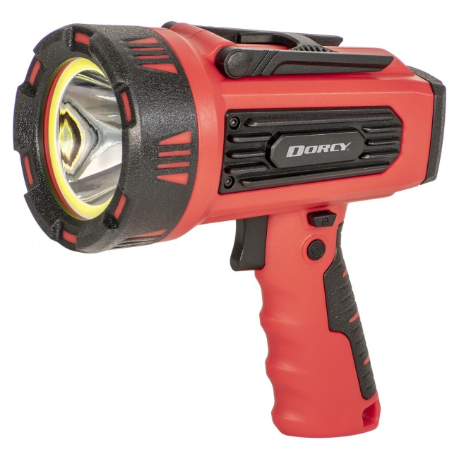
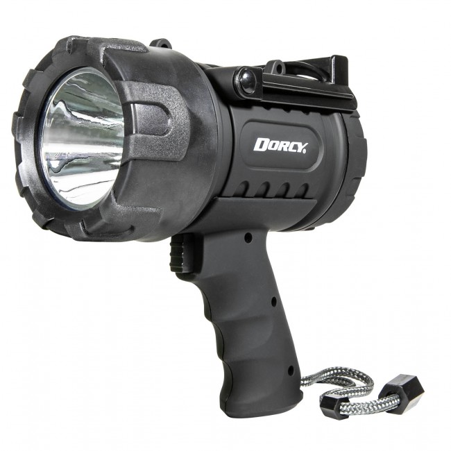
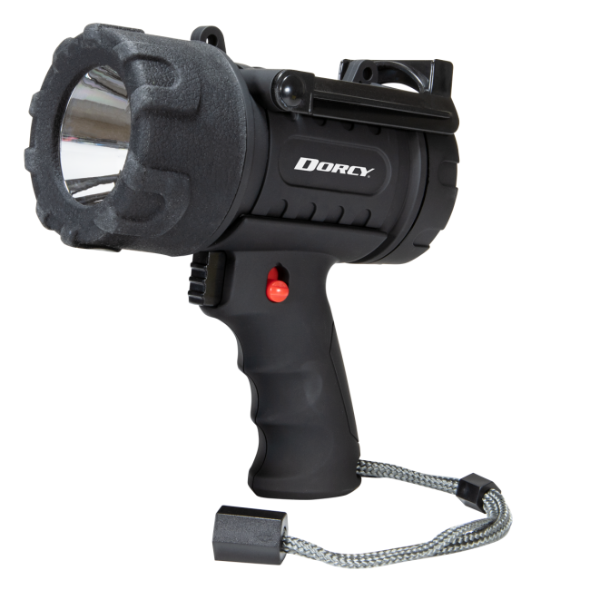
Despite the impressive brightness of Dorcy's LED spotlights, it is possible to use them and not have to continually purchase new batteries for it! Our rechargeable LED spotlights are a great choice for outdoor enthusiasts who are looking to save money on consumables. Further, the portability of our handheld LED spotlights does not equate to sacrifices in durability.
Most bacteria measure in the range of 0.5 to 4 µm (micrometer NOT microns). Mycoplasma and Coxiella are the shortest among bacteria and Spirochetes the longest. Viruses are ultramicroscopic structures; they canât be seen by compound microscopes. They can be visualized by electron microscopes and their measurements are in nm (nanometer). The à (Armstrong units) is the unit of measurement for still smaller particles.
Microscope is a delicate instrument that must be handled with care. All kinds of mechanical shocks must be avoided. The microscope must be lifted by holding its arm in one hard and supporting the base of the microscope with the palm of the other hand. The microscope must be kept in dust free environment. The oil must be wiped clean using a soft tissue paper. After usage the slide must be removed and cleaned before returning to its original place.
Refractive index of oil is 1.5 and that of air is 1.0. NA of dry objective: 1 x sin 90° = 1, but practically the highest practical numerical aperture of a dry lens is 0.95. NA of oil immersion objective: 1.5 x sin 90° = 1.5, however in practice only 1.4 is achieved for apochromatic objective and 1.3 for achromatic objective.
Magnification: This represents the number of times the image of a specimen is amplified. 10x means the size of the image is increased by ten times. The magnifying power of the lens is limited. After a certain point the magnification results in a blurred image and is termed empty magnification. Even with the best optics, 1400x is the highest useful magnification achieved. Magnifying power of an objective is determined by the dividing the optical tube length by the focal length of the lens. Optical tube length is the length of the microscope body tube between the nosepiece opening, where the objective is mounted, and the top edge of the observation tubes where the eyepieces are inserted. In most microscopes, it is fixed at 160mm.
Mar 21, 2016 — How to calculate light therapy dose · Power Density in mW/cm² (milliwatts per centimeter squared) · Time in s (seconds) · Dose in J/cm² (Joules per ...
Catalog · Floor Lamp (46) · Flush Mount (31) · Sconce (72) · Table Lamp (75).
Definedivergentbeam
Condenser: The parallel beam of light from natural or artificial source is condensed into a cone of light that illuminates the specimen or the object (smear on slide) by the sub-stage condenser. If the source of light is sunlight or indoor bulbs, the light is diverted to the condenser using the mirror. Concave mirror is used when low or high power objective lenses are used, whereas plane mirror is used when oil immersion objective lens is used. Mirrors are absent when in-built lamps are used. The condenser consists of series of lenses, which focuses the light to the object placed on the stage. Kohler illumination ensures that the diffuse light of uniform brightness is available without the view of the source of light. Various types of condenser in use are Abbe, Paraboloid and Cardoid condensers. The Abbe condenser (which is named after its inventor Ernst Karl Abbe) is the simplest of condensers that contains two lenses. The condenser can be lowered or raised according to the requirements. The condenser is lowered while using low power dry objectives, and raised while using oil immersion objectives. The amount of light passing on to the specimen from the condenser is regulated by using iris diaphragm. Light is reduced by closing the diaphragm partially for use with dry objectives. Oil immersion objectives require maximum light and this is achieved by keeping the iris diaphragm fully open.
The basic structure of a laser diode is a PN junction diode with a double-hetero-structure, similar to a light-emitting diode (LED), but the following three ...
Limit of resolution or resolving power: In simple words, it is the ability to see two closely placed dots as two separate dots. If the distance between the two points is lessened, it would appear as a single point. It is expressed quantitatively as limit of resolution. The resolution of human unaided eye is 200 µm. This means that human eye can not see objects small than 200 µm. The resolving power of compound microscope is 0.2 µm and that of electron microscope is 1-10 nm. The limit of resolution depends on the wavelength of the light used. Resolution increases with the decreasing wavelength of light. Violet colour light offers more resolution that red coloured light. Electron beams, which have very low wavelength offers maximum resolution. It is calculated by using formula:
Focusing the slide: Before viewing the slide under microscope, it is important to obtain a bright background. For oil immersion objective usage, follow these steps:
Microorganisms are so called because they are so small that they can not be ordinarily seen using unaided eye. The optical instrument that magnifies the image of these organisms that enables us to view their morphological features is a microscope. Antony van Leeuwenhoek is often considered as the father of microscopy, although compound microscopes were actually invented much before. A compound microscope contains several sets of lenses that magnify the image at different levels. Typically, the image is magnified initially by the objective lens and then again by the eye piece before it reaches the eye.
Objective lens: The light passing through the object (specimen) enters the objective lens. Typically, most microscopes have four objective lenses mounted on a revolving nose piece. The dry objectives include scanner (5x), lower power (10x) and high power (40 or 45 x). The oil immersion objective (100x) offers maximum magnification.
Spotlights from Dorcy are a powerful, handheld way to project powerful illumination wherever you need it. Ideal for camping in the darkest of backwoods campsites, many rely on our collection of outdoor led spotlights for late night or early morning hunting/fishing excursions.
The image of a specimen is magnified approximately 1000 times using a compound microscope. With an inclined body, the magnification of the microscope may be improved by another 1.5 times. With the highest practically achievable NA of about 1.4, 1400x is the highest useful magnification that can be achieved.
There are two types of electron microscopes: transmission electron microscopes (TEM) and scanning electron microscopes (SEM).
Low power dry objective: 160/16 = 10x High power dry objective: 160/4 = 40x Oil immersion objective: 160/1.7 = 94x or approximately 100x
Parallel beam oflight
Various types of microscopes are: Simple (dissection) microscope Compound microscope Darkground microscope Phase contrast microscope Fluorescent microscope Interference microscope Electron microscope Atomic Force microscope Polarization microscope
It is defined by the following formula: Numerical Aperture (NA) = n à sin(θ)where n is the refractive index of the medium between the object and the objective θ is one-half the angular aperture (angle of aperture is the angle formed by the two most divergent rays of light which enter the objective, starting from the center of the object).
For example, if green light of wavelength 0.55 µm is used and oil immersion objective with NA 1.4 is used, the maximum resolution obtained is 0.24 µm
whathappens to thelight rayswhen they hit the specimen?
Shift to 100x objective and raise the stage till the oil on the slide touches the objective. This must be done looking sideways and not while looking through the eyepiece or else the slide will break and damage the objective lens.
JavaScript seems to be disabled in your browser. You must have JavaScript enabled in your browser to utilize the functionality of this website.
Light Transmission Rate · On average > 98.5% ; Weight · 14g (62mm) / 15g (67mm) / 18g (72mm) / 22g (77mm) ; Height · 5.5mm (when mounted to lens, the part outside ...
Principle: Phase contrast microscopy is suitable for unstained preparation. A specimen may contain thin and thick areas. Using an annular diaphragm (phase annulus), a hollow cone of light is focused on the specimen. When light travels through thin area, it passes undeviated but when it passes through a thick area, it gets deviated. Such a deviated light is said to have slowed down or out of phase by ¼ of wavelength.
Examples of convergent anddivergentbeam oflight
Since the specimen is kept in perfect vacuum, live specimens can't be viewed. The image generated is actually a shadow of the specimen and colour image is not produced.
Source of light: Brightfield microscope uses visible light for illumination; specimen appears against a bright background. In brightfield microscope, the source of light is sunlight or indoor bulbs, or inbuilt lamps. The inbuilt lamps are made up of low voltage tungsten filament lamp or more recently LED lamp.
Eyepiece: Depending on the type of microscope, the magnified image may travel straight or can be reflected or divided using prism towards the eye piece. Monocular microscopes have single eyepiece where binocular microscopes have two. The magnified image is again magnified in the eyepiece lens and an inverted image is formed on the retina of the viewerâs eye. Older Huyghenian eye pieces were used with achromatic objective lenses but the newer apochromatic objectives require compensating eye pieces. These eye pieces correct the lateral colour errors of their objectives. In a compound microscope, the image is magnified twice; first by the objective lens and then by the eye piece. The eye piece consists of two planoconvex lenses with a circular diaphragm between them. Their magnification can vary form 5x to 10x.
Shop for Trans Globe Lighting Outdoor Wall Lights | Black in Outdoor Lighting Fixtures at Walmart and save.
Simply connect Lily smart outdoor spotlights into any standard wall socket to get millions of shades of white or color light in your yard.
Divergentbeam oflightexample
Resolution of a magnifying system can be improved by decreasing the wavelength of light. Since the wavelength of light is limited, electron beams of wavelength can be used to magnify images up to 1,00,000 times. Depending on the type of electron microscope a resolution of 1-10 nm can be achieved.
Definition: This is the capacity of the objective to render the outline of the image clear and distinct. Definition of an image is disturbed by spherical or chromatic aberrations. The central part of the image is usually well focused but the edges may suffer some aberration, which are of two types; spherical or chromatic. In spherical aberration, the periphery of the image appears out of focus. This happens because all the light passing through the lens doesnât condense at the same point. In chromatic aberration, the light is split into different colours at the peripheral part of the image since the edges of the lens act like a prism. The aberrations can be corrected by using achromatic or apochromatic lenses.
The light rays that pass through a convex lens converge or are brought closer together. There are various uses of a convex lens like in a microscope, magnifying ...
Since the condenser is fully raised for use with oil immersion objectives, light focused on to the specimen are at acute angles. For this reason the objective lens must be as low as possible. Despite this, the light can pass away without entering the objective lens due to diffraction suffered by light when moving from one medium to another (glass to air). Hence, the place between the glass slide and oil immersion objective is filled with immersion oil having same refractive index as that of glass. The light rays leaving the glass do not deviate and enter straight into the objective lens.
5 examples of convergent beam oflight
Place a drop of oil on the stained smear and place it on the stage and center it such a way that the smear is above the source of light and below the objective lens.
ADD LIGHT TO DARK AREAS OF YOUR RIDE VLEDS universal bar lights are made of billet CNC'd aluminum with an exterior grade anodized finish.
Unstained preparation such as saline wet mount is often used to demonstrate living microorganisms and their motility. Visualization of their morphology and internal structures is difficult because they donât have colour of their own and contrast poorly with the background. Phase contrast microscope is used to create contrast between the organism, its structures and the background, thus making its visibility quite clear.
Light, colour, vision & how to see more. Resources for students, educators and researchers of all ages.
This difference is too little for human eye to appreciate; hence the difference is magnified by letting the light rays to pass through a phase plate. A phase plate is placed at the back focal plane of the objective lens. Undeviated rays pass through thin area of the phase plate whereas the deviated rays pass through thick area of the phase plate.


 Ms.Cici
 8618319014500