
Is it possible to convert unpolarized light into linearly ... - polarized and unpolarized light
Light microscopes are used for quality control applications and detailed examination of newly developed materials, electronic devices, metals, and chemicals. Our light microscopes have been designed with modularity in mind—use our optical and digital imaging components to customize your system to attain the level of performance required by today's discerning microscopists. Evident compound light microscopes are used for a variety of applications, from routine inspection to sophisticated analysis. Our light microscopes can be combined with Evident image analysis software to build imaging inspection systems with exceptional optical performance. From basic image capture to image processing, measurement, and report generation, the Evident range sets the standard in light microscopes.
MicroscopeLEDlight
Terms Of Use | Privacy Notice | Cookies | Cookie Settings | About Us | Careers | Careers | Sitemap
"Decollimation" is any mechanism or process which causes a beam with the minimum possible ray divergence to diverge or converge from parallelism. Decollimation may be deliberate for systems reasons, or may be caused by many factors, such as refractive index inhomogeneities, occlusions, scattering, deflection, diffraction, reflection, and refraction. Decollimation must be accounted for to fully treat many systems such as radio, radar, sonar, and optical communications.
Microscope lightprice
Terms Of Use | Privacy Notice | Cookies | Cookie Settings | About Us | Imprint | Careers | Careers | Sitemap
This blog post shares what a digital microscope is, explores how it works, discusses the benefits of using one, and provides some applications.
This video looks at the steel industry and how microscopes in production laboratories can assist with inspections and analysis to ensure that materials will hold up under extreme circumstances.
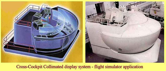
This principle is used in Full Flight Simulators (FFS), that have specially designed systems for displaying imagery of the Outside World (OTW) to the pilots in the replica aircraft cabin. In aircraft where two pilots are seated side by side, if the OTW imagery were projected in front of the pilots on a screen, one pilot would see the correct view but the other would see a distorted view where some objects in the scene would be at incorrect angles. To avoid this, collimated optics are used in the simulator visual display system so that the OTW scene is seen by both pilots at a distant focus rather than at the focal length of a projection screen. This is achieved through an optical system which allows the imagery to be seen by the pilots in a mirror which has a vertical curvature, the curvature enabling the image to be seen at a distant focus by both pilots, who then see essentially the same OTW scene without any distortions. This is shown diagramatically in more detail in the entry on Full Flight Simulators.
GooseneckMicroscope light
Electron microscopes work in a different way than light microscopes: rather than magnifying an image of a sample through light, they do so through a beam of electrons.
Laser light from gas or crystal lasers is naturally collimated because it is formed in an optical cavity between two mirrors, in addition to being coherent. The divergence of high-quality laser beams is commonly less than 1 milliradian, and can be much less for large-diameter beams. It should be noted that diode lasers do not naturally emit collimated light, and therefore collimation into a beam requires a collimating lens
GX series inverted metallurgical microscopes are reliable and high-performance imaging system with our advanced UIS2 optics. GX microscopes are highly efficient when used with our PRECiV image analysis software.
Microscopes are an essential tool for science, with a variety of uses. Light microscopes can be used across a range of disciplines—including medicine, mineralogy, microbiology, and material science—for analysis, inspection, and quality control.
A compound light microscope has two lens systems, the objective lens and the eyepieces, which work together with light to magnify a sample or specimen. A light microscope gathers light from the small area in which a sample sits on the stage. When this light is passed through the sample, it creates an image. This image is then sent up through the microscope’s objective lens and through the eyepieces in order to magnify the sample as the image reaches the user’s eyes.
Microscope lightfunction
Microscoperinglight
The word "collimate" comes from the Latin verb collimare, which originated in a misreading of collineare, "to direct in a straight line".
Microscope lightsource definition
Our modular microscope range offers optical modules that can be integrated into sophisticated inspection systems to optimize performance. Select from our flexible line up of options—from manual to motorized components.
BX series upright metallurgical microscopes meet a wide variety of analysis applications, from routine inspections to sophisticated studies, thanks to superb optical performance and a range of flexible options.
Collimated light is light whose rays are nearly parallel, and therefore will spread slowly as it propagates. The word is related to "collinear" and implies light that does not disperse with distance (ideally), or that will disperse minimally (in reality). A perfectly collimated beam with no divergence cannot be created due to diffraction, but light can be approximately collimated by a number of processes, for instance by means of a collimator. Collimated light is sometimes said to be focused at infinity. Thus as the distance from a point source increases, the spherical wavefronts become flatter and closer to plane waves, which are perfectly collimated.
A perfect parabolic mirror will bring parallel rays to a focus at a single point. Conversely, a point source at the focus of a parabolic mirror will produce a beam of collimated light. Since the source needs to be small, such an optical system cannot produce much optical power. Spherical mirrors are easier to make than parabolic mirrors and they are often used to produce approximately collimated light. Many types of lenses can also produce collimated light from point-like sources.
MicroscopeIlluminator
"Collimation" refers to the process of tweaking an optical instrument for the best possible image quality. With regards to a telescope the term refers to the fact that the optical axes of each optical component should all be centered and parallel, so that collimated light emerges from the eyepiece. Most amateur reflector telescopes need to be re-collimated every few years to maintain optimum performance. Collimation can be done simply via inspection by looking down the drawtube with no eyepiece to make sure the components are lined up, or with the assistance of a simple laser collimator or autocollimator. Collimation can also be tested using a shearing interferometer, which is often used to test laser collimation.

Text from Wikipedia is available under the Creative Commons Attribution/Share-Alike License; additional terms may apply.
LEDMicroscope lightsource
The light from stars (other than the Sun) can be considered collimated for almost any purpose, because they are so far away and have almost no angular size.
A light microscope can also be called a compound light microscope, or simply compound microscope. Compound microscopes are so named because they are designed with a compound lens system.

A light microscope is a microscope that uses lenses and focused light in order to magnify a sample. While a simple light microscope uses a single lens to create an image, a compound light microscope utilizes two lenses—the objective lens and the eyepieces. This provides a much higher magnification of a sample.


 Ms.Cici
 8618319014500