
Different Types of Landscape Lighting - silhouetting lighting
Light sheet microscopyvs confocal
Laser scanning confocal microscopy is a powerful imaging technique that is particularly well-suited for capturing high-resolution images of smaller samples. This technique is compatible with high-NA objectives, which are highly effective at capturing emitted light at the focal plane, resulting in high image resolution.
Light sheet fluorescence microscopycost
This technique enables the camera to capture a fully illuminated plane in a single exposure, allowing for high-speed scanning of the sample through the illumination beam. As a result, static light sheet generation achieves exceptionally high frame rates for volumetric imaging.
The Neo series spectrometer can cover almost any wavelength range in the 250 – 1100 nm range and standard versions, such as VIS and UV-NIR are available. There are various gratings available for use with the Neo series to suit many wavelength ranges.
A conventional laser scanning confocal microscope relies on galvanometric mirrors to raster-scan the illumination beam across the field of view (FOV). This process illuminates each pixel individually in sequence, with the photodetector collecting light from each pixel to assemble the final image.
(Lux is the metric unit for illuminance, measured in lumens per square meter. To convert footcandles to lux, multiply footcandles by 10.76). Lumen: A unit of ...

Light sheet microscopy
In recent years new display technologies have emerged. OLED displays, TFT’s with quantum dot technology or laser projectors offer brighter and more saturated colors than possible before. To achieve consistent colors across different displays, colorimetric calibration of the displays is essential.
Once the light hits the diffraction grating each wavelength of light is reflected under a different angle (similar to a prism). Different diffraction gratings can be used to identify different wavelength ranges. As also this beam is divergent because of the grating’s behavior, a second concave mirror is used to focus light rays of each wavelength towards specific pixels of the detector. At this point, the actual proportions of each individual wavelength are converted into electrons, which are digitized and then output for the operator to read.
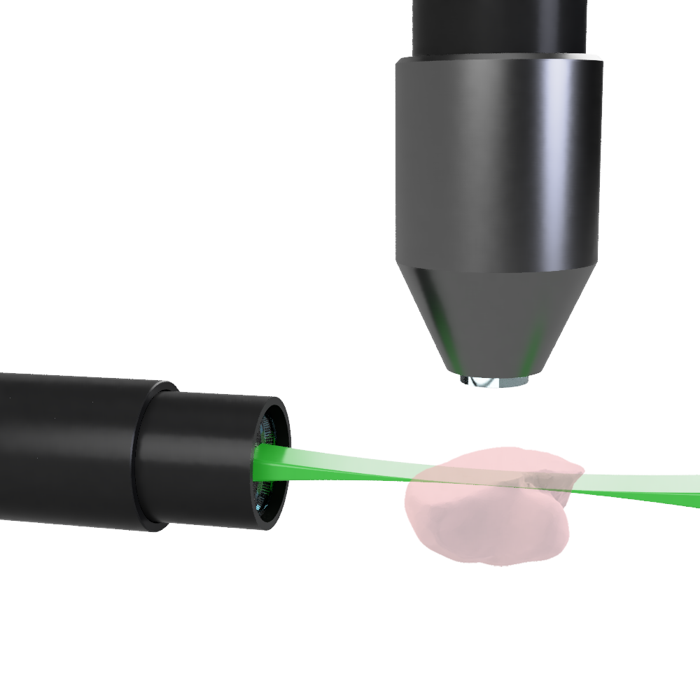
A typical light sheet fluorescence microscope has illumination and detection objectives placed orthogonally. The centers of each focal plane are coincident such that the illumination sheet of light is focused under the detection objective at its focal plane in order to illuminate the z plane at the FOV.
Light sheet fluorescence microscopyPDF
Light sheet fluorescence microscopyuses
"[LSFM] allows for optical sectioning and volumetric imaging with reduced photobleaching and low to zero background from other z planes."
How doeslight sheet microscopywork
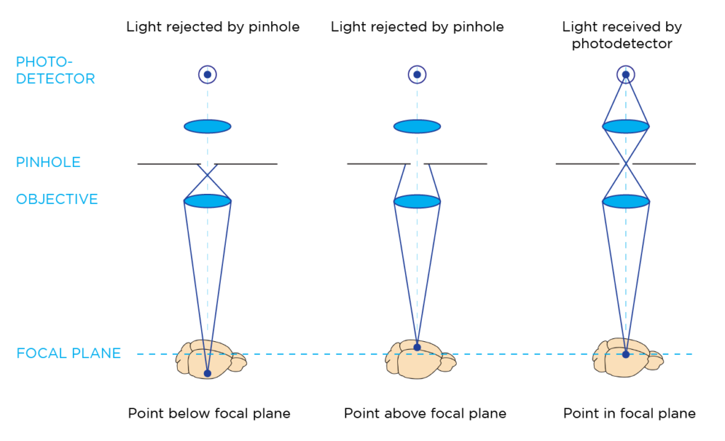
Laser scanning confocal microscopy enables the visualization of biological specimens by using focused illumination and detection optics that converge onto a diffraction-limited spot in the sample. This approach typically employs a single objective lens that serves for both illumination and detection in a confocal setup. A confocal pinhole selectively blocks out-of-focus light, ensuring that the photodetector (usually a photomultiplier tube) only captures fluorescent signals from the focal point, resulting in optical sectioning for 3D imaging.
While increasing the illumination intensity can decrease exposure time and increase the imaging speed, it also leads to a process called photobleaching, which quickly degrades the fluorescent probe signal. Although the pinhole in laser scanning confocal microscopy effectively blocks out-of-focus light from detection to achieve optical sectioning, tissue above and below the focal plane still receives a significant amount of illumination light. This can lead to photobleaching of the sample outside the focal plane, compromising the imaging quality of cleared organs.
How do we ensure that a toy's glowing lights are as safe as they are fun? The answer lies in the power of the spectroradiometer! This case study describes how optical safety testing of toys were improved in efficiency and accuracy of the testing process using the high-end Neo spectrometer.
Picked For You. Sponsored | Top selling items from highly rated sellers with free shipping. Ring Light illuminator 60-LED Adjustable Lamp For STEREO ZOOM ...
Mar 30, 2017 — Light Meters are a useful accessory when using film cameras. They can measure the amount of light and determine the most beneficial shutter ...
Fluorescence microscopy plays an important role in biological studies, allowing scientists to capture detail with high cellular imaging resolution and labeling specificity. However, in its simplest embodiment (epifluorescence microscopy), this technique lacks the ability to optically section samples for true 3D imaging, as it receives both in-focus and out-of-focus signals. To acquire 3D image datasets with optical sectioning, light sheet fluorescence microscopy (LSFM) and laser scanning confocal microscopy (LSCM) are commonly employed. These imaging methods vary in how they acquire fluorescent signals throughout the samples, leading to significant differences in imaging speed and the resulting data quality.
Light sheetmicroscope price
With high-quality fiber optics and a powerful light source, our kit delivers a realistic starry sky that adds a touch of elegance and relaxation to any room.
The Neo series spectrometer is simple to use and provides accurate measurements in a robust instrument. It benefits from high dynamic range and low noise, due to the high-end cooled CCD detector and complete configurability in terms of spectral range and resolution.
... light masterpiece. Special lights are housed in the Illumination Tower, next to Queen Victoria Place; on the roof of Table Rock Centre, at the brink of the ...
Light sheet fluorescence microscopyprice
The Prometheus series offers spectrometers with an integrated viewfinder, especially suited for display measurements, but also useful for many other applications.
A spectrometer is a scientific instrument used to analyze the light properties of a luminous object or reflected light. The instrument measures these properties of light over a specific section of the electromagnetic spectrum.
A light diffuser film is a translucent material that helps soften or scatter light. It is used to evenly diffuse a light source for purposes of photography, ...
Light sheet fluorescence microscopy employs planar illumination to illuminate the entire FOV at once. By effectively decoupling the paths of illumination and detection with separate light paths, only one z plane of the sample is illuminated at a time. This allows for optical sectioning and volumetric imaging with reduced photobleaching and low to zero background from other z planes.
All of the components mentioned above can be configured in different ways to obtain different results. For example, the entrance slit can be altered to allow more or less light enter the spectrometer, and the diffraction grating comes in a variety of types to measure different wavelength ranges.
2013326 — A beam of light of wavelength 606 nm passes through two slabs of material of identical thickness d= 1.40 micrometers, as shown in the figure.
Laser scanning confocal microscopy can offer high-resolution imaging for many applications, but slow imaging speed and the potential for photobleaching can limit feasibility in larger samples. LSCM typically acquires images in a point-scan manner, which can take several seconds for an entire FOV. When imaging whole cleared organs in 3D on the millimeter or centimeter scale, this approach becomes impractical as the imaging time scales up to weeks or even months.
The traditional approach to creating a light sheet is referred to as the static light sheet method. This method utilizes a cylindrical lens to focus the illumination light in one dimension and create a planar illumination.
Light sheet fluorescence microscopy is particularly effective for high-throughput volumetric imaging, with lower potential for photobleaching. Laser scanning confocal microscopy is typically two to three orders of magnitude slower than LSFM, and photobleaching can result from illuminating the entire tissue thickness to acquire only one section of the z-plane.
The entrance slit within the spectrometer is important as it determines the amount of light that is able to enter the instrument to be measured. This does not only affect the speed of the spectrometer engine (more light could typically result in a faster instrument) but also the optical resolution expressed in full width at half maximum (FWHM): the smaller the slit size, the better its resolution.
Commercial LED Lighting & Fixtures - LED Light Experts. NEED HELP? 800 ... Landscape Lighting such as Landscape Spot Lights. Can be solar powered spot ...
2024717 — It's the concept of a continuous, uniform beam of light. If the light is not monochromatic, it is by definition not coherent. But being ...
Light passing through a narrow opening like a slit has the natural behavior to become divergent. By reflecting the divergent beam onto a concave mirror, the light beam becomes collimating: all rays of light are directed parallel towards the diffraction grating. The grating is used as a dispersive element to disperse the wavelengths of light. Properties of the grating do not only include its dispersion range but also influence the optical resolution by the number of grooves. A second parameter of the grating, its blaze wavelength, determines the optimal efficiency at various wavelengths.
M Plöschner · 75 — Light-sheet fluorescence microscopy has emerged as a powerful platform for 3-D volumetric imaging in the life sciences.
The SmartSPIM and MegaSPIM light sheet microscopes are uniquely optimized for cleared tissue imaging. With patented axial sweeping technology, both SmartSPIM and MegaSPIM generate uniform axial resolution across the entire FOV, offering industry-leading image quality and acquisition speed for a wide range of research applications. With our systems, you can easily capture high-quality images of a mouse brain hemisphere or similarly sized intact sample in just 30 minutes. Reach out to an imaging specialist today to learn more!


 Ms.Cici
 8618319014500