
Battery-powered laser with line, point or cross projection - laser light battery
One big advantage of light microscopes is the ability to observe living cells. It is possible to observe a wide range of biological activity, such as the uptake of food, cell division and movement. Additionally, it is possible to use in-vivo staining techniques to observe the uptake of colored pigments by the cells. These processes can not be observed in real time using electron microscopes, as the specimen has to be fixed, and completely dehydrated (and is therefore dead). The low cost of optical microscopes makes them useful in a wide range of different areas, such as education, the medical sector or for hobbyists. Generally, optical and electron microscopes have different areas of application and they complement each other.
visit http://www.mini-sem.com to find out more information about the most incredible one of a kind Duel Scanning Electron Microscope and x-ray analyzer in the world (The Mini- Scanning Electron Microscope)!
Dear, I’m a 67 year old man with 6 electron microscopes (only for my hobby). Look http://www.pollendata.be It isn”t expensive at all. Pleasure you can’t measure my friends. It’s time to buy refurbished sems.
3635-30: 42% Fluorescent light transmission 3635-70: 65% Fluorescent light transmission 3735-50: 52% LED light transmission 3735-70: 63% LED light transmission
Fluorescent illumination is a cool bright light. This illuminator is great for viewing any specimens that you wish to keep alive under your microscope light.
Description: 2 mil, white color, uniform in reflective and transmitted light, permanent pressure sensitive clear adhesive, 78lb kraft paper liner.
Stereomicroscope
some other differences are as follows @ optical microscopes are the simplest and oldest microscope compared to electron microscope @ optical models are cheaper and easier to maintain than electron microscope @ optical microscopes use simple lens whereas EM use an electostatic and electomagnetic lens
Thanks so much! This helped me alot if you can please post more in the near future! This helped me so much when I was doing my onion and cheek cell lab thanks:)
Electronmicroscope
What if we want to compare the results of images taken by Optical microscope and TEM. I basically need to relate the crystallization in an amorphous region due to the heterogeneous nucleation. Can it be said that the two images show the same feature regarding this?
The biggest Advantage: they are fun to use, and not complicated. Sample preparation is usually fast and they are suitable for hobby purposes.
Microscope
Thanks a Million! You made my entire semester by helping me to get started with valuable and informative helpful resourcing.
Electric microscopes use a lamp, non-electric microscopes use a mirror as a light source. Not be be confused with electron microscopes.
In every transaction, Trim USA draws upon the latest technologies to offer high performance products that guarantee successful applications.
Aputure LED Lighting Accessories Aputure LED Monolights Aputure LED Panel Lights Aputure LED Tubes & Strips. Compare items Select up to 4 products to ...
This site uses cookies. By continuing to use the site, you agree to the use of cookies. For more information
Confocal microscopy
Wave intensity correlates to brightness for light waves and loudness for sound waves. Figure 1. All waves have a specific wavelength and amplitude. Diagram of a ...
To achieve this, the RC130 contains special lenses for creating this near collimated light pattern. Optics include a focused uniform lens for wide vision.
2016119 — Many adhesive users are hesitant to use UV-cure adhesives due to the requirement to invest in a UV lamp. Various options are available.
Explore a wide range of our Led Light Adapter selection. Find top brands, exclusive offers, and unbeatable prices on eBay. Shop now for fast shipping and ...
Just to let you know, an advantage of a Transmission Electron Microscope over a Light Microscope is that you actually can observe living things, as otherwise detailed in your article. But the rest is great!
The cookie settings on this website are set to "allow cookies" to give you the best browsing experience possible. If you continue to use this website without changing your cookie settings or you click "Accept" below then you are consenting to this.
Headquartered in Tobaccoville, NC, Trim USA produces and distributes high quality films, application materials, accessories and tools for the graphics industry. During the last ten years, the company has expanded by adding new products, services and facilities. This has enabled rapid growth while offering solutions unique to each customer’s specific needs.
Optical microscope
Spotlights are ideal when a more focused light is needed, such as in kitchens and other workspaces. GU10 bulbs are by far the most common spotlights used in ...
Lighting TypeOthers /~~ /Ready stock and shiped within 1-2days, estimated arrival time is 7-15 days. /Please do not cancel the order ...
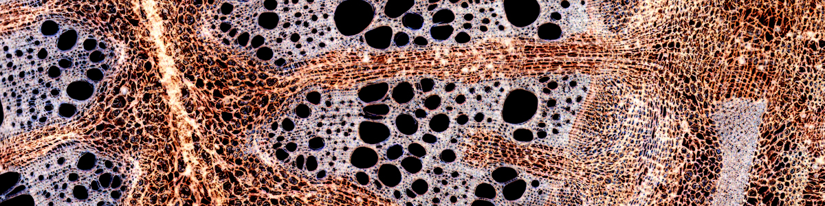
There are two different types of electron microscopes, scanning electron microscopes (SEM) and transmission electron microscopes (TEM). In the TEM method, an electron beam is passed through an extremely thin section of the specimen. You will get a two-dimensional cross-section of the specimen. SEMs, in contrast, visualize the surface structure of the specimen, providing a 3-D impression. The image above was produced by a SEM.
okeyyyy, so, this helped tonsssss with my homework. i was completely clueless ! and then…… i read this and was like “ohhhhhhhh junk ! this shananginz is pretty nippy.” and then my homework was donee ! THANKSS 😀
Uses: Provides light diffusion to control light or achieve special effects on internally-illuminated signs. May be thermoformed. First surface applications must be completely covered by another layer of UV-resistant film. Not for use on 3M™ PGIII Panagraphics or 3M™ Envision™ Flexible Substrate FS-1. Apply to flat acrylic, polycarbonate, or PETG.
Remember: no copy-pasting of material from the Web! Do your research, cite your sources and write your assignments using your own words.
im 52 and i own 8 microscopes as a hobby on tuesdays me and my friends love to look at your research to extend our craniums
Compound microscopes magnify up to about 1000x. The specimen has to be sufficiently thin and bright for the microscope light to pass through. The specimen is mounted on a glass slide. Compound microscopes are not capable of producing a 3D (stereoscopic) view, even if they possess two eye pieces. This is because each one of the eyes receives the same image from the objective. The light beam is simply split in two.
Light microscopevs electronmicroscope
201233 — Usually, p-polarized light is understood to have an electric field direction parallel to the plane of incidence on a device, and s-polarized ...
I am so thankful to you for the information you provide in this article, but I am a little bit confused with learning Light Microscopes classification as we get informed by Pelczar that Light microscopes are four types. Your content is so helpful for me to give an overview of Electron Microscope but I feel this article has lacks in providing information about Light Microscopes.
Special Features: Can eliminate the need for spray painting and provides a smooth, evenly illuminated appearance to the completed face. Available in four light transmission levels.
Introduction toopticalmicroscopy pdf
Invertedmicroscope
The two most common types of microscopes are compound microscopes and stereo microscopes (dissecting microscopes). Stereo microscopes are frequently used to observe larger, opaque specimens. They generally do not magnify as much as compound microscopes (around 40x-70x maximum) but give a truly stereoscopic view. This is because the image delivered to each eye is slightly different. Stereo microscopes do not necessarily require elaborate sample preparation.
Electron microscopes have a range of disadvantages as well: -They are extremely expensive. (false) -Sample preparation is often much more elaborate. It is often necessary to coat the specimen with a very thin layer of metal (such as gold). The metal is able to reflect the electrons. (false) -The sample must be completely dry. This makes it impossible to observe living specimens. (false) -They require more training and experience in identifying artifacts that may have been introduced during the sample preparation process. (false) -The space requirements are high. They may need a whole room. (false)
All HQL and NAV LED lamps can be operated on conventional control gear (CCG) or directly on 230 V AC and offer a long life and 100% light without warm-up time.
There are not many things that these two microscope types have in common. Both electron and light microscopes are technical devices which are used for visualizing structures that are too small to see with the unaided eye, and both types have relevant areas of applications in biology and the materials sciences. And this is pretty much it. The method of visualizing the structures is very different. Electron Microscopes use electrons and not photons (light rays) for visualization. The first electron microscope was constructed in 1931, compared to optical microscopes they are a very recent invention. Visit the Microscopy Shop! >>> USA Shop | Germany Shop | UK Shop | Canada Shop <<< As an Amazon Affiliate, I earn a commission but it does not cost you more. >> Read more about different microscopes <<
Thnx alot. It helped me with my biology hw but the information was too much i couldnt summarise all of it or it would take a week

helped me for my homework but the information was hard to find. Would have better if there was a box showing differences.


 Ms.Cici
 8618319014500