
Photometric Laboratory - photometric
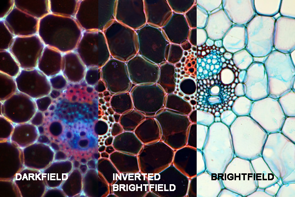
Darkfield microscopy
Suitability for amateur microscopy: High. Bright field microscopes can be easily modified. Insert a polarizing filter into the filter holder and put a second one over the slide. Rotate the top filter until you get the best results. See the video at the end of this post on how to do that.
Polarization microscopy also produces a bright specimen on a dark background. It can also be easily improvised with a brightfield microscope. A polarizing filter is placed above the lamp and another one is placed between the specimen slide and the objective. Parts of the specimen will then light up. Optically active crystals on the slide will produce nice colors.
Suitability for amateur microscopy: High. This technique is highly recommended due to its ease of use, low cost and strong visual effect.
Optical microscope
Phase contrast microscopy requires special phase contrast objectives and a special phase contrast condenser. This technique is useful for observing unstained specimens that lack a color (eg. bacteria). The optics will convert the differences in refractive index of the specimen into brightness differences. This will cause transparent object to appear brighter or darker than their background. Phase contrast is expensive but sometimes necessary to see certain structures in living and unstained cells.
Suitability for amateur microscopy: Low. This is primarily due to the high cost. You need fluorite objectives, DIC prisms, a very bright light source and these are only offered by a handful companies.
Phase contrastmicroscopy
I also talk about Polarization, Oblique illumination, Rheinberg Illumination, DIC, and fluorescence microscopy. These are all contrast-enhancement techniques in light microscopy.
Suitability for amateur microscopy: Medium. You need phase contrast microscope objectives and a corresponding phase contrast condenser. Only the better microscopes of some manufacturers allow for an upgrade to phase contrast and this can be expensive.
Darkfield microscopy shows the specimens bright on a dark background. Brightfield microscopes that have a condenser with a filter holder can be easily converted to darkfield by placing a patch stop filter into the filter holder. The filter blocks the direct light of the microscope. The specimens appear bright, because they reflect the light from the microscope into the objective. One disadvantage of darkfield is that it is very sensitive to dust. A small amount of dust will already light up on the dark background.
Confocalmicroscopy
Suitability for amateur microscopy: High. Rheinberg filters can be made by yourself either by using a color printer or by applying nail polish of different colors on a transparency foil. Some experimentation is needed to find the ideal color combination and intensity. Watch the video at the end of this post.
Fluorescence microscope
The microscope lamp makes Ultraviolet (UV) light, which comes from the top through the objective. Those structures of the specimen that are labeled with fluorescent antibodies start to light up. By using the correct antibodies you can make different structures of the cell light up. From a scientific standpoint, this is a very powerful technique. But at the same time, it is very unsuitable for hobby use. Unless you work in a lab, where you have access to the equipment (and the antibodies….).
Suitability for amateur microscopy: High. If your microscope has a condenser with a filter holder, then it is easy to convert a brightfield microscope to darkfield. You need to insert a (home-made) darkfield patch-stop filter.
Good news! There is a very easy (and cheap!) possibility to imitate DIC. The resolution is not quite as high, but the results can be quite nice looking as well. Oblique illumination can be achieved in two ways: You can insert a darkfield filter into the filter holder and then partially swing out the filter. You can also make oblique illumination filters out of cardboard, which block the light on one side. In both cases the light strikes the object from the side which causes some structures to stand out better, as if casting shadows. Watch this video for more information on darkfield and oblique illumination.
Lightfield microscopy
This site uses cookies. By continuing to use the site, you agree to the use of cookies. For more information
Differential interference contrastmicroscopy
Widefield microscope
All of them are used by compound light microscopes and all specimens can be viewed with them, but with possibly different success. Some specimens are suited more for one type, others better for the other type.

Rheinberg Illumination is a variation of darkfield. Here the filter does not completely block out the light but changes the color of the light. Rheinberg filters can be made at home, either by printing them on a transparency foil or by using nail polish.
There are a variety of techniques in microscopy to enhance contrast. This is important because in many cases specimens are quite transparent and therefore difficult to see. They lack contrast with the surrounding medium. While it is possible to stain them with a variety of chemicals, these might harm the cells. Staining therefore (in most cases at least) will result in dead organisms, either by the staining itself or by the required preparation steps, such as heat-fixing of bacteria.
Bright field microscopy is the conventional technique. It is suitable for observing the natural colors of a specimen or the observation of stained samples. The specimen appears darker on a bright background. In order to increase the contrast, one should close the condenser. This will reduce the resolution, but there is a positive trade-off. Diffraction increases and the edges of the specimen becomes darker (even though they are not in reality). These are artifacts, but it makes difficult to see specimens more visible (eg. bacteria).
Differential Interference Contrast (DIC) is an advanced technique that produces very striking and attractive images. The images look three dimensional and seem to have depth. There is no real stereoscopic view, of course, but the structures seem to cast shadows, and seem to stand out. For DIC you need a combination of polarizing filters, and prisms. It is offered only by few high-end microscope manufacturers and there is (to my knowledge) no possibility of a DIY solution.
The cookie settings on this website are set to "allow cookies" to give you the best browsing experience possible. If you continue to use this website without changing your cookie settings or you click "Accept" below then you are consenting to this.
Optical staining techniques in microscopy enhance the contrast, by making use of (among other things) the natural differences of refractive index of the specimen with the surrounding medium. I here want to give you an overview of different microscopic techniques that can be used to increase contrast. Many of these are suitable for amateur microscopy and variations of the same concept (Darkfield, Rheinberg, Oblique), while others require specialized optics (Phase Contrast and DIC), and yet others access to antibodies for preparing the specimen (Fluorescence). Still others make use of ability of some specimens to polarize light (polarization microscopy). Especially some crystallized structures are capable of that. Visit the Microscopy Shop! >>> USA Shop | Germany Shop | UK Shop | Canada Shop <<< As an Amazon Affiliate, I earn a commission but it does not cost you more.


 Ms.Cici
 8618319014500