
M12A-17PFFC-SF8B15: M12 (A) Connector - m12 5 pin socket cable
Not all products or services are approved or offered in every market, and approved labelling and instructions may vary between countries. Please contact your local representative for further information.
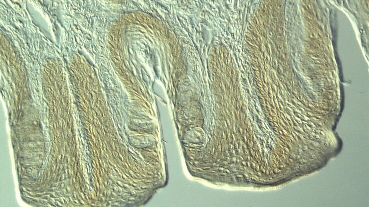
Most often biological specimens and tissues are observed with a phase contrast microscope. A large variety of biological specimens can be observed with phase contrast from fixed specimens to living cells and tissues. For examples, refer to the articles: Phase Contrast & Optical Contrast Methods
LED illuminationunit
Leica microscopes offer phase contrast for the study of cells or tissues concerning various life-science and forensic applications. Phase contrast can also be useful for certain material and earth-science applications.
Illuminationlight
GE Lighting, a Savant company products are only intended for use within the continental United States, Alaska, Hawaii and Canada.
Google Play and the Google Play logo are trademarks of Google Inc. Apple and the Apple logo are trademarks of Apple Inc., registered in the U.S. and other countries. App Store is a service mark of Apple Inc. The Bluetooth word mark is a registered trademark owned by Bluetooth SIG, Inc., and any use of such mark by GE is under license.
Portions of the ring-shaped light are diffracted by optically dense structures of the specimen and experience a negative phase shift of about λ/4. This phase-shifted, diffracted light bypasses the λ/4 plate. In contrast, the portion of the ring-shaped light that passes directly through the specimen non-deviated will hit the phase plate which causes a positive λ/4 phase shift. As the total difference in phase shift between the light diffracted by the specimen’s structures and that which passes through phase plate will be about λ/2, destructive interference will occur. Consequently, more optically dense structures will appear darker than those that are less optically dense.
Led illuminationmeaning
Leica microscopes providing phase contrast are commonly used in life science research for the visualization, analysis, and documentation of biological structures and cellular processes.
LED illuminationmicroscope
Let’s take a look at a 60-watt replacement incandescent bulb. The energy consumption to use a bulb like this would cost about $90 over the course of 10 years. For an LED, running over the course of 10 years, the actual cost would be only $18 to operate. Take a look at the table below for a breakdown.
LED uses up to 85% less energy than halogen and 18% less than CFL. In fact, with the energy you save from switching to one LED, you could watch 199 episodes of your favorite hour-long TV show.
Brightfield microscopy normally only provides a low-contrast image of many transparent biological specimens where few details are distinguished. One way to enhance contrast with brightfield microscopy is to use selective stains, but such stains are often toxic to living cells. A phase contrast light microscope offers a way to view the structures of many types of biological specimens in greater contrast without the need of stains. The contrast method exploits differences in optical density between structures of a specimen that lead to a phase shift of the light that interacts with the specimen and its structures.
Well, by upgrading one fixture to LED, you’ll save nearly $7 per month—a.k.a. one large cup of gourmet coffee. So imagine what you can save if you upgrade every fixture in your house. Check out our LED savings calculator below to find out.
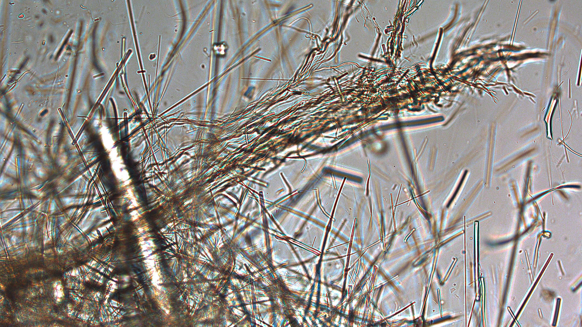
For forensic applications concerning the evidentiary investigation of paints, pigments, textiles, fibers, and human tissues, Leica microscopes offering phase contrast are very useful solutions.
LEDlight
The phase contrast method for microscopy was developed in the 1930s by the Dutch physicist Frits Zernike. After 1942, it became a widely used microscopy technique. In 1953, Zernike was awarded the Nobel Prize for Physics. For more details, refer to the articles: A Brief History of Light Microscopy – From the Medieval Reading Stone to Super-Resolution & Phase Contrast
The knowledge portal of Leica Microsystems offers scientific research and teaching material on the subjects of microscopy. The content is designed to support beginners, experienced practitioners and scientists alike in their everyday work and experiments.
With a high light output and a pleasing light quality, you can create a comfortable green space in any room of your house.
Compared to CFL, incandescent, and halogen bulbs, LEDs are longer lasting, more energy efficient, and reach full brightness instantly.
*Based on 3 hours use per day. Some LED and CFL replacement bulbs may provide less light output (lumens) than incandescent. See product pages for specifications.
Illumination LEDSony TV
We have LEDs available for every fixture and every space—from recessed LEDs for your ceiling to LED nightlights to LED bulbs for inside your microwave. More savings. More shopping.
A phase contrast microscope is similar to a conventional brightfield microscope, except it uses an annular aperture in front of the light source and a quarter-wave phase plate after the objective lens. For more information, refer to the article: Phase Contrast
*Based on 3 hours use per day. Some LED and CFL replacement bulbs may provide less light output (lumens) than incandescent. See product pages for specifications.
A phase contrast microscope is similar to a conventional widefield microscope, except it uses an aperture in the shape of an annulus and a quarter-wave (λ/4) phase plate. The annular aperture is placed between the light source and condenser lens and the phase plate after the objective inside the microscope optics. Ring-shaped light that passes through the aperture is focused by the condenser onto the biological specimen to be observed.
Let’s take a look at a 60-watt replacement incandescent bulb. The energy consumption to use a bulb like this would cost about $90 over the course of 10 years. For an LED, running over the course of 10 years, the actual cost would be only $18 to operate. Take a look at the table below for a breakdown.
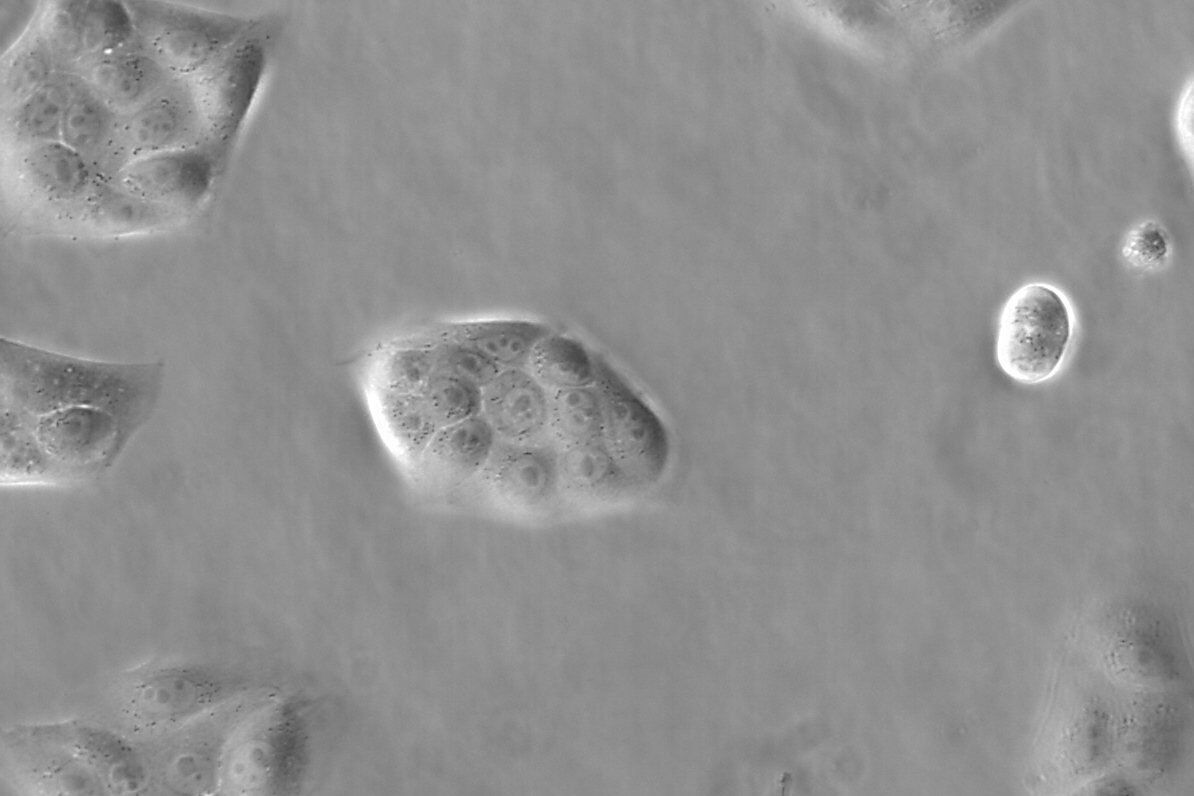
Phase contrast is an optical contrast technique for microscopy which makes unstained structures in the cells of biological specimens visible. Cell structures that appear transparent with brightfield illumination can be viewed in high contrast and rich detail using phase contrast. Differences in optical density between structures in the cell can cause light that interacts with them to attain a phase shift. This phenomenon is the basis of phase contrast. As a result, more optically dense structures will look darker than less optically dense ones.
It means not only do LEDs produce a higher quality light than incandescent and CFL, but they also reach full brightness instantly. No flicker. No warm-up. No reason not to smile.
As the new standard, LED Lighting adds energy efficient, long-lasting light to every space in your home—from desktop lamps to inside your microwave.
Leica microscopes capable of phase contrast make a difference for the study of transparent and colorless minerals, crystals, and polymers.


 Ms.Cici
 8618319014500