
GMK Blue Alert R2 - dcs lam alert
If not (c, d), the DF stop has to be centered. The microscope is now ready for dark field observation. Take care that the glass slide is perfectly clean on both sides. Each finger print, each dust particle will cause stray light and will destroy the aesthetic impression of the dark field image. This is why some people regard dark field as a “dust-detecting method”.
Dark fieldmicroscopy
Pull-out one eyepiece and insert a centering telescope (CT) instead. Loosen the fixing screw and focus the back focal plane of the objective by pulling out the inner part of the centering telescope. A bright disk of light that is partially obscured by the central stop will be visible at the objective rear focal plane. If not, use the centering screws and move the central stop so that the bright disk is completely covered.
Size can be scaled in increments of 100 mm in width and length up to a maximum size of 800 mm x 1000 mm. Choose from three standard colors - other LED wavelengths are available upon request.
MOTICEUROPE, S.L.U. usa cookies propias (necesarias) y de terceros para personalizar el contenido, ofrecer funciones de redes sociales y analizar el tráfico. Además, compartimos información sobre el uso que haga del sitio web con nuestros partners de redes sociales y análisis web, quienes pueden combinarla con otra información que les haya proporcionado o que hayan recopilado a partir del uso que haya hecho de sus servicios. Más información.
Now watch the image of the sample. In case the background is not perfectly dark, carefully raise/lower the condenser until the background becomes dark. Have a closer look on single particles within the sample. They all should have a symmetric brightness all around their periphery. If not, center the DF stop more precisely. The periphery of each single particle should display a symmetric brightness.
How such an image is achieved? We need to eliminate the direct light which ordinarily passes the specimen and which is responsible for the bright background in bright field illumination. For objectives with a Numerical Aperture ≤ 0.65 thiscan be done by inserting a simple “central stop” into the ray path at the height of the condenser. Higher Numerical Apertures require a more sophisticated condenser setup and special objectives with iris diaphragm.
Phase contrast
The Flex Backlight is an indirect, diffuse lighting. This produces a homogeneous, diffuse light that is particularly suitable for assessing and measuring outlines.
Lightfieldmicroscopy
White, red and infrared are usually delivered within 10 working days after receipt of order. For green, blue and yellow please contact our sales team for the delivery time.
MOTICEUROPE, S.L.U. uses own (necessaries) and third cookies on this website are used to personalise content, offer social media features, and analyse traffic. In addition, we share information about your use of our website with our social media, and web analytics partners, who can combine it with other information that you have provided to them or that they have collected from your use of their services. More information.
HAADF-STEM
Flex Series For special light dimensions, also take a look at our Flex series. With the Flex series, you can freely select the lengths and widths of the illuminated area in 100mm increments. You need special lighting with other dimensions or LED colors? No problem, please contact our sales department for an individual solution.
Insert the central stop into the optical path of the microscope. Depending on the microscope model, plug-in the DF attachment, push in the DF slider, or rotate the condenser disc to the respective DF position.
Fluorescence microscope
The data sheet contains an example of the suitable configuration via connection with the MBJ controller CTR-50 for continuous light operation.
Phase contrast microscopy
HandlingFirst prepare the respective microscope for optimal bright field use, best start with a 10X objective. If necessary, perform the KOEHLER setup (focus the sample, center the image of the field diaphragm, adjust the height of the condenser, open the field diaphragm). For dark field now open the condenser (= aperture) diaphragm completely. This allows the condenser to operate at its maximum numerical aperture.
Once done, the sample is illuminated only by light that passes through the slot around the central stop in the form of a hollow cone. Thanks to the front lens of the condenser, this light will miss the front lens of the objective. In case there is no sample placed on the x/y stage, the user will observe a dark field of view.
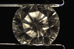
For transparent samples in light microscopy, dark field illumination is a simple and affordable contrast method. The idea of this technique is to display and to emphasize border structures, means abrupt changes of the refractive index within the sample. Especially for single-celled organism in fresh or sweet water environments, with a refractive index close to water, this contrast method gives aesthetic and informative images.
JavaScript seems to be disabled in your browser. For the best experience on our site, be sure to turn on Javascript in your browser.
Only the light which is influenced by sample structures will enter the objective: The result is a bright phenomenon on a dark background. But be careful: We are not talking about an inverted image like “white becomes black, black becomes white”. This would give no additional information, but just a different impression. The dark field emphasizes border structures and outlines. The images of a calibration slide display this focus on borders:
Please note that the product availability and prices in this website might vary depending on the country you are in. The products in your cart will be deleted by this action.
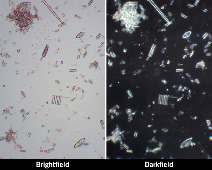
Dark field emphasizes borders and isolated, single structures, flagella of protozoa and tiny particles. The comparison of a diatom sample in bright field and dark field clearly shows this:
Each Flex light is individual and will be manufactured only for you. Reorders can be made easily via your own article number.
Dark fieldscattering spectroscopy
The central stop is a round piece of black metal, mounted into an attachment, a slider or the disc of a turret condenser. The stop has to be aligned so that it is exactly placed in the middle of the ray path. The procedure of alignment is quite similar to phase contrast, where the illumination rings also have to be positioned precisely.
This homogeneous backlight is ideal for inspecting and measuring outlines. The lateral coupling of the LED light ensures a very homogeneous radiation over the surface, which increases the quality of the test result. The MBJ backlight is characterized by its very compact format with a low height of only 12 mm.
If there is no centering telescope at hand, use a sheet of paper as a sample. While slowly raising and lowering the condenser, a bright spot will appear on the paper (a). Below or above this condenser position, a dark spot appears. This spot has to be in central position (b).


 Ms.Cici
 8618319014500