
Contrast Lighting: Definition & Media Effects | Vaia - contrast light
Indirect lighting uses the ceiling, walls or other surfaces as secondary reflectors. If it is sufficiently bright, general illumination with diffuse light is ...
Brightfield microscopy is one of the simplest and most widely used observation method in optical microscopy, generally used with compound microscopes. In brightfield microscopy, illumination light positioned below or above the sample and it is transmitted through the sample and the contrast is generated by the absorption of light in dense areas of the specimen. A typical brightfield illumination image show dark sample with white background. With a conventional bright field microscope, light from a bright source is aimed toward a lens beneath the stage called the condenser, through the specimen, through an objective lens, and to the eye through a second magnifying lens, the ocular or eyepiece.
Diffuse lampfor living room
Widefield exposes whole specimens to light. Brightfield allows you to illuminate the sample from the bottom with white light, and observe the sample from the top.
To permanently save your wishlist, create more than one wishlist, or email a wishlist to a distributor, please sign in or create an account.
Diffuse lampfor sale
ISO/CIE 10916:2024 Light and lighting — Energy performance of ... lighting, in collaboration with the International Commission on Illumination (CIE).
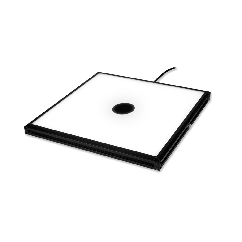
Machine vision dome lights provide diffuse lighting, also known as “cloudy-day illumination”. Reflected light provides non-directional, soft illumination that is free of shadowing. This effect is well suited for inspecting highly specular, and curved objects, but at close working distances. The light I.D. needs to be approximately 50% oversized compared with the object size to be illuminated.
LampShade Diffuser
We work closely with our vendors to provide high-quality LED lighting for machine vision applications. Visit our PRODUCTS section to discover an LED lighting solution for your vision application and choose "CONFIGURE THIS LIGHT" to customize a light to meet your needs.

Diffuse lampamazon
One of the advantages of brighfield microscopy is that not only stained but specimen without staining can also be viewed and the optics used in bright- field technique don’t alter the colour of the specimen. The limitations of brightfield microscopy include low contrast for weakly absorbing samples such as cellular or biological samples and low optical resolution due to the limitation of light's wavelength.
Diffused light for Photography
Traditional light sources tend to have a shorter lifespan the more they're switched on and off, whereas LEDs are unaffected by rapid cycling. In addition to ...
Phase contrast is by far the most frequently used method in biological light microscopy. It is an established microscopy technique in cell culture and live cell imaging. When using this inexpensive technique, living cells can be observed and analysed in their natural state without previous fixation or labelling. A typical phase contrast image has a neutral background and surrounding with varying contrast where light is altered by the specimen.
Find Light bar LED under cabinet lights at Lowe's today. Shop under cabinet lights and a variety of lighting & ceiling fans products online at Lowes.com.
Z Liu · 2014 · 132 — Brightfield, darkfield, and phase contrast are the most common label-free contrast modes used in optical microscopy. Brightfield imaging is most ...
JOBS. Lighthouse offers the opportunity to work on a fascinating range of investigations in partnership with some of the biggest media brands in Europe. We are ...
DIC is a polarization technique rendering contrast in transparent specimens. This method is a good alternative to bright field microscopy producing detailed images of thick unstained samples that often provide poorer images in brightfield. This method also creates pseudo-3D relief shading images making the technique ideal for electrophysiology experiments. The image appearance shows details about colour, optical path boundaries and refractive indices along with whether or not a specimen is isotropic and anisotropic.DIC uses polarized light and additional light-shearing prisms to convert phase delays into intensity changes (contrast). The effect is called differential, because contrast is created only in adjacent structures where differences in thickness and /or refractive indices is present.
Bestdiffuse lamp
Diffuse lampfor home
EDGE® LightEDGE® Light is the missing piece able to drastically improve the quality of your online meetings. Thanks to its built-in battery, you can light ...
Since unstained living cells absorb practically no light this results in extremely small differences in the intensity distribution in the image which are invisible to the human eye. However, using a special adapter (phase plate) which slows down the wavelength of light by ¼ (phase shift) results in the cell having different refractive index than its surroundings. In a phase contrast microscope, these phase shifts are converted into changes in amplitude, which then can be observed as differences in image contrast.
Dark field illumination is a technique in optical microscopy that eliminates scattered light from the sample image. To view a specimen in dark field, an opaque disc is placed underneath the condenser lens, only allowing light to be transmitted around the edges of the condenser, effectively illuminating the sample obliquely. Only light that is scattered by objects on the slide can reach the eye and all transmitted light will be omitted. Instead of coming up through the specimen, the light is reflected by particles on the slide. This yields an image with a dark background around the specimen and is essentially the complete opposite of the brightfield illumination technique. The primary imaging goal of the darkfield illumination technique is to enhance the contrast of an unstained sample.
Flying probe testing; Automated optical inspection (AOI); Burn-in testing; X-Ray inspection; Functional testing; Other functional testing (solderability, ...
ADV/Dual Sport · Street · E-Bike · OEM Parts · My Account · Track Order · Wish List · RM ... Baja Designs Squadron Pro LED Lights$429.95. Rated 5.00 out of five ...
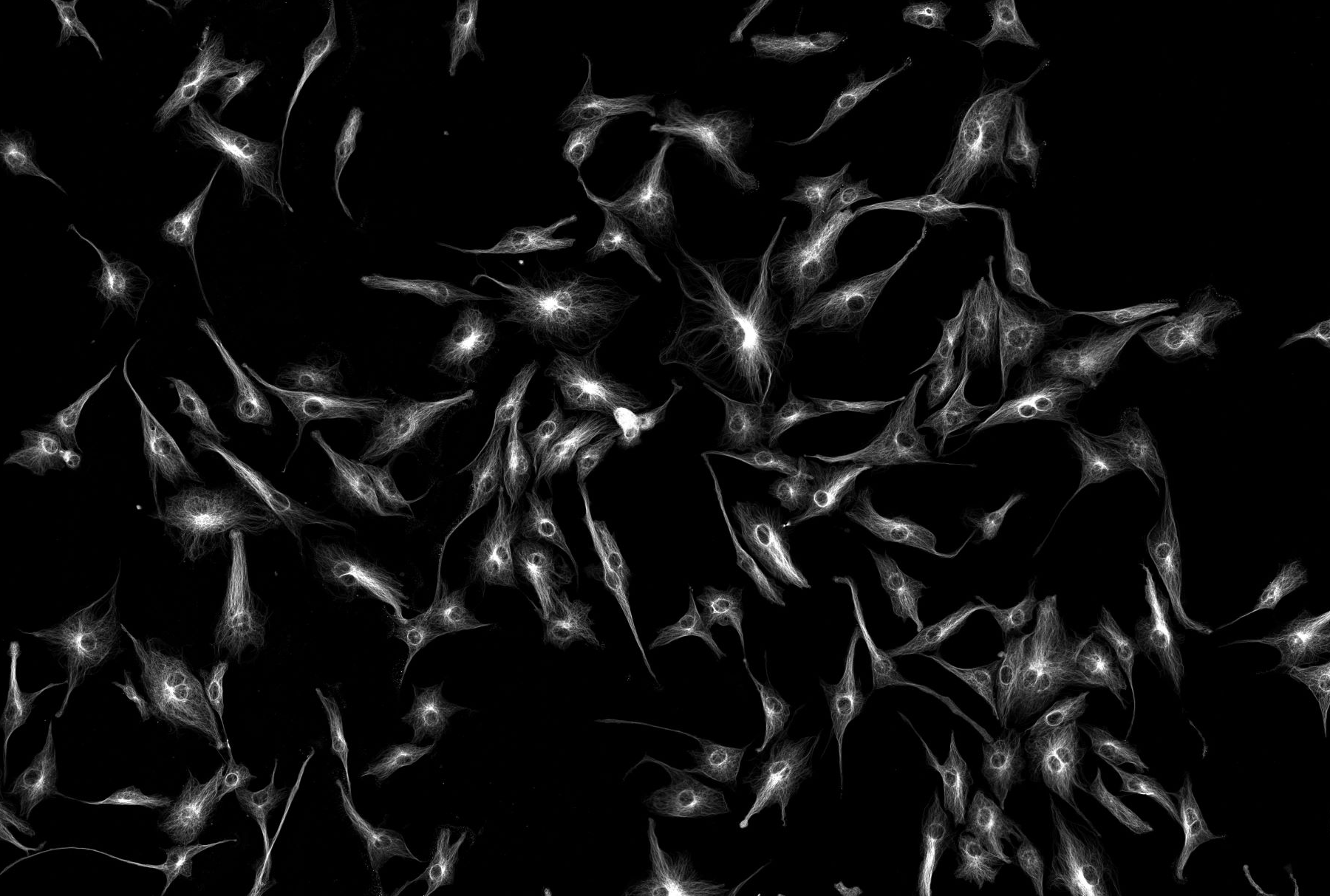
To visualize the molecule of interest, fluorophore-coupled specific antibodies or fluorescent proteins, for example, are transferred into the cell. The specimen is then illuminated at the excitation wavelength and viewed through a filter that allows only the emitted wavelength to pass through. Whereas the background is dark, the structures with a bound fluorophore emit light, indicating the presence of the structure of interest. Widefield illumination means that the whole specimen in the field of view is exposed to the light therefore fluorescent signals from all focal planes are detected. Therefore, widefield microscopy is best applied with thin specimens with low background autofluorescence.
Diffuse lampdiy
The advantage of using darkfield illumination is that unstained specimens can remain alive. The main limitation of dark-field microscopy is the low light levels seen in the final image. This means that the sample must be very strongly illuminated, which can cause damage to the sample. Dark-field microscopy techniques are almost entirely free of artifacts, due to the nature of the process.
Outdoor Collection. Product Type Filter, Area, Bollard, Wall Mount, Buy American Act, DLC, Dark Sky, Field Selectable, Flood, Garage, Highbay & Lowbay ...
Widefield fluorescence microscopy is an optical microscopy technique that utilizes fluorescence, which is induced using fluorophores, as opposed to absorption, scatter, or reflection. This method is mainly applied for the detection of specific structures, molecules or proteins within the cell. Fluorescence microscope systems can range from very simple, such as an epifluorescent microscope, to extremely complex, such as confocal or multiphoton systems. Whether simple or complex, fluorescence microscopes share the same basic concept: excitation energy is used to illuminate a sample containing your fluorophore which then emits lower energy (longer wavelength) light, that, although weak, is quantifiable. The excitation and emission wavelengths do not share the same centre wavelength, and this allows specialized optical filters to increase overall contrast and signal. The three critical filters needed for a fluorescence microscope are the excitation, dichroic, and emission filters which in simple terms separate the excitation and emission wavelengths.
Flat Diffuse Lights are often referred to as “Flat Domes” because of the similar performance characteristics they share with classic domed illuminators. If sufficiently oversized compared with the object of interest, flat diffuse lights can provide an adequate “Cloudy Day” lighting effect at a larger range of working distances than domes.


 Ms.Cici
 8618319014500