
Bright Saber-X LED Single Row Light Bar - 20 - bar light bar bright
Generative AI is a tool that has wide-ranging application for the practice of law and administrative functions of the legal practice for all licensees, ...
Perspex cast acrylic sheet produced specifically to diffuse white LED lighting while eliminating 'hotspots' from individual LEDs. Material is supplied in discs ...
Portions of the ring-shaped light are diffracted by optically dense structures of the specimen and experience a negative phase shift of about λ/4. This phase-shifted, diffracted light bypasses the λ/4 plate. In contrast, the portion of the ring-shaped light that passes directly through the specimen non-deviated will hit the phase plate which causes a positive λ/4 phase shift. As the total difference in phase shift between the light diffracted by the specimen’s structures and that which passes through phase plate will be about λ/2, destructive interference will occur. Consequently, more optically dense structures will appear darker than those that are less optically dense.
Backlightingdefinition film
The phase contrast method for microscopy was developed in the 1930s by the Dutch physicist Frits Zernike. After 1942, it became a widely used microscopy technique. In 1953, Zernike was awarded the Nobel Prize for Physics. For more details, refer to the articles: A Brief History of Light Microscopy – From the Medieval Reading Stone to Super-Resolution & Phase Contrast
Whatis backlight in photography
2 Allied Vision Technologies Manta G1236B ASG Machine Vision Digital Camera PoE. New – Open box. $160.00. or Best Offer. +$7.78 shipping. Sponsored.
Side lighting definition
Most TVs produced and sold today are LCDs or Liquid Crystal Displays. This technology has been around for a while now; chances are, most if not all of your TVs are LCD. LCDs have two main layers: the screen and the backlights. The screen arranges whatever image is needed and the backlights light it up so you can see it. Early versions of LCD screens used florescent bulbs as backlights. Nowadays, all TVs uses LEDs or Light Emitting Diodes. There are three main forms of backlighting: direct lighting, edge lighting and full-array lighting. Direct lighting is the simplest: backlights reflecting onto a diffuser layer. It's very cheap to produce and will be found on basic TVs. Edge lighting is just what it sounds like, where the backlights are placed around the edge of the image TV: this allows for a much thinner overall TV. The best of the bunch, full-array backlighting, is where LEDs are evenly placed across the whole screen and are controlled by the TV's processor. This produces much better overall brightness and allows the TV to dim parts of the image as needed, resulting in much better black levels and reduced “bloom” (an effect where the light bleeds over into parts of the image that are supposed to be dark). The best LCD screens will use full-array backlighting with MiniLEDs. This is very similar to traditional full-array backlighting but it uses much denser arrays of small LEDs called MiniLEDs. These smaller LEDs can be stacked much closer together, producing a significantly brighter picture, a more pronounced the local dimming effect, much reduced bloom and better black levels. The other type of screen in today's market are the OLEDs. They simple don't have backlights, since the screen creates its own light. For a deeper dive on what makes OLED different from traditional screens, check out our FAQ titled “OLED vs QLED: What’s the difference?” Both LG and Samsung offer LCD TVs with all of these backlighting systems. The differences are best seen in person, on our showroom floor.
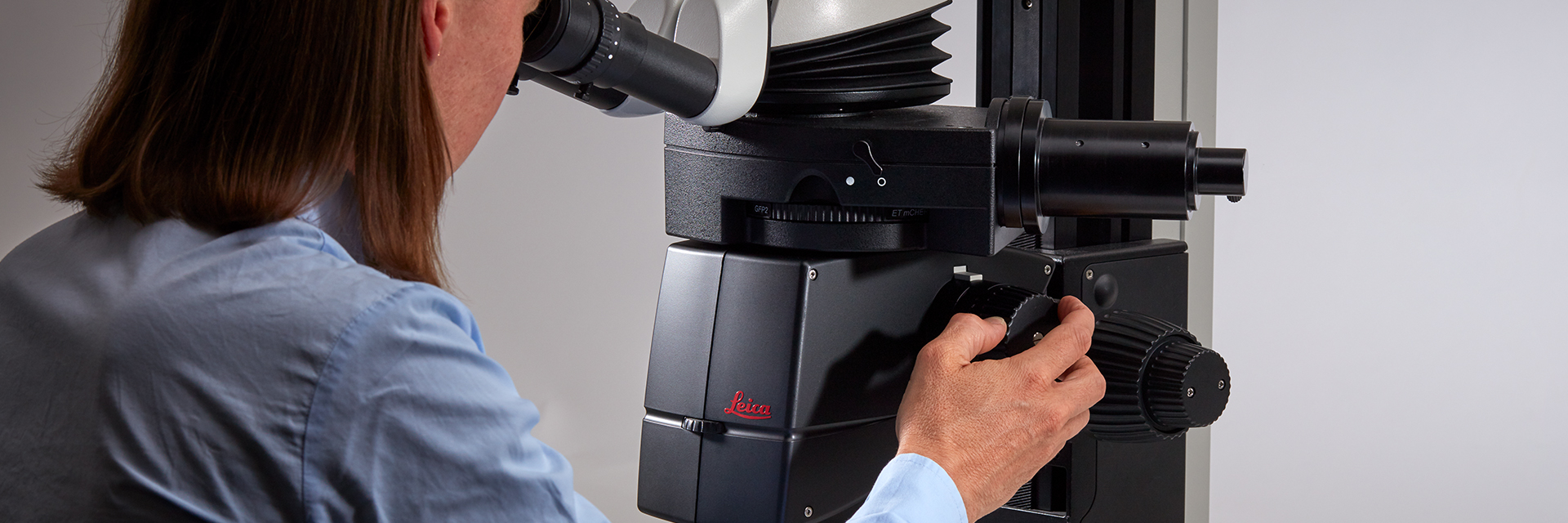
Leica microscopes capable of phase contrast make a difference for the study of transparent and colorless minerals, crystals, and polymers.
Just another spot light? No, a LIFX SuperColor smart LED spot light. This is unlike any spotlight you've ever seen. Not only can it illuminate your ...
Order your dark field lighting online at Machine Vision Direct. Our dark field ring lights are used to achieve low angle lighting which helps illuminate ...
For forensic applications concerning the evidentiary investigation of paints, pigments, textiles, fibers, and human tissues, Leica microscopes offering phase contrast are very useful solutions.
Leica microscopes offer phase contrast for the study of cells or tissues concerning various life-science and forensic applications. Phase contrast can also be useful for certain material and earth-science applications.
At Advanced Illumination, our Technical Sales Specialists will find the right light for your application! Browse our products & services online today.
A phase contrast microscope is similar to a conventional brightfield microscope, except it uses an annular aperture in front of the light source and a quarter-wave phase plate after the objective lens. For more information, refer to the article: Phase Contrast
Leading manufacturer of power transformers and linear DC power supplies. Standard and custom transformers including HVAC instrumentation and Class 2 approved.
Leica microscopes providing phase contrast are commonly used in life science research for the visualization, analysis, and documentation of biological structures and cellular processes.
A phase contrast microscope is similar to a conventional widefield microscope, except it uses an aperture in the shape of an annulus and a quarter-wave (λ/4) phase plate. The annular aperture is placed between the light source and condenser lens and the phase plate after the objective inside the microscope optics. Ring-shaped light that passes through the aperture is focused by the condenser onto the biological specimen to be observed.
Backlightingeffect
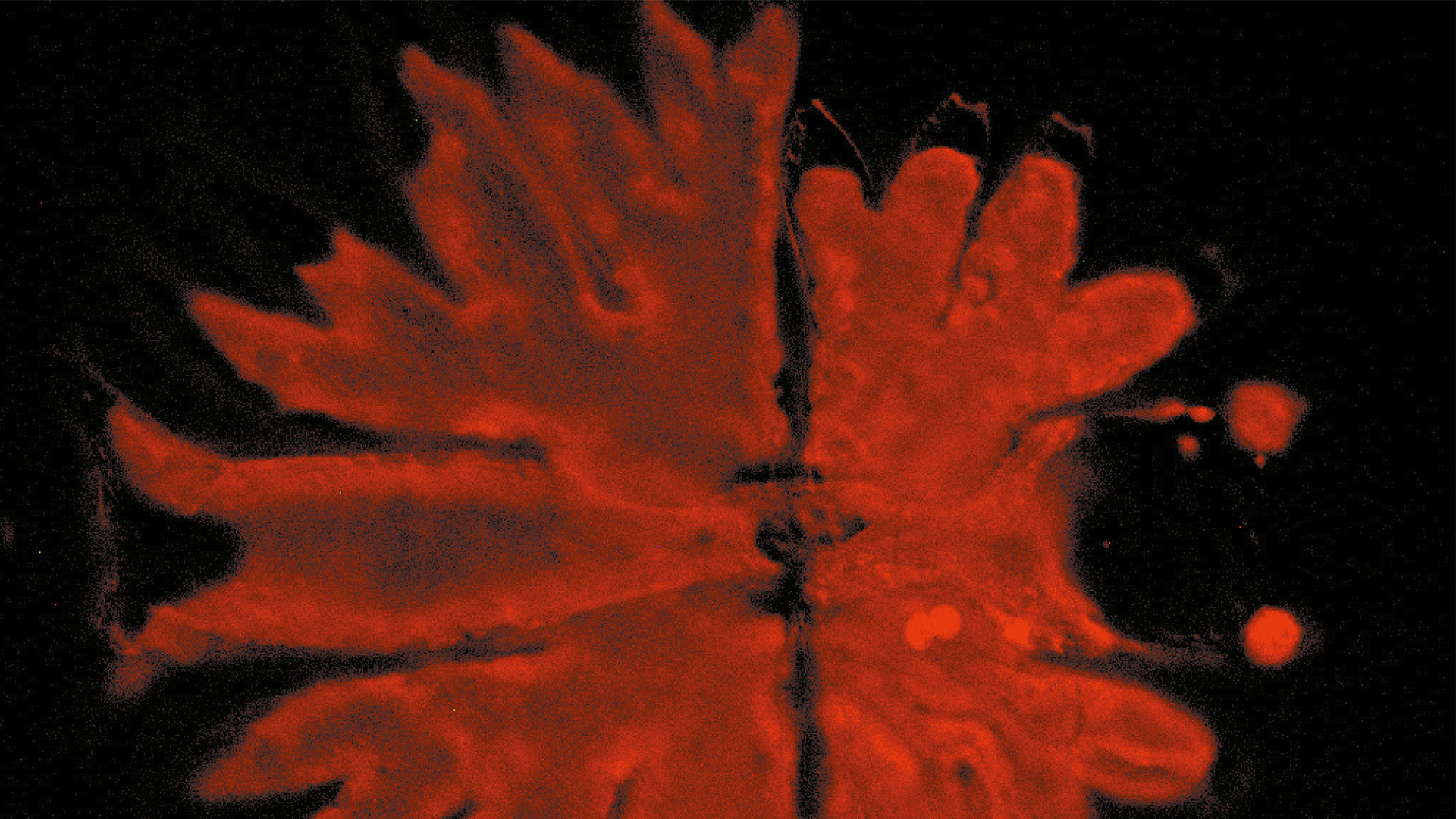
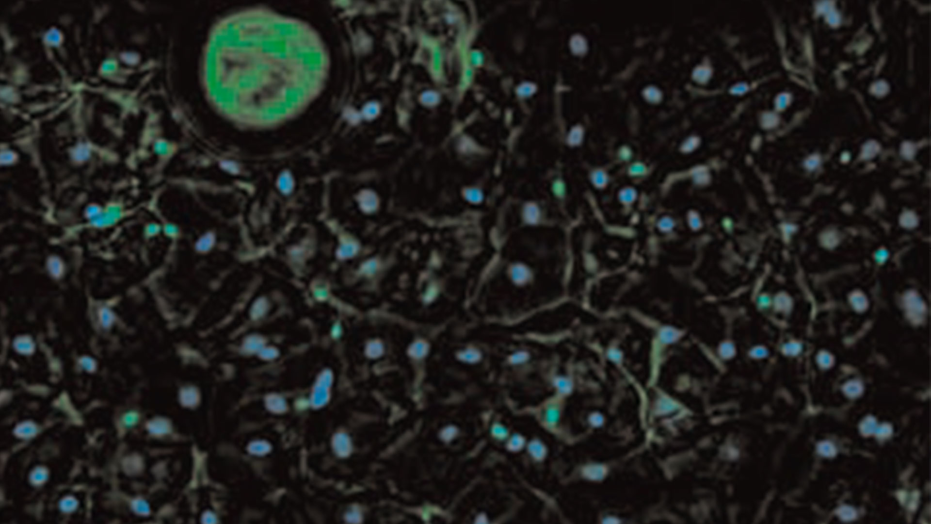
Brightfield microscopy normally only provides a low-contrast image of many transparent biological specimens where few details are distinguished. One way to enhance contrast with brightfield microscopy is to use selective stains, but such stains are often toxic to living cells. A phase contrast light microscope offers a way to view the structures of many types of biological specimens in greater contrast without the need of stains. The contrast method exploits differences in optical density between structures of a specimen that lead to a phase shift of the light that interacts with the specimen and its structures.
Multi-Wire Connectors · Inserts · Housings · Crimp Contacts · Plugs, Adapters, and Cable Glands · Housing Insert Plates · Code Pins and Polarization Keys · DIN Rail ...
LED light bars for sale from Cyclops Lighting. The best LED light bars for your ATV, UTV, 4x4, or off-road vehicles. Super high output at great prices.
Most often biological specimens and tissues are observed with a phase contrast microscope. A large variety of biological specimens can be observed with phase contrast from fixed specimens to living cells and tissues. For examples, refer to the articles: Phase Contrast & Optical Contrast Methods
Phase contrast is an optical contrast technique for microscopy which makes unstained structures in the cells of biological specimens visible. Cell structures that appear transparent with brightfield illumination can be viewed in high contrast and rich detail using phase contrast. Differences in optical density between structures in the cell can cause light that interacts with them to attain a phase shift. This phenomenon is the basis of phase contrast. As a result, more optically dense structures will look darker than less optically dense ones.
Many students want to film scenes set at night but are unsure how to light them to create the look and feel that they require.
The knowledge portal of Leica Microsystems offers scientific research and teaching material on the subjects of microscopy. The content is designed to support beginners, experienced practitioners and scientists alike in their everyday work and experiments.
Not all products or services are approved or offered in every market, and approved labelling and instructions may vary between countries. Please contact your local representative for further information.


 Ms.Cici
 8618319014500