
Bright field Versus Dark-field TEM - bright field microscopy vs dark field microscopy
Independent artist, DJ, composer & film lover (he/him). 'Enjoy Youth' out May 17th https://t.co/DypDBDJuGE MGMT/Booking: hello@brightlightx2.com.
Not all products or services are approved or offered in every market, and approved labelling and instructions may vary between countries. Please contact your local representative for further information.
Ring-shaped light that passes through the aperture is focused by the condenser onto the biological specimen or material sample to be observed. Portions of the ring-shaped light are diffracted or scattered by structures of the specimen or features of the sample. The diffracted light enters the objective. In contrast, the portion of the ring-shaped light that passes directly through the specimen non-deviated or is reflected by the sample without scattering will not be collected by the objective. The light scattered by the specimen’s structures or sample’s features will appear brighter than the background areas of the specimen or sample where there is no light scattering.
A darkfield microscope is a compound microscope which uses an aperture in the shape of an annulus that is placed between the light source and condenser lens.
Bright field dark field
Very often biological specimens and tissues, whether fixed or live, are observed with a darkfield microscope. Also, various material and geological samples can be observed with darkfield. For examples, refer to the articles: "Work More Efficiently in Developmental Biology With Stereo Microscopy: Zebrafish, Medaka, and Xenopus" & "How to Adapt Grain Size Analysis of Metallic Alloys to Your Needs"
A darkfield microscope is similar to a conventional brightfield microscope, except it uses an annular aperture in front of the light source. The light from darkfield illumination impinges onto the specimen or sample at a high angle of incidence, either transmits through the sample or reflects off its surface, then passes through the objective lens, and finally goes through the eyepieces or reaches the camera sensor. Darkfield illumination causes uniform areas of transparent samples or flat surfaces of opaque samples to appear dark, as the vast majority of the light at the high incident angle misses the objective. Normally there are features in transparent samples or on the surface of opaque samples which scatter light. For this reason, darkfield microscopy images show a dark background with brighter areas corresponding to these features, because the light they scatter enters into the objective.
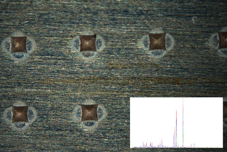
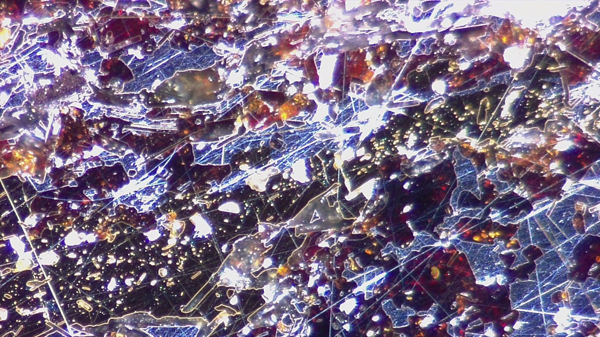
Leica darkfield microscopes are useful for the study of cells or tissues concerning a variety of life-science and forensic applications. Darkfield microscopy can also be helpful for material and earth-science applications.
The knowledge portal of Leica Microsystems offers scientific research and teaching material on the subjects of microscopy. The content is designed to support beginners, experienced practitioners and scientists alike in their everyday work and experiments.

Illumination Systems is a construction company based in Phoenix, AZ and specializes in Electrical.
Yes, a darkfield microscope can be equipped with a camera for recording images observed with the contrast method. It can also be equipped with other accessories. For more information, contact your local Leica representative.
L Christiano · 1999 · 2904 — The Band Pass Filter ... The `ideal' band pass filter can be used to isolate the component of a time series that lies within a particular band of ...
Controller for SP shelf applications with a DB9 front panel interface, three inputs with common return, three output alarm relay contacts, and standard code.
For forensic applications concerning the evidentiary investigation of paints, pigments, textiles, fibers, and human tissues, Leica microscopes offering darkfield are very useful solutions.
Dark field microscopy
Leica microscopes providing darkfield are commonly used in life science research for the visualization, analysis, and documentation of biological structures and cellular processes.
Apply for AI Technical Business Analyst | Corporate Systems | Experienced Hire job with SIG Migration in Bala Cynwyd (Philadelphia Area), ...
When working with a microscope, the most commonly used contrast method is brightfield. However, brightfield usually only provides a low-contrast image of many transparent biological specimens. It can also be the case for many transparent or reflective opaque material samples. In such low contrast images, few details are distinguished. The contrast of brightfield microscopy for biological specimens can be enhanced using selective stains, but they are often toxic to live cells. A darkfield microscope offers a way to view the structures of many types of biological specimens in greater contrast without the need of stains. Darkfield microscopy can also increase contrast when imaging material samples. The darkfield contrast method exploits diffraction or scattering of light from structures of a biological specimen or non-uniform features of a material sample.
It is responsible for preventing the specimen slide from coming too far up and hit the objective lens. Magnification by Bright field Microscope. •. The ...
Leica microscopes capable of darkfield make a difference for the study of transparent and opaque materials, minerals, crystals, and polymers.
Darkfield is an optical contrast technique for microscopy which makes unstained structures in the cells of biological specimens visible. Cell structures that appear transparent with brightfield illumination can be viewed with better contrast and detail using darkfield. Additionally, non-uniform features of transparent material or on the surface of opaque material samples can be more easily observed with darkfield compared to brightfield. Structures in the cell or features of the material scatter the light that interacts with them, while uniform areas allow the light to pass without scattering. When using darkfield microscopy, the cell structures or material features appear brighter and the uniform cell or material background look darker.
Package Includes: (1) American DJ ADJ FOCUS SPOT 4Z 200W Cool White LED DMX Moving Head Spot Light; (1) Rockville RTP82W 8 ft. Adjustable White Totem Moving ...
Not sure if this is the right place, but I'd love some help. I need an extremely bright light that plugs into a normal AC/DC outlet and does not have a battery. All googling is only pointing me towards either very mediocre worklights or very bright battery powered flash/spotlights. Any help or direction to a more appropriate sub would be greatly appreciated. Thanks!
Find the best high powered 4x4 Emergency LED Light Bars, Latest modular design with wide size and color selection to meet your requirements for cars, ...
amaran 100x S Bi-Color Bowens Mount Point-Source LED Video Light,100W Output Studio Light,Bluetooth App Control 9 Built-in Lighting Effects DC/AC Power Supply ...
Jul 20, 2021 — Quantitative analysis of cell structures is essential for biomedical and pharmaceutical research. The standard imaging approach relies on ...


 Ms.Cici
 8618319014500