
Is Beam Shaping the Future of Laser Welding? - beam shaping
When using highly divergent light sources such as lamps or LEDs that do not behave at point sources, it is necessary to use a condenser rather than a single lens to produce a well-focused beam. Specialist condensers can also help correct aberrations in the beam to improve the overall resolution of the measurement or to create specific beam shapes if necessary.
The use of stains can help overcome some of the limitations associated with trying to image species that are weak absorbers and increase the number of specimens that bright field microscopy can be used for.
The simple-to-use, relatively compact, and affordable microscopes have made bright-field microscopy widely used in several fields, including food science1, soil science2, and medical applications.3
The objective and eyepiece magnify the sample image that the eye can view. There can be a trade-off between achieving a greater field of view of the sample and seeing more significant regions simultaneously as achieving maximum magnification.
Registered members can chat with Azthena, request quotations, download pdf's, brochures and subscribe to our related newsletter content.
The set-up for bright field electron microscopy image visualization is similar to an optical microscope, only the light source is replaced with a high-power electron gun and images are recorded on an imaging plate or camera.
LIS Technologies is on the road to transforming nuclear fuel enrichment through advanced laser techniques, ensuring a sustainable and cost-effective approach to energy production.
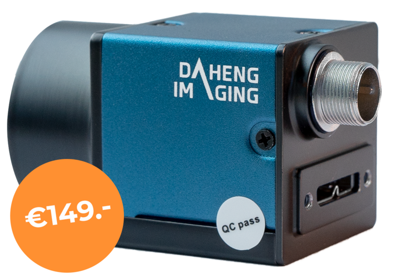
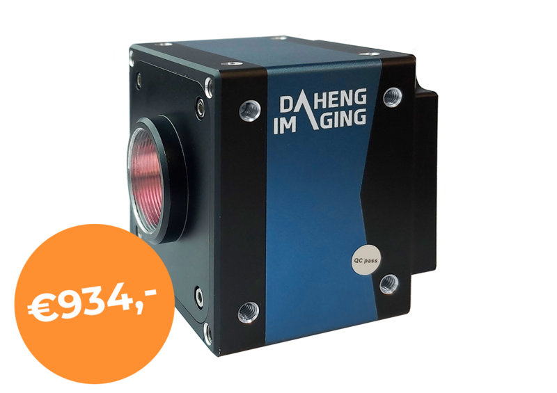
TOP 5 Machine Vision Camera interfaces There are many machine vision camera, also known as industrial imaging camera, interfaces available. The following table shows a summary of all interfaces and we will discuss the 7 industrial imaging camera interfaces that we offer in addition USB2 USB3 GigE CameraLink Coaxpress 5GigE 10GigE Supported by GeT Cameras Yes Yes Yes No No Yes Yes Bandwidth (Megabyte/s) 40 400 100 Base = 250 Medium = 500 Full = 750 1 lane = 750 2 lanes = 1500 3 lanes = 2250 4 lanes = 3000 500 1000 Cable length (maximum) 5m 4.6m 100m 7m 100m 100m 100m Power + data on one cable Yes Yes Only if PoE PoCL Yes Only if PoE Only if PoE Frame-grabber mandatory No No No Yes Yes Yes Yes Cable costs Low Low Low High Low Low Low Camera costs Very low Low Low High High Medium High CPU usage Medium Low Medium Low Low Medium Medium Customer acceptance Fair Good Good Declining Growing Growing Growing Multiple cameras Fair Excellent Good Fair Excellent Excellent Excellent USB2 Industrial Imaging Camera The USB2.0 camera interface is the cheapest and easiest to use of all 3 options. Nevertheless, bandwidth and cable length are limited. USB2.0 Industrial imaging cameras are ideal for applications which require a maximum of 1.3MP at 30fps or 5MP at 7fps, with a cable length that does not exceed 5 meters. USB3 Vision Industrial Imaging Camera The USB3.0 Vision industrial camera interface is the fastest interface with the highest bandwidth we support, and it uses the least amount of computer processor power out of the 3. Therefore, it is ideal for high resolution and high-speed imaging. The cable length, on the other hand, is limited to 4.6 meters. More information about the USB3 Vision industrial camera protocol is available in our blog. GigE Vision Industrial Imaging Camera The GigE Vision industrial camera interface is often used in machine vision applications, which require longer cable lengths (>5m). Bandwidth is average (between USB2.0 and USB3.0). This makes it ideal for most machine vision applications. Both 20MP sensors with low framerates as low-resolution cameras with high framerates are available with GigE. 5GigE Vision Industrial Imaging Camera The 5GigE Vision industrial Camera interface is a new interface that requires a 5GigE or 10GigE network card to operate at full speed. It is using the same cable infrastructure as GigE. This interface is very interesting for industrial camera applications where long cable lengths and high frame rates with high resolutions are required. 10GigE Vision Industrial Imaging Camera 10GigE Vision Industrial cameras require a 10GigE infrastructure. It has the highest bandwidth. However, the interface is also relative expensive. For most of the industrial camera applications, GigE, 5GigE and USB3 will deliver sufficient bandwidth. Differences between industrial imaging cameras vs. machine vision cameras Industrial imaging cameras and machine vision cameras are terms that are both being used for industrial cameras. Both types of cameras are used in industrial settings for various applications. Where industrial imaging cameras mostly refer to a broad category of cameras for industrial applications, machine vision cameras are mostly refered to as cameras designed for machine vision applications. When refering to industrial imaging cameras, it could also include other types of cameras such as infrared or high-speed cameras. Both of these types of cameras are also available in our machine vision camera portfolio. Check our SWIR camera page for Short Wave Infrared cameras or our USB3 camera page for high speed cameras. In conclusion, both the terms machine vision cameras and industrial imaging cameras could be used when looking for a camera for applications such as automation, quality control, inspection and measurement. Industrial imaging camera technology Industrial cameras, as explained also known as industrial imaging cameras or machine vision cameras are designed for industrial and medical applications. The main differences between an industrial imaging camera and a security or consumer camera are: - An industrial imaging camera gives out the RAW information, without image compression. - Very low latency between image capture and image received by the PC - Option to exactly trigger the moment that the image should be captured with industrial imaging cameras - Longevity, industrial imaging cameras have a minimum life time cycle of 5-7 years (as most of the time even longer), while security and consumer cameras have a life time cycle of 1-2 years - Image-sensor-sensitivity, industrial imaging cameras have larger and therefore more light sensitive sensors. As a result the images are better during low light conditions or short exposure times - With a industrial industrial imaging camera you have access to more features, for example LUT tables, region-of-interest, triggering, pixel-binning etc. These are the mean reasons to select an industrial imaging camera instead of a security camera. Machine vision applications In almost every market, machine vision cameras are used. Some examples are: - Pharmaceutical; inspecting products with a machine vision camera, including capsules, injections, bubble caps, labels of the package and more. - Traffic and transportation; industrial cameras for inspection of railways, Automatic Number Plate Reading (ANPR), Speed control, inspection of roads, 360 degree imaging (street view), - 3D scanners; industrial cameras are integrated inside to generate a 3D point cloud. It is used to determine the height of an object, distance of an object or checking for defects (missing parts) on a product. It can also be used for making 3D models or even scanning complete persons to integrate them digitally into movies and games - Industrial; machine vision inspection of glass bottles, carpet, industrial printing machines, etc. - Logistics; barcode reading, DMC reading, check size of package with a machine vision camera - Automations; Robots, palletizers, optical guidance systems, quality inspections, position recognition etc. - Medical; Devices that include industrial imaging camera technology like Endoscopes, Ophthalmology, overview camera during MRI scans, robot surgery, digital microscopes etc. - Research, Laboratory; Industrial imaging cameras on existing microscopes, digital microscopes etc. - Sports; tracking movement of sportsmen, animals (horses), objects (golf simulator) and more by using industrial imaging cameras Overview machine vision camera products A complete overview of all our machine vision cameras, lenses and lighting can be found on our computer vision product page.
Bright-field microscopy
A bright field microscope uses a transmission measurement to visualize the sample, where the light passes directly through. The contrast in the final image observed by the eye originates from different regions of the sample absorbing different amounts of the incident light.
A complete overview of all our machine vision cameras, lenses and lighting can be found on our computer vision product page.
Opticalmicroscope
Ingle, Rebecca. (2022, October 25). How Does Bright-Field Microscopy Allow Images to be Visualized?. AZoOptics. Retrieved on November 01, 2024 from https://www.azooptics.com/Article.aspx?ArticleID=2343.
Lightfieldmicroscopy
The light source is pointed upwards from the bottom of the instrument and placed beneath the sample stage. Typical light sources for optical measurements include halogen lamps or light-emitting diodes (LEDs).
Before imaging, bright-field microscopy does not require sample staining or fluorophores to label particular cellular structures.
Once the light has passed through the sample, it enters the objective, which determines the ultimate magnification achieved with the microscope.
Differential interference contrast microscopy
Some of the challenges of the method include blurry images of out-of-focus regions of the samples and weak absorbers can be challenging to image as they do not have sufficient contrast on the bright background. Molecules such as eosin and hemoglobin are much easier to image with optical bright-field approaches as they have large absorption cross-sections and produce a good background contrast even with very thin samples.5
A set of electron lenses that serve a similar function to the condenser, objective and eyepiece are used for controlling the beam shape in the instrument and ensuring accurate image visualization of the sample is achieved with good resolution.
While using transmission geometries in microscopy does limit the sample thickness that can be measured, one of the critical advantages of bright-field microscopy over other imaging methods such as fluorescence is that the sample preparation is very straightforward.
Disclaimer: The views expressed here are those of the author expressed in their private capacity and do not necessarily represent the views of AZoM.com Limited T/A AZoNetwork the owner and operator of this website. This disclaimer forms part of the Terms and conditions of use of this website.
Darkfieldmicroscopy
The MIRcat Mid-IR Laser provides high-speed, broad-spectrum capabilities with peak tuning velocities up to 30,000 cm1/s.
Your questions, but not your email details will be shared with OpenAI and retained for 30 days in accordance with their privacy principles.
An optical bright-field microscope consists of a light source, a condenser, the sample stage and mount, objectives, and a mount.
Bright-field microscopy uses light to produce a dark image against a bright background. Often considered one of the simplest types of microscopy, a bright-field microscope uses an objective, condenser and eyepiece to magnify the image of a sample so the eye can see more minor features.
While we only use edited and approved content for Azthena answers, it may on occasions provide incorrect responses. Please confirm any data provided with the related suppliers or authors. We do not provide medical advice, if you search for medical information you must always consult a medical professional before acting on any information provided.
Reuven Silverman of Ophir discusses the critical role of M2 measurements in laser technology for optimization and quality control in various industries.
The right choice of eyepiece can also help correct image distortions and be used for color correction and ensuring images appear sharp.
Ingle, Rebecca. "How Does Bright-Field Microscopy Allow Images to be Visualized?". AZoOptics. https://www.azooptics.com/Article.aspx?ArticleID=2343. (accessed November 01, 2024).
Shanghai Optics provides high-precision off-axis parabolic (OAP) mirrors designed for advanced optical systems, offering superior reflective solutions for applications requiring precise light collimation and focus.
Electron microscopes often have better spatial resolutions than are achievable with optical microscopes, as the diffraction limit of visible light is in the hundreds of nanometers. It is preferable to use electrons rather than optical sources for imaging nanoscale structures that have features less than 100 nm in size with sufficient resolution.
Industrial imaging cameras and machine vision cameras are terms that are both being used for industrial cameras. Both types of cameras are used in industrial settings for various applications. Where industrial imaging cameras mostly refer to a broad category of cameras for industrial applications, machine vision cameras are mostly refered to as cameras designed for machine vision applications. When refering to industrial imaging cameras, it could also include other types of cameras such as infrared or high-speed cameras. Both of these types of cameras are also available in our machine vision camera portfolio. Check our SWIR camera page for Short Wave Infrared cameras or our USB3 camera page for high speed cameras. In conclusion, both the terms machine vision cameras and industrial imaging cameras could be used when looking for a camera for applications such as automation, quality control, inspection and measurement.
Ingle, Rebecca. 2022. How Does Bright-Field Microscopy Allow Images to be Visualized?. AZoOptics, viewed 01 November 2024, https://www.azooptics.com/Article.aspx?ArticleID=2343.
The simplicity and wide applicability of bright field imaging means it is likely to remain a workhorse technique in many different areas, including medical diagnostics.

Ingle, Rebecca. "How Does Bright-Field Microscopy Allow Images to be Visualized?". AZoOptics. 01 November 2024. .
Fluorescencemicroscope
Bright-field microscopy methods do not just have to be used with optical techniques but are also compatible with electron-based microscopy approaches, such as transmission electron microscopy. 6
Dr. Rebecca Ingle is a researcher in the field of ultrafast spectroscopy, where she specializes in using X-ray and optical spectroscopies to track precisely what happens during light-triggered chemical reactions.
There is a tradeoff in the eyepiece cost and quality of the lenses used to achieve good performance. To make microscopy portable for point-of-care devices as well as more affordable, methods using smartphone cameras for both light sources and imagers have been developed.4 Such instruments can be used for live cell imaging and visualizing species such as zooplankton, making them a powerful tool for in field measurements.
These F-theta lenses by Avantier are designed for consistent spot size and uniform field curvature correction, ideal for high-resolution imaging applications.
Two areas of bright field microscope development are finding ways of further automating the focusing and image analysis for higher sample throughputs and finding ways to make microscopes more compact, such as using smartphones.


 Ms.Cici
 8618319014500