
λ/2 and λ/4 Wave plate/retarders Knowledge | in Singapore - wave plate
Charge coupled device working principle
Compound microscopes come in various types, each designed for specific applications and offering different features. Here are the main types of compound microscopes:
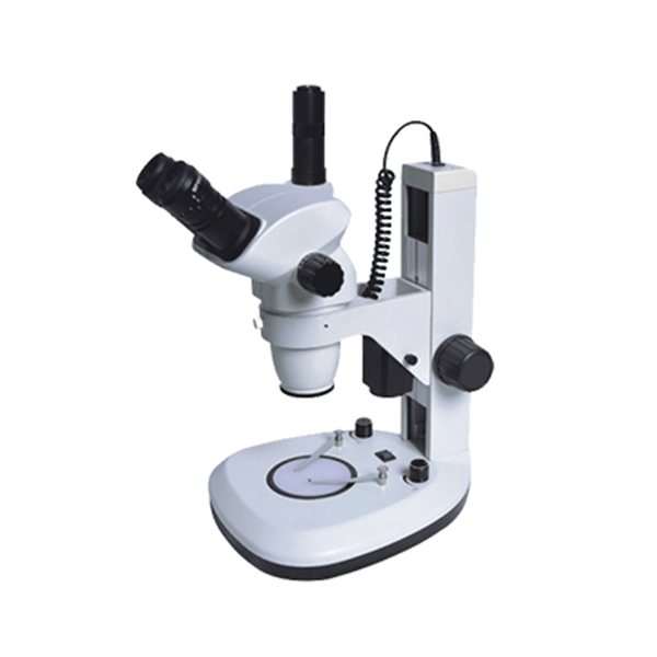
A stereo microscope, also known as a dissecting microscope, is an optical instrument designed for low magnification observation of three-dimensional objects. It typically provides magnification ranging from 10x to 50x. Unlike compound microscopes, stereo microscopes use two separate optical paths to create a three-dimensional image, giving depth perception to the observer. They are used for examining larger, solid specimens that do not require light to pass through them, such as insects, plants, and electronic components.
If you disable this cookie, we will not be able to save your preferences. This means that every time you visit this website you will need to enable or disable cookies again.
Time delay and integrationin ccd
Points to note if considering TDI technology TDI requires more care in synchronization and alignment but in general practice these requirements are not difficult to achieve. A TDI sensor can withstand some misalignment (either translational or rotational) without negative effect on image quality and a total misalignment of one pixel or less across the length of a TDI sensor will not affect image quality. In most applications, a 96-stage TDI device can comfortably tolerate a 2-4% velocity mismatch between inspection web and imager. This is not difficult to achieve using a web-mounted encoder to supply a sync signal to the camera, even with webs that change speed and this approach is used in thousands of successful applications. Designed for light-starved applications, TDI typically lacks antiblooming to subdue "glints", and as well, TDI sensors generally use photogates - surface electrodes covering the entire pixel - and while this ensures 100% fill factor, it can attenuate incoming photons in the deep blue region. One camera manufacturer, Teledyne DALSA has produced an anti-blooming device to solve the first problem and has also developed enhanced blue-response TDI sensors to compensate for the latter issue.
Polarizing Microscopes: These microscopes use polarized light to examine specimens that are birefringent (having different refractive indices in different directions). They have polarizers and analyzers to filter and analyze the polarized light. Applications including Mineralogy, crystallography, and materials science to study crystalline structures and stress patterns.
** High quantum efficiency ** High resolution ** Wide imaging areas ** Efficient thermal packaging ** Efficient management of high-speed circuitry
TDI camera principle
What is TDI? TDI refers to Time Delay Integration, a specialised detector readout mode used for observing a high-speed moving object under low light conditions normally undetectable by classic CCD imaging. With an effect similar to that of a linescan camera, TDI is designed to allow continuous movement of the object past the sensor to produce a continuous video image of a moving, two-dimensional object: a definite advantage over stop-then-start capture of traditional technologies. In the past, certain image-capture deployments traded-off sensitivity in order to capture a fast-moving object. TDI-mode technology preserves sensitivity while not degrading image quality, even given the relative fast movement between the sensor and the objects under image. How does TDI work? Based on the concept of accumulating multiple exposures of the same (moving) object, TDI mode effectively increases the integration time available to collect incident light. The technique senses a change in light pattern and shifts them across the CCD in a manner that is synchronised with the movement of the image, in order to integrate more light from the scene. The number of lines on the sensor corresponds to the increase in sensitivity. For example a 256 line TDI sensor provides 256x more sensitivity than a single linescan camera. Imaging in TDI mode provides dramatically increased responsivity compared to other video scanning methods by permitting much greater scanning speeds in low light, or allows reduced lighting levels (and costs) at conventional speeds and so producing a crisp image.
Microscopes are essential tools in scientific research, education, and various industries, allowing us to observe objects and organisms that are otherwise invisible to the naked eye. Two of the most commonly used types of microscopes are compound microscopes and stereo microscopes. While both serve to magnify small objects, they have distinct differences in design, functionality, and application. This article explores the pros and cons of each type and provides guidance on choosing the right microscope for specific needs.
Fluorescence Microscopes: These microscopes use high-intensity light sources (such as mercury or xenon lamps) to excite fluorescent dyes or proteins in specimens, causing them to emit light of different wavelengths. Filters are used to isolate the emitted light for observation. Applications including Molecular biology, immunology, and medical diagnostics involving fluorescent labeling.
TDI image sensor
Understanding these pros and cons helps in making informed decisions when selecting a compound microscope for specific applications and ensuring that it meets the requirements of the tasks at hand.
What is ccd
Adept Electronic Solutions are "The Machine Vision and Imaging Specialists" and distributor of Machine Vision products in Australia and New Zealand. To find out more about any machine vision product please email us at: adept@adept.net.au or call us at Perth (08) 92425411 / Sydney (02) 99792599 / Melbourne (03) 95555621 or use our online contact us page.
Understanding these pros and cons can help you determine whether a stereo microscope is the right tool for your specific needs and applications, ensuring you selecting the most appropriate equipment for their tasks.
Time delay and integrationtdi

Where can TDI technology be used? TDI CCDs are used in applications that require the ability to operate in extreme lighting conditions, that require both high speed and high sensitivity, for example: ** inline monitoring, inspection and guidance ** sorting ** earth observation satellite (for weather observation, for example)
A compound microscope is an optical instrument that uses multiple lenses to achieve high magnification, typically ranging from 40x to 1000x or more. It consists of an objective lens close to the specimen and an eyepiece lens through which the observer views the magnified image. Compound microscopes are designed for viewing small, thin specimens that light can pass through, such as cells, bacteria, and thin tissue sections.
Time delay and integrationmeaning
Digital Microscopes: These microscopes are equipped with digital cameras to capture images and videos of the specimen. They often connect to computers for image analysis and sharing. Applications including Education, documentation, and any field requiring digital imaging and analysis.
Integration timevs exposuretime

Inverted Microscopes: Unlike traditional compound microscopes, inverted microscopes have the light source and condenser above the stage and the objectives below it. This design allows viewing of specimens from below. Common used in Cell culture studies, live cell imaging, and observing specimens in petri dishes or flasks.
ScopeLab offers an extensive range of microscopes, including compound, stereo, digital, and specialized models, catering to diverse fields such as biology, medicine, material science, and education. If you are not sure how to choose the microscope, please feel free to contact them.
This website uses cookies so that we can provide you with the best user experience possible. Cookie information is stored in your browser and performs functions such as recognising you when you return to our website and helping our team to understand which sections of the website you find most interesting and useful.
Metallurgical Microscopes: Designed for examining the surface structure and composition of metals and other materials, these microscopes use reflected light to view opaque specimens. Applications are Metallurgy, materials science, and quality control in manufacturing.


 Ms.Cici
 8618319014500