
What's The Best Time Of Day To Use ND Filters? - what does a neutral density lens filter do
OCTeye test price
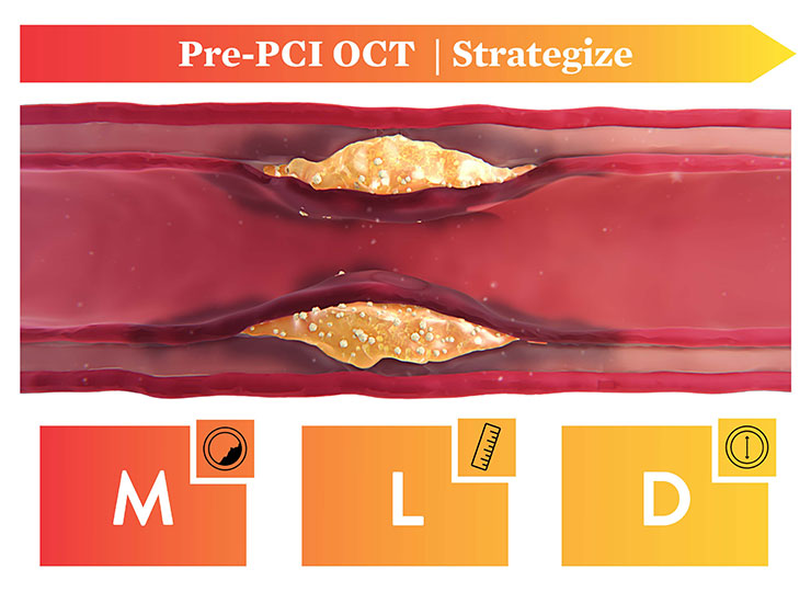
PCI guidance with OCT is easier now with the development of the standardized step-by- step workflow (also referred as algorithm), MLD MAX, which is mnemonic for Morphology, Length, Diameter, Medial Dissection, Apposition and Xpansion. Using OCT with MLD MAX workflow can improve stent expansion4 without additional contrast while reducing radiation exposure compared to angiography-guided PCI.5 Stent expansion is linked to better PCI outcomes.6
The OCT catheter is intended for the imaging of coronary arteries. Available catheters include Dragonfly Opstar™ and Dragonfly™ Optis™ catheter.
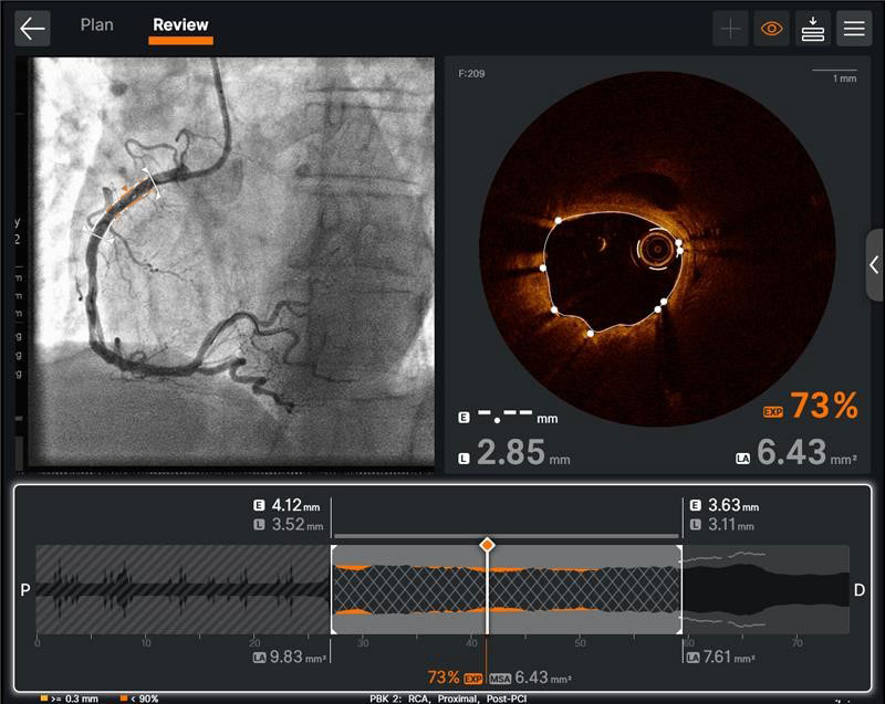
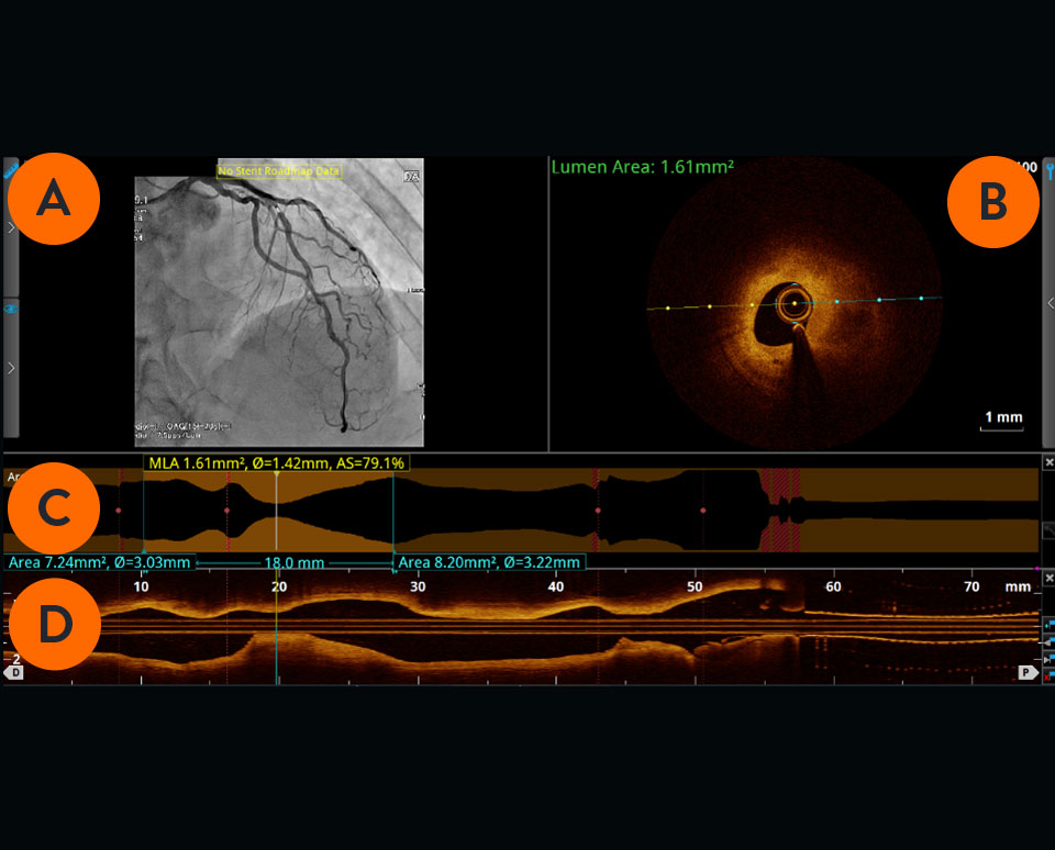
The cross-sectional area of the vessel is visualized along the entire length of the examined vessel segment. It allows precise determination of the vessel diameter (mean reference diameter) and the length of the altered segment. The white marker is located at the position of the cross-section shown.
Achieving optimal expansion is proven to reduce rates of major adverse cardiac events during PCI9. Proper expansion confirmed by imaging results in safety and efficacy benefits.6
OCTprinciple
To create an OCT scan, an OCT catheter is inserted into the vessel and an infrared laser is used to scan the vessel wall in a spiral-like manner. The laser beam penetrates the tissue 2-3mm deep, is reflected from there and returned to the OCT device via the catheter for evaluation.7
Due to the OCT systems’ high acquisition speed, images of pullback can be produced and visualized in a matter of seconds. The system provides precise information about the scanned vessel segment.7
OCTin Cardiology
Information contained herein for distribution outside the U.S. only.CAUTION: These products are intended for use by or under the direction of a physician. Prior to use, reference the Instructions for Use, inside the product carton (when available) or online for more detailed information on Indications, Contraindications, Warnings, Precautions and Adverse Events.Illustrations are artist's representations only and should not be considered as engineering drawings or photographs.Unless otherwise specified, all product names appearing in this Internet site are trademarks owned by or licensed to Abbott, its subsidiaries or affiliates. No use of any Abbott trademark, trade name, or trade dress in this site may be made without the prior written authorization of Abbott, except to identify the product or services of the company. ™ Indicates a trademark of the Abbott group of companies. ‡ Indicates a third party trademark, which is property of its respective owner.© 2024 Abbott. All Rights Reserved.MAT-2001111 v7.0
The following content is intended for Healthcare Professionals except for those in France. Some of the content is not in compliance with the French Advertising law N°2011-2012 dated 29th December 2011, article 34.
OCTfull form
The hardware that runs the OCT software. OPTIS™ Systems use optical imaging catheters that emit near-infrared light to produce high-resolution real-time images. The OPTIS ™ Imaging Systems can be integrated with the cath lab angio systems to display OCT and angio co-registration (ACR) on the same screen.
Watch this video to learn how to initiate a pullback. The step-by-step instructions are also available on the OCT screen.
Optical coherence tomography
These images provide additional information on the degree and characteristics of coronary artery disease compared to angiography which doesn’t delineate the composition of the coronary artery.1 With automated, highly accurate measurements, OCT can guide stent selection, placement, and deployment.1
Note: In the absence of EEL to represent healthy tissue find the largest lumen to avoid areas of TCFA or lipid pools so as to not land your stent edge in these high-risk areas3
Cross-sectional visualization is used to obtain detailed findings, such as structures in the lumen and in the various layers of the vessel wall.
There are three types of dissections: intimal, medial and intramural hematoma. A dissection which penetrates the medial layer and > 1 quadrant arc needs to be treated although various recommended angles exist, including > 60 degrees.9 A simplified approach used by operators is to measure a 1 quadrant arc.
Please be aware that the website you have requested is intended for the residents of a particular country or countries, as noted on that site. As a result, the site may contain information on pharmaceuticals, medical devices and other products or uses of those products that are not approved in other countries or regions
Watch this video to learn how to set up the OCT system. These step-by-step instructions are available on the OCT screen for easy guidance.
Optical Coherence Tomography (OCT) is an intravascular imaging modality that uses near-infrared light to provide high-definition, cross-sectional and three-dimensional images of the vessel microstructure.
OCTmachine
Oct systemcost
Using OCT with MLD MAX workflow, the standardized step-by-step workflow, helps to guide pre- and post-PCI decisions. Use of the workflow resulted in an 88% change in treatment decisions8 compared to angiography alone without a change in contrast usage and a 10% reduction in radiation5, as shown in the LightLab Clinical Initiative.
OCTppt
AHA/ACC 2021 Guidelines recommend intravascular imaging for PCI guidance because of limitations of angiography. OCT is recommended for stent implantation and to determine mechanism of stent underexpansion.
Ultreon™ 1.0 Software is the newest software that includes auto detection of certain components of the vessel based on artificial intelligence. The software is intended to be used only with compatible OPTIS™ Next Imaging Systems. The first generation AptiVue™ Software is intended to be used only with compatible OPTIS™ Imaging Systems.
Si vous êtes un professionnel de santé exerçant en France ou dans les territoires Français, veuillez visiter notre site en français.
Watch these videos to learn more about each step of the MLD MAX workflow: Morphology, Length, Diameter, Medial dissection, Apposition, and Xpansion. If you are already using Ultreon™ 1.0 Software, see how to apply MLD MAX and Ultreon™ Software to an OCT-guided PCI.
Ultreon™ 1.0 Software is powered by artificial intelligence (AI) and automation. It has an improved and intuitive user-interface over the previous generation.


 Ms.Cici
 8618319014500