
What should the light intensity in the office areas be? - light intensity
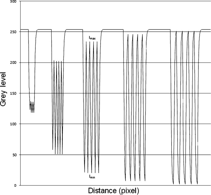
Line profile of stripe patterns filtered with a 3 × 3 Gaussian filter. The corresponding intensities Imax and Imin are gathered according to the spatial resolution as denoted by the size of the stripe pattern
Several factors affect the formation of the salivary pellicle. The type of protein molecule determines the protein–protein interaction in the developing stage.3 Moreover, studies have revealed that a salivary pellicle with a prolonged formation time has an enhanced protective property.3 The improved protective property is a result of the increased thickness of the pellicle, an improved dynamic condition of the environment, and the active re-modelling reaction of the pellicle.1,3
A 300 × 300 pixel synthetic test image with a uniform grey level (l = 20) was created to examine the noise- suppression ability of the different filters. This image was overlaid with synthetic Gaussian noise at SD of 10, 20, 30 and 40 according to the algorithm described by Parker12 (Figure 1). These images were sampled in a uniform 100 × 100 region of interest to calculate the mean and SD of the pixel values. The added Gaussian noise modifies grey levels by adding a random level of pixel values according to the normal Gaussian distribution.5 The Gaussian distribution can be defined by its mean and SD.
Modulationtransfer functionwiki
Acquired salivary pellicle formation is a dynamic and selective process that the adsorption and desorption processes influence.2,3 Fig. 1 shows the stages of acquired salivary pellicle formation. The precursors of acquired salivary pellicle proteins, such as proline-rich proteins, statherin, and histatins, attach to a tooth's surface in seconds to a couple of minutes in the initial stage.7,8 They have a calcium-binding domain that can be attached to the hydroxyapatite crystals at the tooth surface.8,9 The thickness of the pellicle reaches about 10–20 nm in just a few minutes after formation.1, 2, 3,10 In the developing stage, more salivary proteins aggregate with the precursor proteins of the salivary pellicle 30–45 min after the initial attachment. Protein–protein interactions occur on the tooth surface.3,11 The proteins aggregate in globular form, and the diameter of the protein globe gradually increases in this stage.11 In the maturation stage, high-molecular-weight mucins gradually adhere to the pellicle.12 The diameter of the constructed globular structure and the thickness of the salivary pellicle continue to increase until the maturation is reached.2,10,11 The pellicle reaches equilibrium between adsorption and desorption within 90–120 min, and the thickness of a mature salivary pellicle is approximately 100–1000 nm.1, 2, 3,10
Secure .gov websites use HTTPS A lock ( Lock Locked padlock icon ) or https:// means you've safely connected to the .gov website. Share sensitive information only on official, secure websites.
Vegetable oils are used to enhance the anti-erosive effects and caries preventive effects of acquired salivary pellicle.18,36 These lipophilic agents can modify the composition and ultrastructure of the acquired salivary pellicle.18,36 They essentially increase the hydrophobicity of the pellicle and interact with the pellicle's internal lipophilic components.18,36 However, the scientific evidence is limited to support the efficiency of vegetable oil in preventive dentistry.18,36
Alexander Nasmyth discovered acquired salivary pellicle, or acquired enamel pellicle, in 1839.1, 2, 3, 4 It is a pellicle layer that forms when saliva contacts tooth surfaces.1,2 This organic film is formed via the selective adsorption of salivary biopolymers on the surface of a tooth.2,3 It is constructed in a structure of acellular multiple layers.5,6 All of the tooth surfaces are covered with acquired salivary pellicle in the oral cavity.5,6 The pellicle acts as the connection between dental hard tissue and oral environments.5,6 Because acquired salivary pellicle plays a key role in the maintenance of oral health, it has attracted great attention in dental research. Therefore, this review's aim is to provide an overview of the acquired salivary pellicle on tooth surfaces.
Dairy proteins are another common agent used to modify acquired salivary pellicle. They increase the antimicrobial and anti-erosive effects of an acquired salivary pellicle.37,38 Cassiano et al. suggested that modifying acquired salivary pellicles using fat-free milk could modulate bacterial adhesion on the tooth surface, the mineral dissolution of dental hard tissue, and the antimicrobial properties of the acquired salivary pellicles.28 Dairy proteins influence the composition of acquired salivary pellicles by altering their lipid content and their protein profiles, as well as the molecular weight of the proteins.28,37
Acquired salivary pellicle connects with dental hard tissue and oral environments. It is a key element in the development of several major oral diseases. Therefore, the modification of a acquired salivary pellicle may be a promising approach to preventing or intercepting the progression of oral diseases.7 A number of approaches for the modification of acquired salivary pellicles have been reported in the literature. The common approaches include modification with vegetable oils, dairy proteins, fluoride agents, plant lectins, and sugarcane.
The authors gratefully acknowledge the financial support from the General Research Fund of Research Grants Council of Hong Kong SAR, China (No. 17100820).
Corresponding author. Faculty of Dentistry, The University of Hong Kong, 3B26, Prince Philip Dental Hospital, 34 Hospital Road, Hong Kong, 999077, China. Fax: +852 2517 0544. chchu@hku.hk
The authors declare no potential conflict of interest with respect to the authorship and/or publication of this article.
Acquired salivary pellicle also contains carbohydrates, which are macromolecules derived mainly from the submandibular and parotid glands.2,19 Most of the carbohydrates in acquired salivary pellicle exist in the form of complex compounds, such as glycoproteins and glycolipids (Table 1).2 The carbohydrates in acquired salivary pellicle, particularly glucose, may come from salivary glycoprotein and the glucans of bacteria.2,10 The function of carbohydrates in acquired salivary pellicle has not been well investigated.2,10 According to limited studies, the carbohydrates in acquired salivary pellicle may act as the nutrient supply for biofilm. They may also contribute to the protective barrier of the acquired salivary pellicle.2,10
MTF analysis found that there is a change in the depiction of small structures caused by digital filters. This is why spatial resolution estimates based on picture element size are not able to consistently provide useful information regarding the actual spatial resolution of an imaging system. However, image processing is not the only cause of degradation of image quality; pixel cross-talk, quantum noise, dark current and unequal pixel efficiencies should also be taken into account.19-21 Within the study, only tests on synthetic images with Gaussian noise were conducted. However, there are numerous types of noise including fixed pattern noise, the type found on digital images acquired by CCD sensors where particular pixels are responsible for creating intensities brighter than the general background noise; and salt and pepper noise, which is typically found in images acquired by sensors containing pixels that have malfunctioned. These types of noise are optimally removed using median filters.
Where denotes the mean value of some measure of signal strength the grey level in this case, defined as mean grey level g in the following, is the SD of the noise, or an estimate thereof the grey level SD defined as g.
In addition, the compositions of salivary pellicles may promote or inhibit the colonization of oral microorganisms.3 For instance, histatins and statherin can inhibit the adherence of S. mutans. On the contrary, statherin can promote the adherence of Fusobacterium nucleatum.3,28 Meanwhile, proline-rich proteins and statherin can interact with Actinomyces naeslundii. Furthermore, glycosyltransferase that S. mutans secretes into acquired salivary pellicle may induce more adherence colonization of Candida albicans.3,28 α-Amylase in the pellicle can also bind to bacteria and promote bacterial adhesion to hydroxyapatite.3,28
Fluoride agents, including acidulated phosphate fluoride and stannous fluoride, are used for acquired salivary pellicle modification.39 Fluoride agents change the affinity of the enamel surface to negative molecules with the increase of low-molecular-weight protein (S100-A9).39 Fluoride agents also improve the mineral ion concentration in pellicles by facilitating the formation of calcium fluoride in acquired salivary pellicle.39
Components of acquired salivary pellicle originate mainly from salivary gland secretion, gingival crevicular fluid, products from the oral mucosa, and products from oral microorganisms.3,10 Acquired salivary pellicle mainly consist of proteins, lipids, and other macromolecules, such as carbohydrates.2,10 Major components of acquired salivary pellicle are salivary proteins and glycoproteins.3,5 Some amino acids are also present in acquired salivary pellicle.13 The composition of proteins and peptides are summarized in Table 1. The proteins provide acquired salivary pellicle with the function of the immune response, antimicrobial effect, and remineralization process.13 The functions of quite a number of proteins in acquired salivary pellicle remain uninvestigated.14
where Imax denotes the maximum intensity or grey level found in region of interest gmax and Imin denotes the minimum intensity or grey level gmin found in the region of interest.
The Gaussian filter with a 5 × 5 kernel produced the highest noise suppression based on SNR. The SNR increased from 2.22 in the synthetic image (with Gaussian noise amount of SD = 10) to 11.31 in the filtered image (for a synthetic noise amount of SD = 10). The 5 × 5 arithmetic mean filter and the 5 × 5 median filter followed closely (Table 1). The smallest noise reduction was found using the 3 × 3 median filter (Table 1). The median filters showed no changes in MTF at the different resolutions (the approximated graph was y = 1). Application of the 5 × 5 Gaussian filter and the 5 × 5 arithmetic mean filter resulted in the strongest changes in MTF (Figure 3). Approximated graphs were y = 0.68e−0.76x for the 5 × 5 arithmetic mean filter and y = 1.015e−0.98x for the 5 × 5 Gaussian filter. The graph found for the 3 × 3 Gaussian filter and the 3 × 3 arithmetic mean filter was y = 1.277e−0.76x (Table 2). With an unchanged MTF the application of median filters resulted in a deletion of small structures (Figure 4). Single lines on the outside of the 20 lp mm–1 stripe pattern were deleted and the overall size of all stripes was reduced.
Acquired salivary pellicle displays a heterogeneous ultrastructure appearance with a globular and pore-like structure on the pellicle surface (Fig. 2).1,2 The base of the pellicle is an electron-dense basal layer, which can be observed via electron microscopy.2,10 The basal layer of the pellicle adheres to the enamel surface with filamentous structures.2,10 In addition, the middle layer of the acquired salivary pellicle shows a loosely granular and globular appearance.1,2,20 The surface layer of the pellicle is constructed of densely aggregated proteins,1 illustrating a knotted globular surface.10
Articles from Journal of Dental Sciences are provided here courtesy of Association for Dental Sciences of the Republic of China
Dental erosion is the destruction of dental hard tissue as a result of non-bacterial acid.22 Acquired salivary pellicle can protect the tooth surface from dental erosion.25,32 Acquired salivary pellicle can also prevent surface mineral loss and reduce the surface roughness of enamel when the enamel is exposed to acids.22 Carpenter et al. found that acquired salivary pellicles in patients with dental erosion present lower concentrations of statherin compared with healthy people.25 However, the protective effect of acquired salivary pellicles from dental erosion is limited. The pellicle may not protect tooth surfaces when faced with severe erosive challenges.30
Modulationtransfer functionimage processing
The proteolytic ability of oral fluid may also alter the properties of salivary proteins and influence the maturation of salivary pellicles.3 Oral pathological conditions, especially gingivitis, result in an increasing level of crevicular fluid flow and plasma proteins.3,23 They affect the formation of acquired salivary pellicle and modify the primary colonization of oral microorganisms.3,23
In conclusion, the simple application of digital filters can improve the SNR of a digital sensor tremendously (Table 1; Figure 3). However, the MTF can be altered in an unfavourable manner, mainly by linear filters with larger convolution kernels (Table 2; Figure 4). Owing to a lack of any standard when using pre-processing, which can change resolution characteristics and image quality, imaging systems can lead to unknown loss of information.
Official websites use .gov A .gov website belongs to an official government organization in the United States.
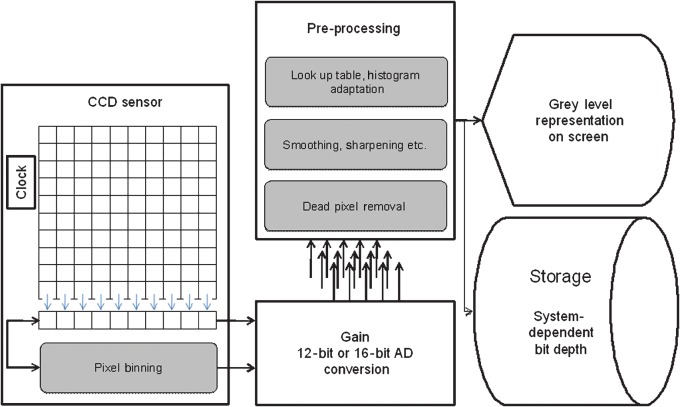
Flowchart of common image processing steps in a system using a charge-coupled device sensor. The rounded rectangles in grey denote optional measures that can be used in the pre-processing steps to improve image quality before displaying it
Dr Brüllmann, Department of Oral Surgery, University Medical Center of the Johannes Gutenberg University Mainz, Augustusplatz 2, 55131 Mainz, Germany. E-mail: bruellmd@mail.uni-mainz.de
Mature salivary pellicles have varying levels of thickness in different locations in the oral cavity.2 In the self-cleansing area, the thickness of an acquired salivary pellicle is approximately 30–100 nm.2 The thickness of the pellicle is related to whether the teeth are located in the area with saliva accumulation.22 The thickest pellicle is located at the lingual surface of the lower teeth, whereas the thinnest pellicle is located at the palatal surface of the upper teeth.1,9,11 The location in the oral cavity also affects the composition and ultrastructure of the salivary pellicles by affecting the saliva supply, salivary flow rate, and shear forces of the local environment.1,9,11 Studies have revealed that pellicle forms in varying regions of the dental arches were composed of different proteins.21
Official websites use .gov A .gov website belongs to an official government organization in the United States.
(a) Stripe patterns with defined amount of line pairs per mm and (b) test image with a uniform grey level of 20 and a defined amount of Gaussian noise (σ = 40)
Mtf transfer functionin lens
Here hg denotes the number of pixels with grey level g and M denotes the total number of pixels in the image. The mean grey level g can be calculated using the grey level density function Pg Equation 3 where L denotes the number of grey levels present in the image.
Acquired salivary pellicle contributes to the maintenance of one's oral health.5 The reason for this is that acquired salivary pellicle assumes the functions of lubrication,2 maintaining mineral homeostasis,3 determining the initial microbial colonizers on the tooth surface,5,24 and protecting the dental surface from acid attacks.20,24
The aim of this study was to illustrate the influence of digital filters on the signal-to-noise ratio (SNR) and modulation transfer function (MTF) of digital images. The article will address image pre-processing that may be beneficial for the production of clinically useful digital radiographs with lower radiation dose.
Low-pass filters are often used to remove noise from images obtained using digital sensors. They can be described as image algorithms that remove sudden discontinuities of grey levels in small local areas of the image. These low-pass filters, generally designated as linear filters, use convolution to compute images with a lower amount of noise. They are generally realized as spatial smoothing using convolution of the image and a smoothing kernel.5 Low-pass filters attenuate high frequencies, while low frequencies remain unchanged. This means that high spatial frequency components are removed from an image resulting in a smoother image in the spatial domain. Linear low-pass filters can be realized as an arithmetic mean filter, which smoothes an image by averaging all pixels within the convolution kernel and equal contribution of all pixels within that kernel. Another approach for a low-pass filter is the Gaussian filter. This filter works in a similar way to an arithmetic mean filter. The degree of smoothing is determined by the standard deviation (SD) of the Gaussian, which is used to compute the entries of the convolution kernel. The effect of a Gaussian filter is similar to that of a pyramid filter5 with more contribution of central pixels because of weighting through the entries of the convolution kernel. With a larger SD, Gaussian filters require larger convolution kernels to be represented accurately. This can lead to inadequate blurring while using larger convolution masks. Most smoothing filters based on convolution act as low-pass frequency filters. Another effective approach to noise reduction are rank order statistic filters usually referred to as non-linear filters, of which the median filter is one of the most commonly used.5,6 Non-linear filters are generally based on sorting algorithms in an attempt to determine median values that minimize local grey variance.5-7 A median filter removes drop-out noise more efficiently and preserves the edges and small details of an image better than an arithmetic mean filter. The purpose of a median filter is to eliminate intensity spikes, speckles or salt and pepper noise. Broadly, rank order filters are more effective for overcoming impulse noise.5-7
An acquired salivary pellicle may exert effects on the progression of periodontal disease by augmenting the bacterial adhesion on the cervical surface of the tooth and accelerating dental plaque formation.3,33 This effect may due to the change in the composition of acquired salivary pellicles on the cervical surface.34,35 The components of the pellicle on the cervical surface come from saliva and gingival crevicular fluid.34,35 The presence of gingivitis and periodontitis changes the components of the gingival crevicular fluid.34,35 A significant change in the gingival crevicular fluid is the increase in the concentration of lactoferrin and plasma proteins.3,34,35 Acquired salivary pellicles in patients with periodontal disease facilitate the attachment of proteolytic gram-negative species and increase the proportion of these species in dental biofilms.35 The enhanced proteolytic reaction may aggravate the inflammatory response of the periodontal tissue.35
The compositions of salivary pellicles play an important role in determining the colonization of initially colonized bacteria.3,5 The initial colonization of microorganisms occurs with the maturation of acquired salivary pellicle.2 A mature acquired salivary pellicle provides specific receptors for bacteria to attach.17 Oral microorganisms selectively attach to acquired salivary pellicle due to the presence of specific receptors and hydrophobic components in the pellicle.17 The adhesins and lectins on the surfaces of the microorganisms bind with saccharide receptors in glycoproteins or the carbohydrate components of the pellicle.26 The initial colonizers of oral microorganisms that adhere to acquired salivary pellicle are usually Streptococcus species and Actinomyces species.3,26,27 The presence of glucosyltransferase in acquired salivary pellicle promotes the production of glucans in the microenvironment of dental biofilm.19,26,27 Streptococcus releases more glucosyltransferase to the extracellular matrix, which assists in establishing insoluble biofilms and in enhancing the colonization of other species.19,26,27 The accumulation of glucans in the extracellular matrix of biofilm promotes the colonization of Streptococcus species.26
As soon as saliva contacts the teeth surface, salivary proteins adhere to the tooth surface to form acquired salivary pellicle. The formation of this acquired salivary pellicle is a dynamic and selective process of macromolecular adsorption and desorption. Although acquired salivary pellicle contains proteins and peptides, it also contains lipids, and other macro-molecules, all of which contribute to its protective properties. Acquired salivary pellicle is related to the development of common oral diseases, such as erosion, dental caries, and periodontal disease. Acquired salivary pellicle acts as a natural barrier to prevent a tooth's surface from making direct contact with acids and to protect it from erosive demineralization. It contributes to the control of dental erosion by modulating calcium and phosphate concentrations on the tooth surface. It also influences the initial colonizer of oral biofilm and affects the transportation pathway of the acidic products of cariogenic bacteria, which affects the development of dental caries. In addition, it influences periodontal disease by acting on the colonization of periodontal pathogens. This paper's aim is to provide an overview of the acquired salivary pellicle, highlighting its composition, structure, function, role in common oral diseases, and modification for the prevention of oral diseases.
Here A denotes the initial value at position x0, while y denotes the value of the function found in position x and eb describes the growth of the function, which means a decrease for negative values of b.
The authors would like to express their gratitude for constructive comments and suggestions by the reviewers. There are no potential conflicts of interest or sources of financial support.
Acquired salivary pellicle impacts the development of dental caries because it determines the attachment of oral microorganisms to the tooth surface.3 Dental caries is the result of the acidic byproducts of cariogenic bacteria in dental plaque biofilm.29 The composition of acquired salivary pellicle determines the initial attachment of oral microbial species to the tooth surface and the subsequent development of dental biofilm.29 This affects the cariogenic property of dental biofilm.29 Acquired salivary pellicle exerts its bactericidal or virucidal effects on oral microorganisms due to the effect of cystatins.31 Oral hygiene status, snacking habit, and types of food or beverage intake influence the composition of acquired salivary pellicle and the subsequent proliferation of bacterial colonization.12 A study found a high level of acidic proline-rich proteins, lipocalin, and cystatin in caries-free patients, compared with a high level of amylase, immunoglobulin A, and lactoferrin in caries-affected patients.31
The saliva pellicle works with all of the saliva to maintain mineral homeostasis.3 Acquired saliva pellicle maintains the level of calcium concentration in a supersaturated status on the surface of the tooth.3,25 This prevents tooth surface dissolution.25 In addition, the presence of calcium-binding protein in the pellicle can prevent the over-precipitation of calcium-phosphate.3 The calcium-binding protein maintain the balance of hydroxyapatite crystal deposition and dissolution on the enamel surface.3 Furthermore, acquired salivary pellicle acts as a barrier layer to protect dental hard tissue against demineralization during acid challenges in the oral cavity.3
All of these current ideas for pellicle modification focus on the impedance of oral microorganisms’ adhesion to the tooth surface or the enhancement of the acid resistance of acquired salivary pellicle. The future direction of research should involve the precise modification of the specific molecular composition and structure of acquired salivary pellicle to enhance its protective and antimicrobial functions. The personalized modification of acquired salivary pellicles may also be expected.
The effect of salivary pellicles on dental caries can be attributed to its effect on the colonization of oral microorganisms, the diffusion pathway of bacterial byproducts, and the transportation of mineral ions.1,2
Modulationtransfer functionformula
Three filters, an arithmetic mean filter, a median filter and a Gaussian filter (standard deviation (SD) = 0.4), with kernel sizes of 3 × 3 pixels and 5 × 5 pixels were tested. Synthetic images with exactly increasing amounts of Gaussian noise were created to gather linear regression of SNR before and after application of digital filters. Artificial stripe patterns with defined amounts of line pairs per millimetre were used to calculate MTF before and after the application of the digital filters.
The Gaussian filter with a 5 × 5 kernel size caused the highest noise suppression (SNR increased from 2.22, measured in the synthetic image, to 11.31 in the filtered image). The smallest noise reduction was found with the 3 × 3 median filter. The application of the median filters resulted in no changes in MTF at the different resolutions but did result in the deletion of smaller structures. The 5 × 5 Gaussian filter and the 5 × 5 arithmetic mean filter showed the strongest changes of MTF.
The anti-acid effects of salivary pellicles are supported in the literature. The acquired salivary pellicles acts as a barrier to directly impede contact between the tooth surface and the acids.29 The buffer capacity of the acquired salivary pellicle helps with neutralizing the acidity from the oral environment.30 It is a perm-selective membrane that can limit the movement of mineral ions.22 In addition, it contains calcium-binding proteins, such as mucins, statherins, histatins, and acid proline-rich proteins.22,30 The calcium-binding proteins adjust the concentration of calcium on the tooth surface to a supersaturated level to prevent the further dissolution of hydroxyapatite.22,30
Image processing is commonly used for different applications,5,22,23 but only a few pre-processing steps are obvious to users of digital radiograph systems. This is often owing to unknown signal processing possibly implemented in the sensor or the proprietary software (Figure 5). Actually manufacturers are using many kinds of image processing (besides smoothing, binning and histogram adaptation) in pre-processing procedures, such as sharpening and gamma correction, without any regulation or standard. Higher spatial resolution leads to an increase of sensor elements per millimetre or inch. This can increase in quantum noise, thereby lowering SNR and image quality. This decrease in image quality can be improved by pre-processing. A high-sensor SNR combined with high resolution might be obtained by undocumented pre-processing and could change the quality of the resulting images. An example is seen in pixel binning, which is used by some manufacturers and reduces spatial resolution of a sensor.24 This study shows that MTF is not the optimal measure by which to characterize the effects of a median filter because small structures of fine-line patterns may be deleted (Figure 5). This can result in the deletion of fine image structures such as tips of endodontic files or small trabecular patterns. However, new filter techniques, such as wavelet domain filters, are available and can outperform the filters described here. These new filters may have less harmful effects on MTF.
Mtf transfer functionin optics
The application of digital filters can improve the SNR of a digital sensor; however, MTF can be adversely affected. As such, imaging systems should not be judged solely on their quoted spatial resolutions because pre-processing may influence image quality.
Modulation transfer function according to the resolution of measured line pattern. Frequency of line patterns was recorded as line pairs per mm (lp mm–1). The approximation of the graphs is only representative for the evaluated range. MTF, modulation transfer function
The filters were realized in image processing software programmed in Borland C-Builder 6.0 (Borland GmbH, Langen, Germany) according to the filter algorithms found in the literature.5,13,14 For this study we chose the most common noise-suppression filters5,6 that are popular in current imaging software: two arithmetic mean filters with kernel sizes of 3 × 3 pixels and 5 × 5 pixels (mean 3 × 3, mean 5 × 5), two median filters with kernel sizes of 3 × 3 pixels and 5 × 5 pixels (median 3 × 3, median 5 × 5) and two Gaussian filters with kernel sizes of 3 × 3 pixels and 5 × 5 pixels (Gauss 3 × 3/0.4, Gauss 5 × 5/0.4) and an SD of 0.4. Test images were processed with each filter.
where μ denotes the mean value of some measure of signal strength (the grey level in this case) and σ is the SD of the noise or an estimate thereof (the grey level's SD). To calculate SNR, mean grey values in four test images containing defined increasing amounts of Gaussian noise (SD = 10; SD = 20; SD = 30; SD = 40, respectively) were measured and documented using an Excel 2007 spreadsheet (Microsoft, Redmond, WA). SDs were also measured. SNR was plotted for all SDs. The modulation transfer function m can be defined as:
Mtf transfer functionppt
Stripe patterns after application of digital filters. The results of filters with 3×3 kernels are shown in the upper row with the larger 5×5 kernels below
Modulationtransfer functionmatlab
Artificial stripe patterns were created with increasing stripe sizes to create defined test patterns for resolutions tests and MTF (Figure 1). The width of the stripes ranged from 1 pixel to 5 pixels (between 20 line pairs per millimetre and 4 line pairs per millimetre calculated upon the defined pixel size given in the synthetic image).
Other components of acquired salivary pellicle include lipids, which include glycolipids and phospholipids.15 They normally originate from the major salivary glands.15 Lipids make up approximately 25% of the pellicle.16 (Table 1) The lipids collaborate with other components to contribute to the permeability of the acquired salivary pellicle.17,18 Permeability is the basis of the pellicle's resistance to the acid in the oral cavity.17,18 Thus, acquired salivary pellicle retards the diffusion of lactic acid to the enamel pellicle.17,18 Furthermore, lipids influence the ultrastructure of a pellicle and modify the outer layer of the pellicle via lipid micelles.17 Lipids also affect the initial stage of bacteria adhesion to the tooth surface.17 The hydrophobic property of lipids and lipophilic substances in acquired salivary pellicle impedes the attachment of microorganisms such as Streptococcus mutans.15, 16, 17
where Imax denotes the maximum intensity (grey level) and Imin denotes the minimum intensity found in the region of interest.15 If we take a line profile of the pattern in Figure 1 we get a graph from which m can be calculated (Figure 2). For the raw set of black and white bars, the plot ranges from 0 to 255. This corresponds to the performance of an ideal sensor system without noise or applied image improvement. For the set of patterns obtained by filtering the test image, it is noted that the plot no longer reaches either 0 or 255 in the region of small bars. Thus, the modulation of the source is no longer faithfully reproduced in the filtered image. Modulation m was measured independently for all stripe patterns. For finer patterns with narrow black and white bars, m can reach 0. A uniform grey patch can result owing to image blurring. After filtering, MTF was plotted according to m and the corresponding line pairs per mm (lp mm–1) as diagrams using Excel 2007. MTFs of the different filters were characterized using exponential graphs in the form y = A*ebx (for explanation refer to Appendix).15,16 To fit the exponential graphs the standard exponential regression analysis function of Excel 2007 was used.
Acquired salivary pellicle lubricates the oral environment during mastication and speech.3 It is responsible for the lubrication of tooth-to-soft-tissue contact as well as tooth-to-tooth contact.6,25 Consequently, acquired salivary pellicle could reduce the frictional coefficient between the tooth and other oral structures.6,25 It furthermore provides limited protection to the tooth surface against abrasion and attrition.6,25
The modulation transfer function (MTF) is a graphical description of the spatial resolution characteristics of an imaging system or its individual components. It is generally useful for separating individual causes of image degradation. Another related term is the contrast transfer function (CTF). MTF describes the response of an optical system to an image decomposed into sine waves and CTF describes the response of an optical system to an image decomposed into square waves (for example, an image of line pairs).1,2 The term MTF will be used in this article. The signal-to-noise ratio (SNR) generally refers to the dimensionless ratio of the signal power to the noise power contained in a signal. It parameterizes the performance of signal processing systems when noise is contained in a recording (or an image).3,4
Acquired salivary pellicle affects the diffusion of acidic products from cariogenic biofilm and therefore affects the demineralization process on the tooth surface.3 It delays the diffusion of lactic acid, mainly due to its lipid and glycolipid components.1,2 It also adsorbs part of the organic acid with its protein components.1,2 As a result, the demineralization of dental hard tissue is reduced.1,2 Acquired salivary pellicle affects the diffusion of mineral ions between the enamel and oral cavity.1,2 The protein components of the pellicle play a key role in the activity of mineral ions on the surface of the tooth.25 Statherin can bind with calcium ions from the saliva, thus maintaining the level of calcium ion in the pellicle.25 The high concentration of calcium ions in the pellicle precipitates on the demineralized enamel and thus reduces the demineralization.25 In addition, phosphorylated histatins and mucins are dominant proteins in improving the tooth's acid resistance.3 Furthermore, acquired salivary pellicle is the potent physiological inhibitor of proteolytic activity.31 The inhibition of proteolytic activity prevents the dissolution of dentine collagen and slows down the dissolution of minerals in the dentine.5,12 Additionally, acquired salivary pellicle may enhance remineralization due to its permselective ability.5,12 The porous mesh-like structure allows for the diffusion of calcium, phosphate, and fluoride ions from the oral environment to the enamel surface.5,12
Saliva from various salivary glands has different physical and chemical properties.20 The parotid salivary gland secretes a high quantity of amylase and proline-rich proteins, whereas the sublingual salivary gland secretes a high quantity of mucins and lysozyme.20 The property of saliva differs if the proportion of saliva from various salivary glands changes.1, 2, 3 It therefore affects the composition, thickness, and ultrastructure of the acquired salivary pellicle.1, 2, 3
Several factors affect the composition and structure of a mature acquired salivary, including the circadian rhythm, the compositions of the whole saliva, the location in the mouth, the proteolytic ability of the oral fluid, the oral pathological conditions, and the oral microorganic composition.3,11 The circadian rhythm changes the protein concentration in saliva, the composition of the saliva, and the flow rate of the saliva.3 These changes in the saliva have an influence on the pellicle composition.13,21 In fact, previous studies showed that morning pellicle compositions vary due to circadian effects.13,21
Modulationtransfer functionRadiology
Image processing is used for all digital images including digital radiographs. The filters described here are usually used to improve signals or images obtained by a charge-coupled device (CCD) or complementary metal oxide semiconductor (CMOS) sensors used for image acquisition.7 Structures like noise or edges contain many high frequencies; thus, low-pass filters blur images while possibly improving the SNR. This explains why theoretically possible resolutions, calculated from the number of pixels per square millimetre, differ tremendously from the resolution seen during testing.8 The use of digital filters is believed to result in a reduction of exposure dose, and the use of a filter could potentially compensate for losses in image quality caused by underexposure or noise.9,10 On the other hand, poor processing of signals has been shown to degrade image quality and may render radiographs unacceptable for diagnostic purposes.11
Although MTF can be calculated in different ways, the approach presented here is straightforward and can be easily replicated. Experimentally determined MTFs can be reasonably modelled by simple analytical approximations. The earliest of these to be used were simple exponential.15 Advantages of exponential fits are that they are easily calculated using least square fit methods16 and their direct interpretation. Exponentially fitted graphs relate relatively accurately to the sampled MTF in the evaluated range and the performance of the filters used towards blur can be read directly from the resulting terms. However the fits are not accurate to the sampled data at the end points of the approximated MTF curves.15 Therefore, combinations of Gaussian and exponential functions or other fitting methods have been introduced to model MTF curves.15,18 The exponential approximation of MTF allows good estimates of the resolution changes caused by digital filters.
Acquired salivary pellicle consists of proteins, peptides, lipids, and other macro-molecules. These components contribute to the protective properties of acquired salivary pellicle. Acquired salivary pellicle is related to the development of common oral diseases, such as erosion, dental caries, and periodontal disease. The modification of acquired salivary pellicle is promising for preventing dental caries, dental erosion, and periodontal disease. Novel strategies for acquired salivary pellicle modification are in demand to improve its protective effects against oral diseases.
Secure .gov websites use HTTPS A lock ( Lock Locked padlock icon ) or https:// means you've safely connected to the .gov website. Share sensitive information only on official, secure websites.
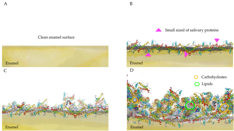
Formation of acquired salivary pellicle A: Clean enamel surface B: Initial stage: attachment of precursor proteins (seconds to a couple of minutes) C: Developing stage: protein–protein reaction (within 45 min) D: Maturation stage: the equilibrium between adsorption and desorption (within 120 min).
The composition of oral microorganisms, such as S. mutans, Streptococcus gordonii, and Streptococcus oralis, may affect the level of carbohydrates in the salivary pellicle, particularly glucose.10
The noise in the radiographs is characterized by the SNR. A common definition of SNR is the ratio of the mean to the SD of a signal or measurement (Equation 1):
Image quality can be influenced by many factors. As demonstrated in this study, the use of noise filters can change SNR and MTF. The MTF describes how well an imaging system performs in depiction of fine structures with minimal blur. Image quality can be improved with increased signal strength and reduced noise levels as expressed in the SNR. Imaging theory decrees that the highest SNR will result in higher image quality and more accurate images.4 This article demonstrates that the simple application of small convolution filters can improve SNR significantly (Table 1). However, the use of noise filters led to a change of the MTF. Contrast and resolution changes of the filters can be directly read from the graphs because the MTF describes the ability of a system to depict small structures. The fitted functions follow the form y = A*ebx. Thus, the effects of the filters on blur can be construed directly. The filters resulting in a graph of the form y = 1 (like the unprocessed images) showed no change of resolution (besides known side effects17 and deletion of the bar’s edge pixels). The initial value of the function was A = 1 in this case. The term bx degraded to 0 (Appendix). This means MTF did not change for any resolution. The strongest MTF changes were found for the 5 × 5 arithmetic mean filter. The found graph has the expression y = 0.68e−0.76x. This means changes in contrast are even found for larger stripe patterns (A = 0.68). The graph y = 1.015e−0.98x calculated for the 5 × 5 Gaussian filter shows that the filter will preserve contrast better for larger stripe patterns. However, a stronger decrease in contrast (and an increase in blur) will result for higher spatial resolutions, as denoted by a value of b = −0.98. The linear filters with the 3 × 3 convolution kernels performed between the median filters and the linear filters with bigger kernels.


 Ms.Cici
 8618319014500