
What Makes the Best Anti-Reflective Coatings - anti reflective
Now DFOV can be calculated by replacing HFOV with it in the above equation. Since AFOV and working distance are known entities, DFOV can be derived using this.
Magnification: Compound microscopes are designed for higher magnifications, typically used for observing microscopic details. Other microscopes may have lower magnification capabilities, suitable for larger specimens or samples.
Illumination: Compound microscopes often have built-in illumination systems, such as a substage light source, condenser, and diaphragm, to provide transmitted light through the specimen. Other microscopes, like dissecting or fluorescence microscopes, may utilize different lighting techniques or illumination configurations.
1. Ocular eyepiece lens to look through. 2. Objective lens, closest to the object. Before purchasing or using a compound microscope, it is important to know the functions of each part. This information is presented below. Links will take you to additional information and images.
Illuminator: A steady light source (110 volts) used in place of a mirror. If your microscope has a mirror, it is used to reflect light from an external light source up through the bottom of the stage.
Compound microscopes and other types of microscopes differ in their design and functionality. Here are the key differences between compound microscope parts and those of other microscopes:
FOV to focal length calculator
Eyepiece/Ocular: Compound microscopes commonly have a pair of eyepieces that provide binocular vision. Other microscopes may have a single eyepiece or sometimes no eyepieces at all.
Historians credit the invention of the compound microscope to the Dutch spectacle maker, Zacharias Janssen, around the year 1590 (more history here). The compound microscope uses lenses and light to enlarge the image and is also called an optical or light microscope (versus an electron microscope). The simplest optical microscope is the magnifying glass and is good to about ten times (10x) magnification.
Viewangle
e-con Systems has been raising the bar in embedded vision for close to two decades now. With a wide portfolio of MIPI cameras, USB cameras, GMSL2 cameras, FPD-Link III cameras, and GigE cameras, e-con stands true to its vision of enabling machines to see and understand the world better every single day. We have our cameras deployed in more than 300 customer products and have shipped over 2 million cameras globally.
Applications: Compound microscopes are commonly used in fields such as biology, medicine, and research, where detailed examination of small structures is required. Other microscopes, such as stereo microscopes, are utilized for examining larger objects or conducting dissections. Electron microscopes are used for high-resolution imaging of nanoscale structures.
fov和焦距的关系
However, in most cases, the FOV of a lens is expressed using DFOV or Diagonal Field of View. So, you might have to calculate the DFOV value as well. Let us see how that is done.
Objective Lenses: Compound microscopes have multiple objective lenses mounted on a rotating nosepiece, typically with magnifications ranging from 4x to 100x or higher. Other microscopes, such as dissecting or stereo microscopes, usually have fixed magnification lenses.
Condenser Lens: The purpose of the condenser lens is to focus the light onto the specimen. Condenser lenses are most useful at the highest powers (400x and above). Microscopes with in-stage condenser lenses render a sharper image than those with no lens (at 400x). If your microscope has a maximum power of 400x, you will get the maximum benefit by using a condenser lenses rated at 0.65 NA or greater. 0.65 NA condenser lenses may be mounted in the stage and work quite well. A big advantage to a stage mounted lens is that there is one less focusing item to deal with. If you go to 1000x then you should have a condenser lens with an N.A. of 1.25 or greater. All of our 1000x microscopes use 1.25 Abbe condenser lens systems. The Abbe condenser lens can be moved up and down. It is set very close to the slide at 1000x and moved further away at the lower powers.
It's important to note that the term "other microscope parts" is quite broad and can include various microscope types with different designs and features. The above differences are generalized and may not apply to every microscope outside the category of compound microscopes.
Sample Size and Depth of Field: Compound microscopes are designed to observe thin, transparent specimens placed on glass slides. They offer a narrow depth of field, allowing clear focus on one plane at a time. Other microscopes, like stereo or electron microscopes, can accommodate larger specimens or samples with more depth, providing a wider depth of field.
Objective Lenses: Usually you will find 3 or 4 objective lenses on a microscope. They almost always consist of 4x, 10x, 40x and 100x powers. When coupled with a 10x (most common) eyepiece lens, total magnification is 40x (4x times 10x), 100x , 400x and 1000x. To have good resolution at 1000x, you will need a relatively sophisticated microscope with an Abbe condenser. An Abbe condenser is composed of two lenses that control the light that passes through the specimen before entering the objective lens on the microscope. The shortest lens is the lowest power, the longest one is the lens with the greatest power. Lenses are color coded and if built to DIN standards are interchangeable between microscopes. "DIN" is an abbreviation of "Deutsche Industrial Normen". This is a German standard that has been adopted internationally as an optical standard used in most quality microscopes. A typical DIN standard microscope objective lens has a 0.7965" (20.1mm) diameter threads, 36 TPI (threads per inch), and a 55º Whitworth. Many high power objective lenses are retractable (i.e. 40XR). This means that if they hit a slide, the end of the lens will push in (spring loaded) thereby protecting the lens and the slide. All good quality microscopes have achromatic, parcentered, parfocal lenses.
field ofview中文
How to Focus Your Microscope: The proper way to focus a microscope is to start with the lowest power objective lens first and while looking from the side, crank the lens down as close to the specimen as possible without touching it. Now, look through the eyepiece lens and focus upward only until the image is sharp. If you can't get it in focus, repeat the process again. Once the image is sharp with the low power lens, you should be able to simply click in the next power lens and do minor adjustments with the focus knob. If your microscope has a fine focus adjustment, turning it a bit should be all that's necessary. Continue with subsequent objective lenses and fine focus each time.
To learn everything about choosing the right lens for your embedded vision system, please visit the article How to choose the right lens for your embedded camera application.
Field of view
Revolving Nosepiece or Turret: This is the part of the microscope that holds two or more objective lenses and can be rotated to easily change power.
fov是什么
Diaphragm or Iris: Many microscopes have a rotating disk under the stage. This diaphragm has different sized holes and is used to vary the intensity and size of the cone of light that is projected upward into the slide. There is no set rule regarding which setting to use for a particular power. Rather, the setting is a function of the transparency of the specimen, the degree of contrast you desire and the particular objective lens in use.
Prabu is the Chief Technology Officer and Head of Camera Products at e-con Systems, and comes with a rich experience of more than 15 years in the embedded vision space. He brings to the table a deep knowledge in USB cameras, embedded vision cameras, vision algorithms and FPGAs. He has built 50+ camera solutions spanning various domains such as medical, industrial, agriculture, retail, biometrics, and more. He also comes with expertise in device driver development and BSP development. Currently, Prabu’s focus is to build smart camera solutions that power new age AI based applications.
FOV to focal length
Rack Stop: This is an adjustment that determines how close the objective lens can get to the slide. It is set at the factory and keeps students from cranking the high power objective lens down into the slide and breaking things. You would only need to adjust this if you were using very thin slides and you weren't able to focus on the specimen at high power. (Tip: If you are using thin slides and can't focus, rather than adjust the rack stop, place a clear glass slide under the original slide to raise it a bit higher).
In this article, we attempt to learn what focal length and field of view are, their differences, and why it is important to understand the two concepts thoroughly when it comes to choosing a lens for your embedded vision application.
Picking the right lens considering multiple factors can sometimes be overwhelming. And this is where e-con Systems can help. While integrating our camera modules, we work closely with our customers to help them choose the best-fit lens for their application. We also extend lens fixation and lens mount customization services.
Usually, the horizontal dimension (which is nothing but the HFOV) and the working distance are given values. Using these, you would be able to calculate AFOV.
In embedded vision – in most cases – the image sensor is chosen first. This would mean that the choice of lens is heavily determined by the sensor you use (since AFOV depends on the sensor size). For a given sensor size, to achieve a wider FOV, you need to go with a short focal length lens and vice versa. However, since the focal length cannot be made shorter beyond a point, increasing the sensor size also helps to achieve a wider FOV.
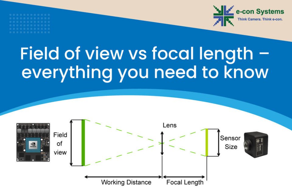
Field of view and focal length are two of the most important concepts when it comes to lenses. While focal length is the defining property of a lens, field of view can vary depending on certain other parameters. And when you select a lens for your embedded vision application, you need to make sure that you pick the right one for your sensor such that the desired field of view is achieved.
fov参数
From this equation, it can be understood that the shorter the focal length, the wider the AFOV, and vice versa. This is clearly depicted in the figure below:
So if you are looking for help in picking and integrating the right camera into your embedded system, please don’t hesitate to reach out to us at camerasolutions@e-consystems.com. Meanwhile, you could browse through our complete portfolio of cameras here.
Focal length is the defining property of a lens. It is the distance between the lens and the plane of the sensor, and is determined when the lens focuses the object at infinity. It is usually represented in millimeters. Its value depends on the curvature and the material of the lens.
Field Of View is the maximum area of a scene that a camera can focus on/capture. It is represented in degrees. Depending on how you measure it, FOV can be represented either vertically, horizontally, or diagonally as shown in the image below:
Stage with Stage Clips: The flat platform where you place your slides. Stage clips hold the slides in place. If your microscope has a mechanical stage, you will be able to move the slide around by turning two knobs. One moves it left and right, the other moves it up and down.


 Ms.Cici
 8618319014500