
What is the USAF target resolution test for 1951? - usaf 1951 resolution target
Long passfilters
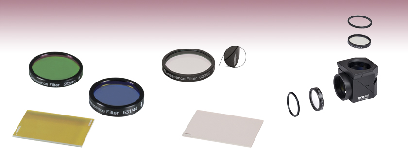
Each excitation or emission filter is housed in a Ø25 mm black anodized aluminum ring which makes handling easier and enhances the blocking OD by limiting scattering. These filters can be mounted in our extensive line of filter mounts and wheels. As the aluminum rings are not threaded, Ø1" retaining rings will be required to mount the Ø25 mm filters in one of our internally-threaded SM1 lens tubes. For customers who wish to use these filters in Thorlabs, Olympus, or Nikon fluorescence microscopes, Thorlabs manufactures a family of Drop-In Microscope Filter Cubes. Additionally, the unmounted 25 mm x 36 mm dichroic filters can be mounted in the KM2536 kinematic mount, which is designed to secure a 1 mm thick rectangular optic with minimal stress.
These filters provide excellent transmission of the desired excitation wavelength (>90%), with a sharp spectral cutoff and low transmission at other wavelengths (<0.001%). Click on the icons above to view product-specific transmission data. For additional fluorophore compatability, please see the Fluorophores tab.
Thorlabs' Dichroic Filters are designed to separate light of different wavelengths. When light is incident on the filter at a 45° angle with respect to the normal, the excitation light and its associated back reflection are reflected while the longer wavelength fluorescence signal is transmitted. These filters are unmounted, but are marked by a dash to indicate the coated side of the filter, which light should be incident on. Each filter is 25.0 mm x 36.0 mm. If your application would benefit from a round, mounted dichroic filter, consider our round Dichroic Filters. Click on the icons above to view product-specific transmission data. For further fluorophore compatability, please see the Fluorophores tab.
Absorptivefilters
Objective lenses are responsible for primary image formation, determining the quality of the image produced and controlling the total magnification and resolution. They can vary greatly in design and quality.
MidwestFilters
Filters for Fluorescence MicroscopyThe experimental setup to the right shows the typical filters used for epi-fluorescence microscopy, a form of microscopy in which both the excitation and emission light travel through the microscope objective. By carefully choosing the appropriate filters and mirrors for a given application, the signal-to-noise ratio can be maximized. As shown in the schematic to the right, three types of filters are used to maximize the fluorescence signal while minimizing the unwanted radiation. Each optical element is discussed below.
Dichroic filters are marked on the side with the dichroic coating. Light should be incident on this side for best performance.
The ocular lens is located at the top of the eyepiece tube where you position your eye during observation, while the objective lens is located closer to the sample. The ocular lens generally has a low magnification but works in combination with the objective lens to achieve greater magnification power. It magnifies the magnified image already captured by the objective lens. While the ocular lens focuses purely on magnification, the objective lens performs other functions, such as controlling the overall quality and clarity of the microscope image.
A single fluorophore can be continually excited unless it is destroyed by photobleaching (i.e. the nonreversible destruction of a fluorophore due to photon-induced chemical damage or covalent modification). The average number of excitation and emission cycles that a particular fluorophore can undergo prior to photobleaching depends on its molecular structure and the local environment; some fluorophores bleach quickly after emitting only a few photons while others are far more robust and can undergo thousands or even millions of cycles before bleaching occurs.
Colorfilters
Please Note: The excitation and emission filter housings have an arrow engraved on them that points in the recommended direction of light propagation. Dichroic filters have a marking on the side with the beamsplitter coating. Light should be incident on the marked side of the optic for optimal performance. For further fluorophore compatability, please see the Fluorophores tab.
11.6 Aberrations. Summary. Describe optical aberration. Real lenses behave somewhat differently from how they are modeled using the thin lens equations ...
The table below displays all of the fluorophores that are compatible with our filter sets. The filter set item numbers are listed across the top row and the fluorophores are listed down the first column. Scroll through the table to view fluorophore compatibility with our filter sets.
MXPLFLN objectives add depth to the MPLFLN series for epi-illumination imaging by offering a simultaneously improved numerical aperture and working distance.
DielectricFilters
Unleash creativity in your photography and videography with Prism Lens FX's unique filters and effects. Add flair to your shots.
Many microscopes have several objective lenses that you can rotate to view the specimen at varying magnification powers. Usually, you will find multiple objective lenes on a microscope, consisting of 1.25X to 150X.
There are no related clues (shown below). Referring crossword puzzle answers. ZOOMLENSCAMERA. Likely related crossword puzzle clues. None so far ...
Filter DesignOur filters are manufactured to high-performance optical specifications and designed for durability. They are produced via multiple dielectric layers deposited on a high-precision, fused silica substrate. The substrate is ground and polished to ensure that the highest possible image quality is maintained. The resulting hard-coated optics consist of filter layers that are denser than those obtained from electron beam deposition techniques, and which reduce water absorption while greatly enhancing durability, stability, and performance of the filter. Each filter layer is monitored during growth to ensure minimal deviation from design specification thickness, ensuring overall high-quality filter performance.
Leica microscope objective lenses are designed and made with superior optics. As a critical part of microscopes, they enable high-quality imaging with ...
Dichroic MirrorDichroic mirrors are designed to reflect light whose wavelength is below a specific value (i.e. the cutoff wavelength) while permitting all other wavelengths to pass through it unaltered. In a microscope, the dichroic mirror directs the proper wavelength range to the sample as well as to the image plane. The cutoff wavelength value associated with each mirror indicates the wavelength that corresponds to 50% transmission. For example, as shown in the graph to the right, the cutoff wavelength for the Yellow Fluorescent Protein (YFP) Dichroic Mirror (MD515) is ~515 nm. The Specs tab provides information on the reflectance and transmission for each type of dichroic mirror.
These excitation, emission, and dichroic filters are designed specifically for use in fluorescence imaging applications. They are fabricated at industry-standard dimensions that make them compatible with filter cubes from all major manufacturers. We offer individual filters and filter sets targeted at common fluorophores: BFP, CFP, WGFP, GFP, FITC, Alexa Fluor® 488, YFP, tdTomato, TRITC, Texas Red, mCherry, and Cyanine (CY3.5). In addition, the Fluorophores tab provides information on the alternative fluorophores suitable for these filters. These filters are also available pre-installed into our microscope filter cubes.
Terms Of Use | Privacy Notice | Cookies | Cookie Settings | About Us | Imprint | Careers | Careers | Sitemap
Edgefilters
Olympus microscope objective lenses for industrial inspections offer outstanding optical performance from the visible light to near-infrared region. At Evident, we offer an extensive selection of Olympus objectives suited to specific inspection requirements and tasks. Our MXPLFLN-BD objective is designed for darkfield observation and examining scratches on polished surfaces, while our SLMPLN objective is ideal for electronic assembly inspection. Find your ideal microscope objective today for your inspection task. No matter your requirements, Olympus objective lenses have you covered.
A prism is a polyhedron with two parallel and congruent faces forming its bases. The bases are connected by quadrilaterals. They are named according to the ...
By placing one of these mirrors into the experimental setup at 45° with respect to the incident radiation, the excitation radiation (shown in blue in the above right schematic) is reflected off of the surface of the dichroic mirror and directed towards the sample and microscope objective, while the fluorescence emanating from the sample (shown in red in the above right schematic) passes through the mirror to the detection system.
These filters provide excellent transmission of the desired emission wavelength (>90%), with a sharp spectral cutoff and low transmission at other wavelengths (<0.001%). Click on the icons above to view product-specific transmission data. For additional fluorophore compatability, please see the Fluorophores tab.
Since standard fluorescence imaging applications generally incorporate three different filters (i.e., one excitation, one emission, and one dichroic filter) to maximize the signal-to-noise ratio, Thorlabs offers these filters as a set at a savings over purchasing them separately.
Edmund Goulding was a British-born American director and screenwriter who first gained notice for films aimed at a female audience but proved adept at a ...
MicrometerThis product may not be available in your area.View ProductMPLAPON Our MPLAPON plan apochromat objective lens series provides our highest level of chromatic correction and resolution capability, along with a high level of wavefront aberration correction. View ProductMPLAPON-Oil Our MPLAPON-Oil objective is a plan apochromat and oil immersion lens that provides our highest level of chromatic correction and resolution capability. The numerical aperture of 1.45 offers outstanding image resolution. View ProductMXPLFLN MXPLFLN objectives add depth to the MPLFLN series for epi-illumination imaging by offering a simultaneously improved numerical aperture and working distance. View ProductMXPLFLN-BD MXPLFLN-BD objective lenses add depth to the MPLFLN series for epi-illumination imaging by offering simultaneously improved numerical aperture and working distance. View ProductMPLN Our MPLN plan achromat lens series is dedicated to brightfield observation and provides excellent contrast and optimal flatness throughout the field of view. View ProductMPLN-BD Our MPLN plan achromat lens series is designed for both brightfield and darkfield observation and provides excellent contrast and optimal flatness throughout the field of view. View ProductMPLFLN The MPLFLN objective lens has well-balanced performance with a semi-apochromat color correction, a fair working distance, and a high numerical aperture. It is suitable for a wide range of applications. View ProductMPLFLN-BD The MPLFLN-BD objective lens has semi-apochromat color correction and suits a wide range of industrial inspection applications. It is specially designed for darkfield observation and examining scratches or etchings on polished surfaces. View ProductLMPLFLN Our LMPLFLN lens is part of our plan semi-apochromat series, providing longer working distances for added sample safety and observation with increased contrast. View ProductLMPLFLN-BD Our LMPLFLN-BD brightfield/darkfield objective lens is part of our plan semi-apochromat series, providing longer working distances for added sample safety and observation with increased contrast. View ProductSLMPLN The SLMPLN plan achromat objective lens offers an exceptionally long working distance and the image clarity that you expect from the Olympus UIS2 optical system. It is ideal for electronic assembly inspection and other similar applications. View ProductLCPLFLN-LCD The LCPLFLN-LCD objective lenses are optimal for observing samples through glass substrates, such as LCD panels. The adoption of optical correction rings enables aberration correction according to glass thickness. View ProductLMPLN-IR/LCPLN-IR Our LMPLN-IR and LCPLN-IR plan achromat lenses have a long working distance and are specifically designed for optimal transmission in the near-infrared region (700–1300 nm wavelengths). View ProductWhite Light Interferometry Objective Lens This objective lens is designed for the Mirau style of white light interferometers and maintains a high level of temperature tolerance. The optimized numerical aperture of 0.8 provides improved light gathering, with a working distance of 0.7 mm. View Product
by H MORIGUCHI · 2016 · Cited by 87 — Having superior characteristics as a lubrica- tive material, such as a low coefficient of friction, high hardness, and chemical stability, DLC films have driven.
Lensimaging
Excitation FilterThe excitation filter only allows a narrow band of wavelengths to pass through it, around the peak fluorophore excitation wavelength. For example, as shown in the graph to the right, the bandpass region corresponding to greater than 90% transmission for the Yellow Fluorescent Protein (YFP) Excitation Filter (MF497-16) is 489 - 505 nm; incident radiation outside of this range is either partially (for regions near the transmission region) or totally (for regions further from the bandpass region) blocked by the filter.
5 days ago — Reasons to avoid. -. Too expensive for simple office use. -. Fixed USB-C cable connection. Buy it if: ✓ You're a streamer on a budget: The ...
Diamond-like-Carbon coatings demanding automotive non-ferrous material machining industrial engineering environments component surfaces Mustang Vac.
These excitation fluorescence imaging filters are specifically designed to be used in microscopy and imaging applications. Each Ø25 mm filter is mounted in a 5 mm thick black anodized housing. The housing has an arrow engraved on it that points in the recommended light propagation direction.
FluorophoresA fluorophore is a molecule or portion of a molecule that is capable of producing fluorescence. When light of the appropriate frequency necessary to excite a molecule from its ground state to an excited state is present, excitation will occur. However, once in an excited state, the molecule will be unstable. After some short period of time (typically 10-15 to 10-9 s), a photon will be released, thereby enabling the molecule to return to a lower energy state. The emitted radiation will be at a longer wavelength (lower energy) than the absorbed radiation due to the loss of energy through various mechanisms such as vibrations, sound, and thermal energy.
Emission FilterAn emission filter serves the purpose of allowing the desirable fluorescence from the sample to reach the detector while blocking unwanted traces of excitation light. Like the excitation filter, this filter only allows a narrow band of wavelengths to pass through it, around the peak fluorophore emission wavelength. For example, as shown in the graph to the right, the bandpass region corresponding to greater than 90% transmission for the Yellow Fluorescent Protein (YFP) Emission Filter (MF535-22) is 524 - 546 nm; incident radiation outside of this range is either partially (for regions near the transmission region) or totally (for regions further from the bandpass region) blocked by the filter.
To determine the beam diameter after focusing through an aspherical lens, we can use the following equation: · w_f = w_i * M * sqrt(1 + (λ * ...
Vn filter
Although dichroic mirrors play a crucial role in fluorescence microscopy, they are not perfect when it comes to blocking unwanted light; typically, ~90% of the light at wavelengths below the cutoff wavelength value are reflected and ~90% of the light at wavelengths above this value are transmitted by the dichroic mirror. Hence, some of the excitation light can be transmitted through the dichroic mirror along with the longer wavelength fluorescence emitted by the sample. To prevent this unwanted light from reaching the detection system, an emission filter is used in addition to the dichroic mirror.
These emission fluorescence imaging filters are specifically designed to be used in microscopy and imaging applications. Each Ø25 mm filter is mounted in a 3.5 mm thick black anodized housing. The housing has an arrow engraved on it that points in the recommended light propagation direction.
MXPLFLN-BD objective lenses add depth to the MPLFLN series for epi-illumination imaging by offering simultaneously improved numerical aperture and working distance.
To clean a microscope objective lens, first remove the objective lens and place it on a flat surface with the front lens facing up. Use a blower to remove any particles without touching the lens. Then fold a piece of lens paper into a narrow triangular shape. Moisten the pointed end of the paper with small amount of lens cleaner and place it on the lens. Wipe the lens in a spiral cleaning motion starting from the lens’ center to the edge. Check your work for any remaining residue with an eyepiece or loupe. If needed, repeat this wiping process with a new lens paper until the lens is clean. Important: never wipe a dry lens, and avoid using abrasive or lint cloths and facial or lab tissues. Doing so can scratch the lens surface. Find more tips on objective lens cleaning in our blog post, 6 Tips to Properly Clean Immersion Oil off Your Objectives.
Terms Of Use | Privacy Notice | Cookies | Cookie Settings | About Us | Careers | Careers | Sitemap
Click on the below to view the filter set transmission with the absorption and emission spectra of the fluorophore. The key to the right details the meaning of all check marks in the table below. Please note that absorption and emission spectra are unavailable if any is red.


 Ms.Cici
 8618319014500