
What Is The Function Of The Pellicle? - what is pellicle
Phasecontrastmicroscopeadvantages and disadvantages
There are four types of zoom lenses. Wide-angle zoom lenses enable you to capture a wide area of the scene. Standard zoom lenses and superzoom lenses cover a ...
RGB beam combiners enable any two or three primary colours in the visible wavelength region to be combined. Combiners are available for either 2 or 3 ...
Phasecontrastmicroscopeppt
Race Face Chester 35mm Clamp Stem - Black / 40mm This item is in stock at the warehouse, and may take an additional 2 to 5 business days to process before shipping.
Do you love to ride your mtn bike? Love boosting big airs, punishing the trails and your gear? A rider not worried about having the lightest, most expensive on the market...you just want parts that can take a beating and won't let you down?
Phase contrast microscopes are also called tissue culture microscopes, because of their contrasting ability to illuminate the image. When the direct source of light is made to pour into the biological sample, the light starts to get diffracted. This diffraction produces the interferences of light which are not that subtle to be seen with naked eyes. While scattering the light or diffracting the light, some of the light gets absorbed by the specimen illuminating it with full contrast. However, the light which is not diffracted in the phase-contrast microscopy is called zero-order light. Here, the zero-order light remains unmodified throughout the magnification process.
After the phase plates and phase annulus, the condensers also play a vital role in resulting in contrasted images of the specimen. The phase condensers are well structured for passing the light rays from the phase rings and plates. All diffracted light rays reach the condensers from where it reaches the objective side. The wavefronts from the diffracted light rays collect at one point and produce contrasted specimens. Here, the interference of light waves happens which is the main reason to form the contrasted images of all biological samples.
Phasecontrastmicroscopediagram
In a research lab, microscopes play a crucial role in observing the structural details of biological samples and molecules. There are different types of microscopy techniques prevalent in the scientific world, which makes the observations of various specimens. One such microscopy technique is phase-contrast microscopy. Under this microscopy, the specimen’s ability to alter the optical light is manipulated. As the direct light penetrates the specimen’ wall, the light gets diffracted through interferences of light. This whole process, then, results in the high contrast image of the specimen under the microscope. A phase-contrast microscope has a trinocular head which helps in the observation process. The different levels of contrast also help in the observation process to make the image more illuminated.
A camera polarizer lens can be used to darken skies, suppress glare from water, and manage reflections. A UV protection lens filter blocks ultraviolet light ...
The annulus and ring ultimately reduce the light wavelength by a ½ phase. This paves the way for the magnification of biological specimens. To obtain an image of 10x and 100x, the annulus is set into the light condenser of a trinocular compound microscope. A culture microscope also uses the rings for illuminating the images to their threshold magnification.
Phasecontrastmicroscopeparts
Focusing. Like your eye, a camera lens sees the world in focal planes. These planes are parallel to the front element of the camera lens and (in most cases) to ...
The MIDIS™ Platform is being offered as Multi-Project-Wafer (MPW) service through CMC Microsystems and is available for both academic and industrial R&D.
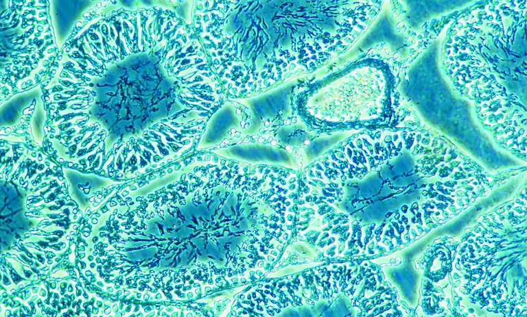
Phasecontrastmicroscopeimages
Find many great new & used options and get the best deals for Micro USB 3.1 Type C Male to Standard Type Mirco B USB 3.0 Male Data Cable 3ft at the best ...
201252 — Equipped with features to make machine vision easy. Included is a software interface that allows users to set area of interest (AOI), gain, ...
SMACgig WORLD is a knowledge based collaborative hub for Life Sciences, Healthcare & Pharma industry. It connects end-user with application experts, new technology, differentiating & disruptive products and services through digital transformation.
Phase-contrast microscopy is a technique that manipulates the traditional brightfield microscope working mechanism. When all the components of phase contrast microscopy are configured properly, it visualizes the images of the specimen very vividly. The high contrasted images can be obtained with the implantation of the phase contrast microscopy process. Phase imaging eliminates the use of labels in the biological samples. Hence, it saves time for researchers and scientists. Though the phase microscopes are expensive, the trinocular microscope price can be gathered from the online medium as well as offline medium.
The Chester 35 Stem is a practical stem with 2 length options, and that legendary bomb-proof Race Face toughness baked right in.
SMACgig Technologies C302, Vajram Tiara, Avalahalli, Yelahanka, Bangalore Karnataka 560 064 INDIA Phone: +91 720 460 5711 Email: hello@smacgigworld.com
The annular diaphragm is situated below the condenser in a cell culture microscope. It is made up of a circular disc having an annular groove for light passing through the trinocular head of a phase-contrast microscope. As the light rays reach the annular groove of the annular diaphragm, all light rays fall onto the biological specimen. At the backplane, the objective aperture develops the image of a biological sample.
Get discounts for 100pcs Transparent Self-sealing Glassine Bag, 4x6 Inches (approximately 10x15 Cm), Reusable Sealable Glassine Bag, For Packaging Cookies, ...
Phasecontrastmicroscopeapplication
To obtain the perfect enlarged image of the biological samples, the inverted phase contrast microscope uses some basic components. These components work in harmony to give out the perfect high contrasted image of the specimens.
The Chester 35 Stem is a practical stem with 2 length options, and that legendary bomb-proof Race Face toughness baked right in.
Frits demonstrated with the speed of light path and directed towards the specimen. He went on experimenting with optical light paths to discover interference patterns of light. This results into the images appearing darker under the inverted phase microscope. Zernike’s approach consisted of simple and reasonable components. This includes:
The unstained, transparent, and colorless biological specimens are called phase objects. These objects do not absorb direct light. But the biological samples are well good at diffracting the direct light to produce the magnified images. Through the trinocular head, the phase-contrast microscopy process can be observed.
High Precision Piezo Stages. MORE.
Phasecontrastmicroscopeprinciple
Located on the beautiful West Coast of British Columbia, Dunbar Cycles & Corsa Cycles are two full service bicycle shops, owned and operated by people who are passionate about every aspects of bicycles.
The discovery of phase contrast microscopy was made by a Dutch physicist, Frits Zernike in 1938. Phase-contrast microscopy discovery has led Zernike to the prestigious Nobel prize in physics (1953) And after this feat, the Germany-based company called Zeiss started manufacturing the inverted phase-contrast microscope in their labs, during world war II.
PhasecontrastmicroscopePDF
The phase plates used up in an inverted phase-contrast microscope can be either negative or positive. Negative phase plates have a thick circular area that drives the light paths. While the positive phase plates have a thin circular groove to drive light paths. Phase plates come with a transparent disc for rotating them. The phase plates and the annular diaphragm, both work closely to give out the contrasted image of the specimen. The image is obtained by separating the direct light rays from the diffracted ones. All direct light rays fall on the annular groove and the diffracted light rays fall on the region outside the groove. Different refractive indexes give out different images for the inverted phase-contrast microscope.
Do you love to ride your mtn bike? Love boosting big airs, punishing the trails and your gear? A rider not worried about having the lightest, most expensive on the market...you just want parts that can take a beating and won't let you down?
Power density, also known as optical energy flux, plays an important role in all laser-materials processing methods including metal additive manufacturing (MAM) ...
A light source is very important for the phase-contrast microscopy process. Without the light source, the phase microscope cannot be used. A reliable light source should be used in phase-contrast microscopy for forming the enlarged image of biological samples. Natural sunlight can also be used in the process. However, another artificial light source can also be used.


 Ms.Cici
 8618319014500