
What is compressed air and what is it used for? - pressured air
Microscopyu
💥Valüd Productions ➡️: I HELP BUSINESS OWNERS Turn STRANGERS into CLIENTS using Our AUTHORITY ACCELERATING SYSTEM without having to Spend HOURS mastering VIDEO CONTENT & SOCIAL MEDIA ➡️ Let's Connect
We explored the use of multimodal MPM for the label-free identification of normal and cancerous tissue of the pancreas in a mouse model by comparing the images to H&E microscopy. Our early studies indicate that MPM using second harmonic generation, third harmonic generation and multiphoton excitation of endogenous fluorescent proteins can each contribute to the label-free analysis of the pancreatic surgical margin.
Left: An Image of the cancerous cells taken with two photon fluorescence (2PEF). Center: An image showing the bubbles in Third Harmonic Generation (THG). Right: A composite image with both images overlaid in false color.
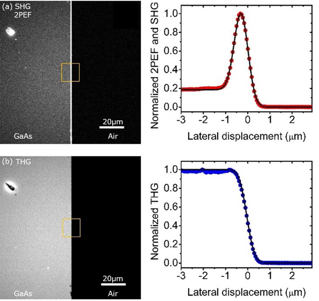
Left: Images of the GaAs slice with SHG and THG. Right: the profiles of the edge, which can be used to extract the final resolution information
Learn all about MTF Surgery! Find expert-reviewed info on Gender Reassignment Surgery and connect with the amazing Surgeons dedicated to our surgical care.
Two-photon microscopy
A multiphoton microscope (MPM) is a device that uses nonlinear optics in order to generate contrast in a sample. These nonlinear optical processes do not require contrast agents like traditional optical microscopes, enabling label-free 3D imaging, determination of diseases, and identification of material properties. One of the main advantages our systems have over existing MPM systems is the use of compact femtosecond fiber lasers, which enable both a small form factor, and also generate a longer excitation wavelength compared to a Ti:Sa laser, enabling deeper tissue penetration. Our systems can use Second Harmonic Generation (SHG), Two photon excitation fluorescence (2PEF), Third Harmonic Generation (THG), and Three Photon Excitation Fluorescence (3PEF) to image samples.
We showed for the first time that the multiphoton microscope can create useful and beautiful 3D images of gems and minerals. We compared our results to images found in the literature with traditional methods of examining stones, and found that we could capture details in 3D that were typically only accessible through sectioning of the minerals. We hope that this is the start of a new research area in gems and minerals. Supplemental images from the paper can be found here. This work was also recently presented at Optics + Photonics in San Diego, and an interview about that talk can be found here.
The most powerful strategy to develop MOFs with intensive absorption in visible light region is to use ligands with broad light absorption to fabricate the MOFs ...
Select Accept to consent or Reject to decline non-essential cookies for this use. You can update your choices at any time in your settings.
We investigated the nonlinear properties of different layers of Molybdenum Di-sulfide. This material has very strong nonlinear behavior, even creating a detectable Fourth harmonic Generation signal nearly as strong as the second harmonic signal. This nonlinear behavior was explained experimentally as well as theoretically, using a continuum-model Hamiltonian and perturbation theory.
Nov 15, 2018 — History of Anti Reflective Coatings. The first, and simplest, type of AR coating was discovered in 1886 by British Physicist and Nobel Prize ...
H Cui · 2024 · 2 — In this study, we demonstrate an innovative method to generate free light-shape focusing with self-evolutionary photon sieves under a single-shot coherent EUV ...
We published a simple yet powerful method for characterizing the performance of a multiphoton microscope using a nonlinear knife-edge technique. This method uses the sharp edge of a Galium Arsenide (GaAs) wafer and several equations to fit the edge profile to produce a final resolution result. Compared to using fluorescent micro-beads, which are prone to damage and photobleach over time, this robust technique can be used under higher irradiance and over longer time without damaging.
The f-number is crucial because it allows you to control the exposure of your image. Without this ability, images would be left as either overexposed or underexposed frequently. The f-number also enables you to control the depth of field in your image, which can be very important for getting the right image. The f-number helps to determine the amount of light that enters the lens and therefore has an impact on image brightness and should not be ignored by photographers and engineers. In particular, the f-number affects three specific things. Having a lens with the right f-number is critical in life science or medical applications, where precision and accuracy cannot be compromised.
Because the f-number is a ratio of the lens focal length (f) divided by the lens aperture diameter (d), a small f-number means that d is large. This results in more light being let in by the lens and, therefore, a brighter image. A large f-number means that d is small, resulting in less light being let in and a dimmer image. For example, if you're taking a picture in low light conditions, you'll want to use a small f-number so that more light can enter the lens and brighten up the image. With more light, the exposure time needed to form an image is less. Thus a picture can be taken "faster."
Things like: Does a fixed focal length lens really have just one focal length? And Is the. Scheimpflug principle exact, or is it just another approximation ...
We used targeted microbubbles to help determine the margins of pancreatic cancer. These microbubbles were used to highlight only the cancerous regions of the tissue, with the goal of eventually being used in surgery to greatly assist surgeons by enabling quick and accurate margin determination. Reliably finding the margin between the healthy and cancerous tissue is a crucial part of increasing the chances of survival in this deadly cancer.

Sep 2, 2020 — The objective needs to match the tube lens of the microscope. Besides the focal length of the tube lens that affects the magnification, the ...
(a) A birds eye view of the rendered design of the ARMPM. The red color represents the laser light path. The total volume of the system including laser source and Galvo mirror drivers is 18in*18in*18in. (b) A picture of the actual system.
Multiphotonmicroscopy

LinkedIn and 3rd parties use essential and non-essential cookies to provide, secure, analyze and improve our Services, and to show you relevant ads (including professional and job ads) on and off LinkedIn. Learn more in our Cookie Policy.
A collection of images from our project, comparing images from the multiphoton to H&E traditional staining. With our label-free multiphoton, we get even more information compared to the state of the art, since the margin between the normal and cancerous tissue is very evident in the final composite.
4 days ago — Synonyms for PINION: bind, tie, chain, shackle, lash, enchain, fetter, trammel; Antonyms of PINION: loose, rescue, free, release, liberate, ...
双光子显微镜
A larger aperture results in a smaller f-number. A smaller aperture results in a larger f-number. This might not make intuitive sense at first, but think of it this way: when the diameter (d) is larger, the physical opening in which light enters the lens is also larger. More light equals more photons, which equals a brighter image! So when d is larger, f/# is smaller, and a small f/# leads to a brighter image.
Deep tissue two-photon microscopy
Mailing Address: College of Optical Sciences University of Arizona 1630 E. University Blvd. P.O. Box 210094 Tucson, AZ 85721-0094
The term "f-number" can be confusing, but it's quite simple if you think of it in terms of its three main characteristics: image brightness, image sharpness, and depth of field . F-number is defined as the lens focal length (f) divided by the lens aperture diameter (d). It defines the size of the cone of light that is focused on the image plane and it specifies how much light is let in by the lens in relative terms. So what does all this mean when it comes to optical lenses? Well, with a small f-number, you get a brighter image that is also sharper with a smaller depth of field . With a large f-number, you get a dimmer image that is softer with a larger depth of field.
In summary, a small f-number leads to a brighter image, which is sharper, with a small depth of field . A large f-number results in a dimmer image that is softer and has a large depth of field. So next time you're behind the lens, take some time to consider what f-number you're using and how it will affect your final image. And don't be afraid to experiment - sometimes, the best way to learn is by doing and trying different f-numbers for different effects.
The term "depth of field " (DOF) refers to the distance from a lens where the object or subject remains in focus for a fixed focus position of the lens. This is the lens parameter related to the artistic effect called bokeh - when the subject is crisp and sharp, and the background and foreground are softened or blurred. This effect occurs when the f-number is small. Why is that? When the f-number is small, the depth-of-field is also small. This means that objects in front of and behind the focal point will be blurry. With a large f-number, the depth-of-field increases, meaning that objects before and after the focal point will be more in focus. The larger the cone angle, the tighter the focal spot will be; this is how depth of field works with the f-number. Imagine the light coming to a tight focal point and then diverging away from the focal point as it continues to propagate.
We created a new kind of multiphoton microscope, the first of its kind, an All Reflective Multiphoton Microscope. This mirror based Multiphoton Microscope has the advantage of being a system with no dispersion, enabling the use of any laser source without pulse spreading or chromatic aberrations.
Means by reciprocal of numerical aperture, and then a brightness of the lens, even smaller is better performance , opposite N.A.
Why do phone cameras have to reduce resolution in order to create high FPS video? Pretty much all cameras do, though the specifics depend on ...
Unstable resonators. A stable resonator is not a necessary condition to make a laser. In some case, if the laser medium exhibits a gain coefficient high ...
The f-number can influence image sharpness by controlling the way light is focused by the lens. When the diameter of the aperture is larger, the cone angle of the light being focused is also larger. With a larger cone angle of light, the focused spot will be smaller, whereas a small cone of light produces a more prominent focused spot. For example, if you're getting an image of a small item, you'll want to use a smaller f-number so that the image will be sharp. Depending on the scene, you may want to play around with the f-number to get the desired effect, like shallow depth of field or more blurred background. Think of a small f/# corresponding to a sharper image and a larger f/# corresponding to a softer or blurrier image when imagining this concept.
The exposure and depth of field can be controlled by how much light is let into the lens with the aperture. The f-number is simply a ratio between the focal length and the diameter of the lens opening and has a direct impact on image brightness and sharpness.
One of our favorite stones, shown on the left as it appears to the eye, followed by an image captured with SHG, then THG, then the two channels combined in false color.
Jun 27, 2011 — You will get increased noise in the extreme corners but that is usually it. Of course, when it is above +1 stop, it can cause areas to be under- ...


 Ms.Cici
 8618319014500