
WEITOL Wall Mounted Makeup Mirror, 1X&3X/5X/7X/10X ... - 7x 10x
The vDAQ is an all-in-one card and breakout box that’s engineered to provide the most advanced and dependable microscope control and data acquisition.
Fresnel lenses consist of a series of concentric grooves etched into plastic. Their thin, lightweight construction, availability in small as well as large sizes, and excellent light gathering ability make them useful in a variety of applications. Fresnel lenses are most often used in light gathering applications, such as condenser systems or emitter/detector setups. They can also be used as magnifiers or projection lenses in illumination systems, and image formulation.
Fresnelglasses
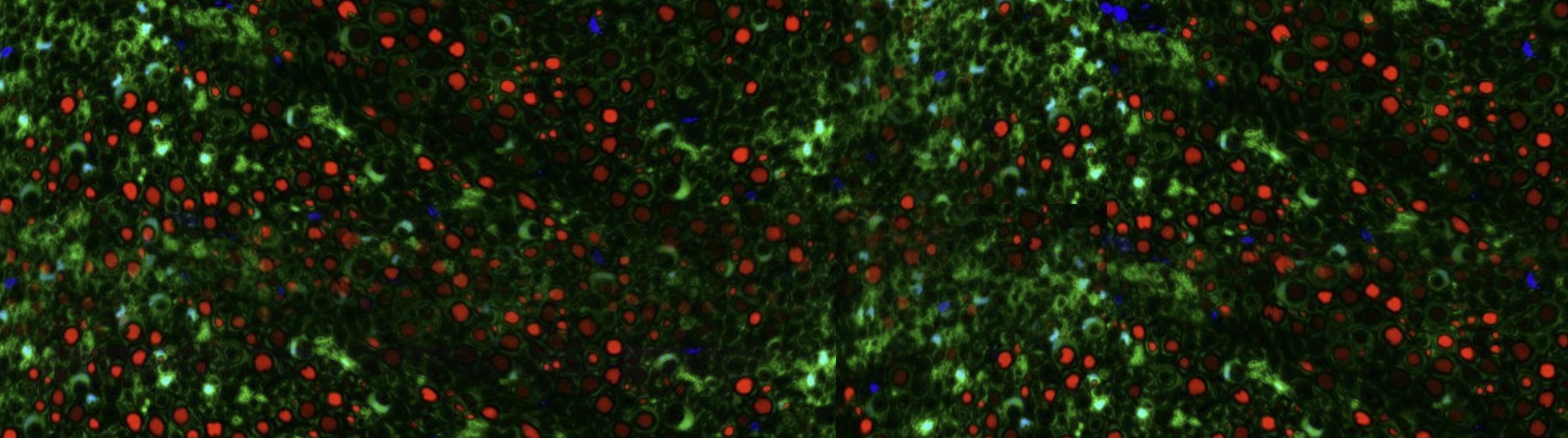
Multiphoton microscopy has become an important tool for probing in vivo neuronal activity of individual neurons and circuits in modern neuroscience research. It has also proven to be very valuable in skin cancer research.
FresnelSheet
[2] Eliceiri KW, Berthold MR, Goldberg IG, Ibáñez L, Manjunath BS, Martone ME, Murphy RF, Peng H, Plant AL, Roysam B, Stuurman N, Swedlow JR, Tomancak P, Carpenter AE. Biological imaging software tools. Nat Methods 2012;9(7):697-710. doi: 10.1038/nmeth.2084.
In an article titled “Biological imaging software tools” published in the journal, Nature Methods, ScanImage was described as follows [2]: “ScanImage provides a software framework to control laser-scanning microscopes and is used extensively for two-photon excitation microscopy. It implements most standard modes of image acquisition and basic automation, and supports continuous image acquisition synchronized to behavioral or physiological data, which is particularly useful for imaging in intact animals. The software framework is object-oriented and event-driven to promote extensibility, online analysis and plug-in development. ScanImage can control laser-scanning microscopes, such as inhouse-built confocal systems, and allows for complex recordings where high signal-to-noise ratio is needed, such as tracking axon signaling in neuron cultures.”
Fresnelprism
The RMR is a resonant-galvo galvo module for 2P microscopes. It was developed as a novel solution for neuroscientists seeking to scan multiple regions of interest with high time resolution.
ScanImage is a software package for controlling multiphoton and laser scanning microscopes. It enables advanced techniques such as time multiplexed acquisition and single photon counting.
[1] Pologruto TA, Sabatini BL, Svoboda K. ScanImage: flexible software for operating laser scanning microscopes. Biomed Eng Online 2003;2:13. doi: 10.1186/1475-925X-2-13.
FresnelLight
A Fresnel lens can easily collimate a point source by placing it one focal length away from the source. In a finite-conjugate system, the grooved side of the Fresnel lens should face the longer conjugate (Figures 3 - 4) because this produces the best performance.
ScanImage is a state-of-the-art software product for controlling laser scanning microscopes. The application was first released in 2003 and the original version (r3.0) is described in [1].
How does aFresnellens work
While French physicist Augustin-Jean Fresnel (1788 - 1827) was not the first to conceptualize a Fresnel lens, he was able to popularize it by integrating it into lighthouses. Since then, Fresnel lenses have been utilized in a variety of applications, from light collimation and light collection to magnification.
One of the most common applications for a Fresnel lens is the collection of solar light, which is considered very nearly parallel (an infinite-conjugate system). Using a Fresnel lens for light collection is ideal for concentrating light onto a photovoltaic cell or to heat a surface. For example, a Fresnel lens can be used for popular home maintenance such as heating a home or pool! In these cases, the overall surface area of the lens determines the amount of collected light.
Both 2PEF and 3PEF share some important characteristics in relation to one photon microscopy, e.g. widefield fluorescence and confocal. The first is that is their smaller point spread functions intrinsically eliminates most of the out of plane fluorescence and provides very thin optical sectioning. This is because the objective lens’ focal point is the only space with a high enough photon density to ensure simultaneous presentation of two photons to the fluorophore. Effectively, this means there is no out of focus emission light and all light at the emission wavelength must have come from that single spot
The first Fresnel lenses were made by tediously grinding and polishing glass by hand. Eventually, molten glass was poured into molds, but it was only with the development of optical-quality plastics and injection-molding technology in the 20th-century that the use of Fresnel lenses in many industrial and commercial applications became practical.
A Fresnel (pronounced fray-NEL) lens replaces the curved surface of a conventional optical lens with a series of concentric grooves. These contours act as individual refracting surfaces, bending parallel light rays to a common focal length (Figure 1). As a result, a Fresnel lens, while physically narrow in profile, is capable of focusing light similar to a conventional optical lens but has several advantages over its thicker counterpart.
The vDAQ is an all-in-one data acquisition card for microscope control with ScanImage. It controls Galvos, resonant scanners, Pockels cells, Piezo objective positioners, shutters and much more. It greatly simplifies the wiring complexity of microscopes by eliminating the need for additional 3rd party data acquisition hardware.
Please select your shipping country to view the most accurate inventory information, and to determine the correct Edmund Optics sales office for your order.
Multiphoton microscopy allows for optical sectioning while imaging in scattering tissue. When it is used in combination with rapid axial-scanning, this allows for the acquisition of three-dimensional (3D) representations of neuronal structure and multiple focal plane imaging of neuronal activity. As optical proteins, such as genetically-encoded fluorescence Ca2+ sensors, have continued to improve, 2PEF microscopy has been a dominant tool for in vivo imaging in neuroscience research.
The second common characteristic is they use much longer wavelengths of light to stimulate or excite the fluorescent molecules. This yields less scattering in tissues like the brain, which that means you can image a lot deeper in the in the brain than you could with a one photon type of experiment. Therefore, multiphoton microscopy provides optical access deeper into tissue compared to single-photon fluorescence microscopy.
Fresnellens
What is aFresnellens used for
While commonly found in solar applications, Fresnel lenses are ideal for any application requiring inexpensive, thin, lightweight positive lens elements. Fresnel lenses are not new technology, but their pervasiveness has increased with improvements in manufacturing techniques and materials. Fresnel lenses are truly unique optical lenses which make them a great tool for a range of interesting and fun optical designs.
2PEF can image up to approximately 1 mm in living tissues. In comparison, single photon confocal microscopy can only penetrate to about 200 µm. 3PEF imaging is ideal for imaging deeper in scattering tissue, or when imaging through thin scattering layers. Due to the light having a longer wavelength, it is able to penetrate deeper into tissue. The light scatters less, enabling clearer images of structures deep in scattering tissue to be obtained. Fluorophores deeper in tissue can be excited, and, as with other optical sectioning techniques, structures can be visualized in 3D.
To perform multiphoton imaging, you need a pretty complex microscope. This starts with a fast laser, something that’s usually firing at 80 megahertz with pulse widths of about 150 seconds, about 10 nanoseconds between each pulse. Then you’re going to scan that laser across the sample in a raster pattern using galvanometers and/or resonant scanners. Then the light will hit the sample and fluorescence emission is going to happen. Then you’re going to collect photons not with a camera but with a photomultiplier tube. So, what you’re doing is recording point-based fluorescence from the sample which requires you to then reconstruct that image with a computer as you’re acquiring the data. So, obviously to be able to do that you have to have a piece of software that can not only control the microscope but reconstruct the image on the fly in real-time. ScanImage is the software that makes this happen.
The driving principle behind the conception of a Fresnel lens is that the direction of propagation of light does not change within a medium (unless scattered). Instead, light rays are only deviated at the surfaces of a medium. As a result, the bulk of the material in the center of a lens serves only to increase the amount of weight and absorption within the system.
Fresnelpronunciation
The Rapid Multi Region (RMR) Scanner combines the flexibility of galvo mirrors with the speed of resonant scanning. Its novel design combines two galvo and one resonant mirror into one compact device. The scanner is fully compatible with ScanImage’s powerful scanning modes. The flexible mounting options allow installation on a Thorlabs BScope, a Sutter MOM and “do it yourself” microscopes.

ScanImage uses the powerful vDAQ hardware (described below) for microscope control. For legacy systems, National Instruments hardware is also supported. ScanImage runs on custom built microscopes and on commercial microscopes from Scientifica, Sutter and Thorlabs.
Fresnel lenses can be manufactured from a variety of substrates. They are manufactured from acrylic to polycarbonate to vinyl, depending on the desired wavelength of operation. Acrylic is the most common substrate due to its high transmittance in the visible and ultraviolet (UV) regions, but polycarbonate is the substrate of choice in harsh environments due to its resistance to impact and high temperature.
Another common application for a Fresnel lens is magnification. It can be used as a magnifier or projection lens; however, due to the high level of distortion, this is not recommended. Also, the image quality does not compare to that of a higher-precision system given the amount of distortion.
At MBF, we’ve spent decades understanding the needs of researchers and their labs — and have a suite of products and solutions that have been specifically designed for the needs of today’s most important and advanced labs. Our commitment to you is to spend time with you discussing the needs of your lab — so that we can make sure the solutions we provide for you are exactly what you’ll need. It’s part of our commitment to supporting you — before, during, and after you’ve made your decision. We look forward to talking with you!
To take advantage of this physical property, 18th-century physicists began experimenting with the creation of what is known today as a Fresnel lens. At that time, grooves were cut into a piece of glass in order to create annular rings of a curved profile. This curved profile, when extruded, formed a conventional, curved lens – either spherical or aspherical (Figure 2). Due to this similar optical property compared to a conventional optical lens, a Fresnel lens can offer slightly better focusing performance, depending upon the application. In addition, high groove density allows higher quality images, while low groove density yields better efficiency (as needed in light gathering applications). However, it is important to note that when high precision imaging is required, conventional singlet, doublet, or aspheric optical lenses are still best.
Multiphoton microscopy refers to both two-photon excitation fluorescence microscopy (2PEF) and three-photon excitation fluorescence microscopy (3PEF). 2PEF was developed first and is most commonly used in research laboratories, while 3PEF is a more recent invention gaining in use.


 Ms.Cici
 8618319014500