
Understanding Rare Earth Materials - rare earth materials
Whatare the importance of amicroscope
Microscopy is a varied and valuable science that has many different forms and uses several different techniques. Whatever the object to be studied, there will be a type of microscopy that is suitable. Some techniques are very expensive, so the type of microscopy and sample preparation chosen for a particular project will always depend on the objectives and the budget available.
Microscopy is continually developing as science and modern manufacturing techniques allow for better and better equipment to be produced and higher image quality and greater magnification.
Note: This is an updated and expanded version of an article that appeared in the October 2013 issue of Sky&Telescope magazine.
Polarizing Microscopy incorporates a polarizing filter into the microscopes so that only a single wavelength of light is transmitted. This type of microscopy finds use in studying crystals and detecting asbestos fibers
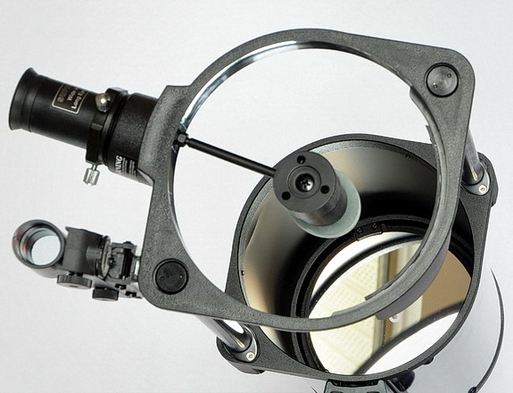
If you’re working with a Newtonian reflector, it helps to have someone else make the adjustments while you watch and give instructions. If you’re collimating a SCT, you can probably do the necessary adjustments to the secondary mirror yourself. Proceed slowly and methodically. Remember — if your scope has three adjustment screws, it should be possible to collimate it using only two of them, which makes the task somewhat simpler.
Registered members can chat with Azthena, request quotations, download pdf's, brochures and subscribe to our related newsletter content.
Trevelyan, Oliver. "What is Microscopy?". AZoLifeSciences. 24 November 2024. .
Explore the groundbreaking work of Mark Bear, a leading figure in neuroscience, as he shares insights on synaptic plasticity, learning, and the future of neurological research.
Compoundmicroscope
Inverted Microscopy is where the light source is above the sample and the lenses are below it. This type of microscopy is particularly useful in biological research
Stereo microscopy involves a microscope that has two matched microscopes side by side so that each eye has an individual view of the sample. They are used for dissection, moving microscopic tools, and examing electronic components
What ismicroscopes
In addition to different types of microscopes, different treatments can be applied to samples such as dye and fluorescent substances to enhance the image or to highlight certain components that need to be studied.
Deflection can be caused by a mechanical, electrostatic, magnetic, chemical bond, Vander Waals, or capillary forces. The probe can be in continuous contact with the surface or, if it is very soft, sometimes the probe will rapidly tap the surface continually.
Metallographic microscopy is used in forensics and diagnostic microscopy where a light that shines onto an opaque surface and is reflected back into the microscope for examination.
Scanning Probe Microscopy (SPM) can analyze from a nanoscale down to individual atoms. The instrument consists of a sharp tip (as small as one atom) on a cantilever which is moved across a surface to measure deflections. These deflections are recorded and used to produce an image by deflecting a laser off the top of the cantilever.
What isamicroscopeused for

5. When you’re think you’re done, centre the star, defocus it, then slowly refocus while paying close attention to the dark hole at the centre of the star image. If your scope is well collimated, the bright rings in the defocused star image will collapse down concentrically around the shrinking black centre.
Atomic Form SPM measures electrostatic forces and Magnetic Form Microscopes using Magnetic forces and Scanning Tunnelling Microscopes which measure the current between the probe and the cantilever.
Scanning Electron Microscopy where high-energy electrons are scanned across a sample and various emissions are emitted and recorded. This type of microscopy can magnify five to 500,000 times
Microscope
While we only use edited and approved content for Azthena answers, it may on occasions provide incorrect responses. Please confirm any data provided with the related suppliers or authors. We do not provide medical advice, if you search for medical information you must always consult a medical professional before acting on any information provided.
The word microscope is derived from the Greek “mikros”, meaning small and, “skopein” meaning to see. A microscope is an instrument used for looking at objects that cannot be seen with the naked eye and microscopy is the science of using a microscope.
3. Move the out-of-focus star around the field of view by re-aiming the scope until you find the location where the black hole in the star image is centred, or most nearly so. That’s the sweet spot. Your task is to move the defocused star from there to the centre of the eyepiece field by using your scope’s collimation screws.
There are several variations of optical microscopy, one of which is the Compound Microscope. This is the most well-known type of microscope and consists of a tube containing an eyepiece lens at one end, and one or more objective lenses at the other end with different strengths that can be interchanged dependent on what is being studied. There will also be a focussing mechanism and stage to mount the sample and a light source below the sample shining through it. Optical microscopy is usually limited to about 1000 times magnification.
2. Adjust the focus (in or out, it doesn’t matter) until the star is no longer a sharp point, but rather, a disk of light with dark hole (the secondary mirror’s silhouette) near its centre. If your scope is out of collimation, that hole will not be exactly in the middle, indicating that the primary mirror’s zone of sharpest imagery isn’t centred in the eyepiece. Your task is to locate that zone of sharpness, and shift it to the centre of the eyepiece’s field of view.
This is not an exhaustive list of types of optical microscopy, there are interference techniques that measure the interference to the light as it passes through a sample along with other types.
Trevelyan, Oliver. 2021. What is Microscopy?. AZoLifeSciences, viewed 24 November 2024, https://www.azolifesciences.com/article/What-is-Microscopy.aspx.
The Nobel Prize has been awarded to microscopy work twice; In 1986 it was awarded jointly to Ruske for work on the electron microscope and Binig and Rohrer for work on scanning and tunneling microscopy. In 2014, the prize was awarded to Betzig, Hell, and Moernerfor the development of super fluorescent microscopy which allows for resolution down to two micrometers.
4. Once you’ve moved the defocused star to the centre of the eyepiece field of view, adjust the scope’s focus to shrink the star image down into a smaller circle of light. This step ups the sensitivity. Repeat the previous step, then focus down tighter still, and repeat again. After one or two iterations, you will be looking at a star image that’s just slightly out of focus, which is where this method is most accurate.
In 1874, Ernst Abbe developed a formula that allowed the maximum resolution of a microscope to be calculated. In 1931, Ruske and Knoll built the first Transmission Electron Microscope using an idea from Sziland. Throughout the 20th and early 21st Century, there have been continued innovations in all branches of microscopy.
This method works very well, but there are a couple of provisos. First, if you’re collimating a Newtonian, you have to make sure your secondary mirror is already correctly positioned. (See this article for the procedure.) Second, collimation accuracy depends on the seeing conditions. But since the seeing also limits the quality of the telescopic views anyway, this is a saw-off. Finally, the star-test collimation method works best for a quick touch-up in the field after the scope has already been roughly aligned — but most of the time that’s all the collimation that’s needed.
Transmission Electron Microscope equipment is similar to an optical microscope but uses high-energy electrons instead of light. They are used for study in life sciences, medicine, forensics, and metallurgy amongst many applications.
The method outlined here is essentially a star test, but with a twist. It can be performed in the dark and requires no tools — only a clear night sky. Here’s how to proceed:
microscope中文
Your questions, but not your email details will be shared with OpenAI and retained for 30 days in accordance with their privacy principles.
Trevelyan, Oliver. "What is Microscopy?". AZoLifeSciences. https://www.azolifesciences.com/article/What-is-Microscopy.aspx. (accessed November 24, 2024).
What is microscopein science
Give the no-tools method a try. I’m confident that with a little practice, it’ll take you only a few moments to fine-tune your scope’s optical alignment in the field.
Did you find this article interesting or helpful? If so, consider using this link the next time you shop at Amazon.com. Better yet, bookmark it for future use. Thanks to Amazon’s associates program, doing so costs you nothing yet helps keep this site up and running. Thanks!
Trevelyan, Oliver. (2021, March 24). What is Microscopy?. AZoLifeSciences. Retrieved on November 24, 2024 from https://www.azolifesciences.com/article/What-is-Microscopy.aspx.
Oliver is a graduate in Chemical Engineering from the University of Surrey and has 25 years of experience in industrial water treatment in the UK and abroad. He has worked extensively in steam system controls and energy management. Oliver writes on science, engineering, and the environment.
Transmission electron microscopy where electrons pass through a thin sample and are recorded. This type of system can magnify up to 50,000,000 times. They can produce detailed three-dimensional images of a sample which allow the topography, structure, and composition of a sample to be examined. This type of microscopy has biological uses as well as industrial uses where they can be used to detect fractures or impurities in minute products such as microelectronics.
How to usemicroscope
In simple terms, it is looking at small things and making them appear bigger so that we can study them. This simple idea has lead to a huge number of techniques and methods of observing small things.
The origins of microscopy can be traced to around 1000 AD when a glass sphere was used to magnify text. In 1021 Iqbal al Haytham wrote a book on “Optics” which increased the understanding of how light behaved but it wasn’t until 1590 that Hans and Zacharia Janssen placed lenses in a tube to create the forerunner of modern microscopes. In 1609, Galileo famously developed the compound microscope which was not named until 1625 by Giovanni Faber.
1. Select a star that’s around 2nd magnitude, and centre it in your scope. For Dobsonian users, Polaris is the ideal choice — it’s the right brightness and essentially stationary. Of course, if your scope has a tracking mount, you have more options. Next, choose an eyepiece that provides the right amount of magnification. How much is “right?” The ideal power is around 25× per inch of aperture, which is what Dick Suiter recommends in his classic book, Star Testing Astronomical Telescopes (Willmann-Bell). So, for an 8-inch scope, you want to be operating around 200×. A simple, math-free way to get 25× per inch is to choose an eyepiece with a focal length that matches the f/ratio of your scope. For example, if your scope is f/6, use a 6mm eyepiece. If it’s f/10, a 10mm eyepiece will get you about 25× per inch. Easy.
Several variations on the optical microscope exist, with some variations having single or binocular eyepieces and different light sources, either light emitting or reflection. It is also possible to have camera attachments or even a digital microscope that displays the image on a computer screen. The traditional method is to shine a light source through the sample, meaning that sample preparation is critical.
Most telescope users know that the only way to get every last drop of performance from a reflector telescope is to ensure that the optics are in good collimation. Here’s a method that’s simple and doesn’t require tools or even a centre-dotted primary mirror. Most of the time when we need to align the optics in our scope, we reach for devices such as a Cheshire eyepiece or laser collimator. But in spite of their utility and usefulness, tools like these do have their shortcomings. For instance, most collimation devices depend on the centre of the primary mirror being marked in some way. That’s fine for Newtonian reflectors, but not for SCTs and Maks. In addition collimation accuracy, depends to some extent on how precisely the tools were made, and how carefully the centre mark was positioned. And of course, tools cost money. Luckily, there’s a way of achieving optical alignment that bypasses these considerations.
In this interview, Kyle James from ERWEKA highlights the company's commitment to supporting pharmaceutical sciences through advanced equipment and continuous innovation.


 Ms.Cici
 8618319014500