
Understanding Focal Length and Field of View - define focal distance
High-powered microscopes allow scientists to study cells and discover microorganisms. These microscopes are most popular as they are usually seen in schools, hospitals, and laboratories.
A fluorescence microscope uses a mercury or xenon lamp to produce ultraviolet light. The light comes into the microscope and hits a dichroic mirror — a mirror that reflects one range of wavelengths and allows another range to pass through. The dichroic mirror reflects the ultraviolet light up to the specimen. Some specimens fluoresce naturally under ultraviolet light because they contain fluorescent substances such as chlorophyll. If the specimen to be viewed does not naturally fluoresce, it can be stained with fluorescent dyes called fluorochromes.
Scanningobjectivelens
Fluorescent dyes are organic compounds that possess a property of fluorescence, by which they can form a fluorescent image by emitting highly contrast visible green light after getting excited by the highly illuminating ultraviolet light. Commonly used fluorescent dyes are; DAPI (49,6-diamidino-2-phenylindole), acridine orange, auramine-rhodamine, Alexa Fluors, or DyLight 488.
The total magnification of a microscope is equal to an objective lens magnification multiplied by the eyepiece magnification. For example, if your eyepiece has 10x magnification and your high-power objective lens is 40x, the total magnification will be 400x.
High power objective functionmicroscope
This kind of microscope’s light source and condenser are located on the top, facing downwards. The angle of illumination must be at 90 degrees with respect to the surface of the specimen being examined.
Be careful to buy a microscope with the magnification that you need. High-power does not necessarily mean it is better in quality. Many microscopes provide fuzzy images if the magnification is more than 1000x.
A high-powered microscope is just an ordinary compound microscope. It is considered high powered because of its two-stage magnification.
Oil immersionobjectivemicroscopefunction
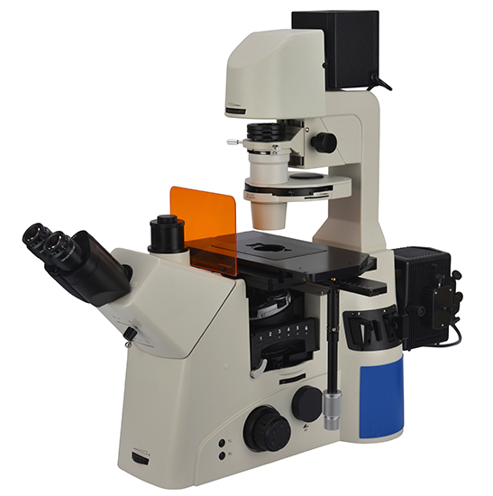
A fluorescence microscope is a type of optical microscope that uses a high-intensity light source to illuminate the specimen and excite fluorochromes in the sample. The illumination of the specimen is usually done with a light source that emits ultraviolet light. They are widely used in biological, medical and industrial fields.
We use cookies to enhance your browsing experience, serve personalized ads or content, and analyze our traffic. By clicking "Accept", you consent to our use of cookies.
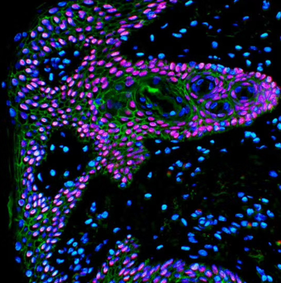
2. In the medical field, the fluorescence microscope can use fluorescent reagents to detect the presence and distribution of bacteria and viruses, or to assist in labeling surgical targets to facilitate surgery.
The condenser is located below the stage. It collects wavefronts from the light source and focuses them to illuminate the specimen evenly across the field of view.
Types ofobjectivelenses
High-powered microscopes are mostly used in schools. They are used to examine small samples, such as bacteria, blood cells or other tiny life forms. They can also be used to view items that you cannot see with your naked eye.
Confocal Fluorescence Microscope: This type of fluorescence microscope combines laser scanning with fluorescent illumination to produce an image. It can be used in wide range of applications, such as studying cells and tissues, detecting proteins and other substances within cells, and measuring the thickness of materials.
Objectivelens magnification
The resources are collected and organized on the Internet, and are only used for learning and communication. If there is any infringement, please contact us to delete.
-The cells are susceptible to the phototoxic effect after staining with fluorescent dyes, as the fluorophore molecules absorb the high energy photons from the short-wavelength light.
The emission filter passes only the wavelengths emitted by the fluorophore and blocks all undesired light outside this band – especially the excitation wavelengths.
Place a microscope slide on the stage underneath the low power objective lens for more effortless adjustment under the high power objective.
The most common light sources are mercury, xenon, and LEDs. Mercury provides the best quality of light for fluorescence microscope. LEDs are becoming more popular because they are less expensive than other sources and they consume less power.
Mediumpower objectivemicroscopefunction
The excitation filter is essential for the operation of a fluorescence microscope. It passes the light of a shorter wavelength, which the fluorescent dye could absorb. Also, it blocks the other sources of exciting light.
Stage microscopefunction
Objective lenses are attached to the revolving nosepiece that you can turn to choose which lens you need. When focusing, you always start with low-powered objectives.
The dichroic mirror is a type of optical filter that reflects light at certain wavelengths while transmitting others. It is used in fluorescence microscopes to separate the excitation and emission wavelengths.
When buying a compound microscope, there are many things you should consider. The budget is one. Commonly, microscopes that belong to a higher price range have better optics. That's why they are excellent investments.
3. In the field of mineralogy, the fluorescence microscope is often used to study substances with spontaneous fluorescence properties, such as asphalt, petroleum, coal, graphene oxide and other minerals.
It is also essential to choose the correct magnification. The most useful objective lens magnifications are x4, 10, 20, and x40.
Oil immersionobjective
4. In materials science, the fluorescence microscope can be used in the textile industry or the paper industry to analyze fiber-based materials.
-The photobleaching due to the electron excitation during the process of fluorescence may affect reactive molecules of the fluorescent dyes. As a result, the reactive dyes might lose their chemical property of fluorescence emission intensity.
Fine focus is a small knob that allows you to adjust the lens to make your image clearer. You can achieve a clear focus more often under a high-power magnification by adjusting the fine focus.
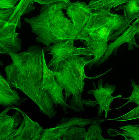
1. In the field of biology, the fluorescence microscope enables accurate and detailed identification of cellular and submicroscopic cellular components and activities with the help of fluorescent dye labeling.
Because a high-powered microscope is also a compound microscope, it is vital to learn its different parts, functions, and jargon before setting them up.
Fluorescence microscopes are widely used in various fields of research and application including biochemistry, cell biology, microbiology, immunology, and medicine.
It is the most common type of fluorescence microscope. The excitation of the fluorophore and detection of the fluorescence are done through the same light path (i.e. through the objective). The majority of fluorescence microscopes, especially those used in the life sciences, are of the epifluorescence design.


 Ms.Cici
 8618319014500