
The Math of camera Field of View Calculations (FOV) - calculate fov
Given these limitations and biases, how do we decide that a polarization pattern is a signal? The minimal criteria are these: (1) the intended receiver must be capable of perceiving polarized objects; (2) the signal must be used on a body part or region prominently displayed when the sender is interacting with a conspecific; (3) the signal must be used in a behaviorally relevant context (e.g., aggression, deterrence, mate selection); (4) detection of the signal by the receiver must be associated with a clear response; and (5) the signaling should occur in a natural setting (particularly regarding lighting, polarization environment, and reasonable freedom of movement). In fact, no hypothetical polarization signal has ever met all 5 of these criteria. It is difficult to control the polarized-light environment (Foster et al. 2018), very difficult to separate the polarization content of a possible signal from other features (e.g., movement, brightness, color), and hard to know whether a receiver’s subsequent behavior is a response or some irrelevant action. Nevertheless, there are some compelling cases that strongly suggest the use of polarization signaling.
To this point, I have only covered a type of polarization properly called “linear polarization,” since the e-vector, viewed along the axis of the polarized beam, is a straight line. In the laboratory, this light is usually produced by passing it through a linear polarizer (Figure 1A,B). However, light can also be circularly polarized, in which case the view along the beam axis is of an e-vector that rotates in a full circle, either clockwise or counterclockwise (Figure 1; see also Johnsen 2012). The e-vector makes this circle once for each wavelength travelled by the beam, so seen from the side the tip of the vector traces a helical path (Figure 1B,C). In the laboratory, circular polarization is produced by passing fully linearly polarized light through a quarter-wave plate (an optical device that delays one axis of linear polarization by ¼ of a wavelength relative to the orthogonal axis; Figure 1A,B). Much light in nature is elliptically polarized, having a mix of linear and circular components (Johnsen 2012; see also Cronin et al. 2014). In such cases, the long axis of the ellipse traced out by successive positions of the e-vector is at the e-vector angle of the linear component.
Marshall J, Roberts N, Cronin T, 2014. Polarisation signals. In: Horváth G, editor. Polarized Light and Polarization Vision in Animal Sciences. Berlin: Springer, 407–442.
Horváth G, Kriska G, Robertson B, 2014b. Anthropogenic polarization and polarized light pollution inducing polarized ecological traps. In: G Horváth, editor. Polarized Light and Polarization Vision in Animal Sciences. Berlin: Springer, 443–513.
The very low refractive index of air (n = 1.0) compared with almost any solid or liquid material, together with the scattering of sunlight in the atmosphere, means that most terrestrial scenes have a complex pattern of polarized light, visible in almost any direction during the day. Underwater, the situation is much simpler. With water’s elevated refractive index (n=1.33), there is far less reflection of light from submerged objects than there is in air. This leaves scattering as the primary means of producing polarization. Near the surface (perhaps to depths of 20 or 30 m in very clear water, less in turbid locations) the polarization pattern of the sky can be seen, although multiple-path scattering in water quickly obliterates it. On the other hand, water itself is a scattering medium. Since light generally enters water from nearly overhead, and becomes even more centered on the zenith as depth increases, the tendency to produce greatest polarization at scattering angles of 90° means that underwater scenes are dominated by a field of horizontally polarized light visible in all directions (Figure 2C; see also Ivanoff and Waterman 1958; Waterman 1981; Cronin and Shashar 2001; Cronin and Marshall 2011; Marshall and Cronin 2011; Cronin et al. 2014). If an animal is interested in seeing nearby objects or other creatures as silhouettes against this bright, horizontally polarized background, it is advantageous to view the world through a visual system that is able to detect this horizontal axis of polarization. On the other hand, animals may wish to penetrate this obscuring haze to see things that are further away and washed out by the intervening veil of horizontally polarized light. In this case, vision could favor enhancing vertical polarization sensitivity. Of course, depending on downstream neural processing, the same visual system could handle both vertically polarized and horizontally polarized objects or scenes.
As of 2023, the company had 26,622 employees. Their stock is listed at the New York Stock Exchange under the ticker symbol COHR. In 2022, II-VI acquired laser ...
Chiou TH, Marshall NJ, Caldwell RL, Cronin TW, 2011. Changes in light reflecting properties of signaling appendages alter mate choice behaviour in a stomatopod crustacean, Haptosquilla trispinosa. Mar Freshw Behav Physiol 44:1–11.
Seliger HH, Lall AB, Lloyd JE, Biggley WH, 1982b. On the colors of firefly bioluminescence. II. Experimental evidence for the optimization model. Photochem Photobiol 36:681–688.
How MJ, Pignatelli V, Temple SE, Marshall NJ, Hemmi JM, 2012. High e-vector acuity in the polarisation vision system of the fiddler crab Uca vomeris. J Exp Biol 215:2128–2134.
Marshall J, Cronin TW, Wehling MF, 2011. Introduction: new directions in the detection of polarized light. Phil Trans R Soc B 366:615–616.
The circularly polarized signal is also not difficult to produce, as many arthropods besides mantis shrimps and dung beetles reflect circularly polarized light produced by several distinct mechanisms (Neville and Caveney 1969; Neville and Luke 1971). In the case of stomatopods, the chitin of the cuticle often is fortified with calcite, an optically active form of calcium carbonate. In combination with the linear polarization passing through astaxanthin-based dichroic polarizers, this can form a circularly polarized signal. So it appears that the preconditions for circularly polarized signaling probably existed in these creatures, and signals evolved more or less by the fortuitous coincidence of there being an accidental sensory modality and a strange optical structure in the cuticle available to build a unique signaling system.
All of LASER COMPONENTS' AR coatings are optimized for high power lasers and are available for the wavelength range from 193 nm to. 5000 nm. Both the bandwidth ...
Brady P, Cummings M, 2010. Differential response to circularly polarized light by the jewel scarab beetle Chrysina gloriosa. Am Nat 175:614–620.
Animals use the same processes as environments to generate linearly polarized light from depolarized light sources. Most animals have surfaces that reflect polarized light, a process that is generally unavoidable in air. Some animals, such as many tropical butterflies (Figure 4A), have specialized features that enhance polarization reflections and use these to produce signals (Douglas et al. 2007). Butterflies use specially formed scales to produce bright, often spectrally narrow, reflections (Douglas et al. 2007; Vukusic and Stavenga 2009; Giraldo and Stavenga 2016). Linearly polarized reflections are also possible underwater, but because of the elevated refractive index of water, dense organic structures must be used. Cephalopod molluscs use specialized cells called iridophores containing a very high refractive-index protein, reflectin, to produce patterns of polarization on their bodies (Figure 4B;Shashar et al. 1996; Crookes et al. 2004; Chiou et al. 2007; Izumi et al. 2009).
Address. 1220 N Robinson Ave., Ste B Oklahoma City, OK 73103. Phone. 405.260.2265. Norman. Address. 2122 W. Main St. Norman, OK 73069. Phone. 405.260.2265.
Temple SE, McGregor JE, Miles C, Graham L, Miller J et al. , 2015. Perceiving polarization with the naked eye: characterization of human polarization sensitivity. Proc R Soc B 282:20150338.
Douglas JM, Cronin TW, Chiou TH, Dominy NJ, 2007. Light habitats and the role of polarized iridescence in the sensory ecology of Neotropical nymphalid butterflies (Lepidoptera: Nymphalidae). J Exp Biol 210:788–799.
Gagnon YL, Templin RM, How MJ, Marshall NJ, 2015. Circularly polarized light as a communication signal in mantis shrimps. Curr Biol 25:1–5.
Underwater, the polarized world is much simpler than in air, since underwater surfaces are poorly reflective. This leaves the omnidirectional spacelight (which is horizontally polarized; Figure 2C) as the primary source of underwater polarization, against which objects (and signals) can be viewed. How et al. (2014) demonstrated that individuals of the protosquillid mantis shrimp H. trispinosa are significantly more likely to respond to a looming, horizontally polarized stimulus than to a vertically polarized one. This species and several close relatives have the bright blue scattering polarizers on their first maxillipeds (Figure 4C; see Cronin et al. 2003; Chiou et al. 2008a, 2011; How et al. 2014; Jordan et al. 2016). How et al. (2014) argue that the use of horizontal polarization in signaling evolved under the influence of the innate response to horizontally polarized objects. Significantly, the elevated responses to horizontally polarized looming stimuli are seen in several protosquillid stomatopods, including those lacking polarization signaling, which implies that the response is innate and available for signal evolution. Many aquatic animals, including crabs, crayfishes, and cephalopods, respond to polarized looming stimuli (Tuthill and Johnsen 2006; How et al. 2012; Temple et al. 2012), but biases toward particular e-vector angles have not yet been investigated.
Schwind R, 1984a. Evidence for true polarization vision based on a two-channel analyzer system in the eye of the water bug Notonecta glauca. J Comp Physiol A 154:55–57.
Thomas W Cronin, A different view: sensory drive in the polarized-light realm, Current Zoology, Volume 64, Issue 4, August 2018, Pages 513–523, https://doi.org/10.1093/cz/zoy040
Roberts NW, Porter ML, Cronin TW, 2011. The molecular basis of mechanisms underlying polarization vision. Phil Trans R Soc B 366:627–637.
Because humans are nearly insensitive to light’s polarization and are incapable of resolving polarization patterns in nature or in animal signals, this aspect of visual signaling was ignored until Shashar et al. (1996) reported that cuttlefish produce rapidly varying patterns of polarized stripes that appear to act as visual signals. Since then, many examples of polarized-light signals (or potential signals) have been discovered (Cronin et al. 2003, 2014; Marshall et al. 2014). Here, we consider how sensory drive might have structured these polarization signals. We also report a type of polarized-light signal that appears to have evolved to a sensory modality that may have been lying dormant until an appropriate signal evolved.
Horváth G, Csabai Z, 2014. Polarization vision of aquatic insects. In: Horváth G, editor. Polarized Light and Polarization Vision in Animal Sciences. Berlin: Springer, 113–145.
Modern microscopes feature vastly improved plan-corrected objectives in which the primary image has much less curvature of field than older objectives. In addition, most microscopes now feature much wider body tubes that have greatly increased the size of intermediate images. To address these new features, manufacturers now produce wide-eyefield eyepieces that increase the viewable area of the specimen by as much as 40 percent. Because the strategies of eyepiece-objective correction techniques vary from manufacturer to manufacturer, it is very important to use only eyepieces recommended by a specific manufacturer for use with their objectives. Additionally, most eyepieces on research-level microscopes have a focusing ring, which makes it possible to precisely focus on reticles that are mounted into the space where the intermediate image resides. The focusing ring also makes it possible to establish a condition on the microscope that is referred to as being parfocal, where operators with different eyesights can configure the microscope in such a manner that the images produced by the objective remain in focus when a new objective is inserted into the optical path.
Circularlypolarized light
Boal JG, Shashar N, Grable MM, Vaughan KH, Loew ER et al. , 2004. Behavioral evidence for intraspecific signaling with achromatic and polarized light by cuttlefish (Mollusca: Cephalopoda). Behaviour 141:837–861.
One of the most important criteria to be considered in the purchase of an optical microscope is the required field of application. Another, perhaps equally important, is the state of (aberration) correction of the optical components, in particular, the objectives. Microscope objectives are perhaps the most important components of an optical microscope because they are responsible for primary image formation and play a central role in determining the quality of images that the microscope is capable of producing. Objectives are also instrumental in determining the magnification of a particular specimen and the resolution under which fine specimen detail can be observed and recorded using the microscope. The objective is the most difficult component of an optical microscope to design and manufacture, and is the first component that light encounters as it proceeds from the specimen to the image plane.
Horváth G, 2014. Polarization patterns of freshwater bodies with biological implications. In: G Horváth, editor. Polarized Light and Polarization Vision in Animal Sciences. Berlin: Springer, 333–344.
Diagrammatic representations of linearly and circularly polarized light as produced by an unpolarized light beam entering from the right and propagating through first a linear polarizer oriented at 45° and then a quarter wave plate with its fast axis oriented vertically. The linear polarizer transmits fully linearly polarized light, while the quarter wave plate delays the horizontal component of this light by ¼ wave relative to the vertical component, producing left-handed circularly polarized light. Panels A and B illustrate 2 ways of portraying polarization (as axes or vectors). Panel C shows the successive peaks of the horizontal (green) and vertical (blue) components of the same circularly polarized wave, as well as the helical path traced by their resultant vectors (red). (Figure modified from Wikipedia.).
Aperture adjustment and proper focusing of the condenser (with regard to height in relation to the objective) are of critical importance in realizing the full potential of the objective. Specifically, appropriate use of the adjustable aperture iris diaphragm (incorporated into the condenser or just below it) is of significant importance in securing correct illumination, contrast, and depth of field. The opening size of this iris diaphragm controls the angles of illuminating wavefronts (and thus the aperture size) that bathe the specimen. Condenser height is controlled by a rack and pinion gear system that allows the condenser focus to be adjusted for proper illumination of the specimen. Correct positioning of the condenser with relation to the cone of illumination and focus (a step in establishing Köhler illumination) is critical to quantitative microscopy and to ensure the best digital images.
Condensers are divided primarily into classifications of imaging modality (such as brightfield, darkfield, and phase contrast), but also according to their degree of optical correction. There are four principle types of condensers with respect to correction of optical aberrations, as listed in Table 1. The simplest and least corrected (also the least expensive) condenser is the Abbe condenser that can have a numerical aperture up to 1.4 in the best models with three or more internal lens elements. Although the Abbe condenser is capable of passing bright light, it is not corrected for either chromatic or spherical optical aberrations. A typical Abbe condenser is illustrated in Figure 2. In its simplest form, the Abbe condenser has two optical lens elements that produce an image of the illuminated field diaphragm that is not sharp and is surrounded by blue and red color at the edges, characteristic of chromatic aberration.
(A) Absorption of polarized light by vertebrate rod photoreceptors. Light arrives normal to the flat surfaces of rod disks and is absorbed by chromophores of visual pigment in the disk membranes (double-headed arrows show preferred e-vector orientations). Since the absorption preference is random, there is no overall polarization signal (top). Light aimed from the side of the disk encounters chromophores roughly parallel to the membrane, favoring the absorption of horizontally polarized light (bottom). (B) Absorption of polarized light by microvillar photoreceptors. Light arrives orthogonal to the long axis of each microvillus, and encounters visual pigment chromophores that are oriented roughly parallel to the axis of the microvillus (top). Thus, the microvillus as a whole preferentially absorbs light polarized parallel to its axis. If light were to arrive from the end of the microvillus (bottom), it would encounter chromophore orientations at all angles, so there would be no preferred polarization absorption. (C) Diagrammatic view of typical polarization-sensitive photoreceptors in compound eyes of insects, seen in cross section (crustaceans and cephalopods have analogous arrangements). Light arrives perpendicular to the plane of the page, and passes through many layers of microvilli. Cells on opposite sides of the receptor extend parallel microvilli toward the junction in the center, and thus have parallel polarization sensitivity. Two orthogonal sets of receptors (Cells 1 and 3 vs. Cells 2 and 4) can provide information for 2-axis polarization analysis.
Elliptical polarization
Many surfaces reflect polarized light, and some of these may be modified to form visual signals. Such modifications are only useful if they can be viewed and interpreted. As a consequence, the polarization vision systems of animals have the potential to drive the evolution of polarized-light signals. We now turn to the properties of such visual systems.
Every signal described in this review is horizontally polarized in normal viewing conditions. In natural illumination in either terrestrial or aquatic environments, the geometry of the situation is such that reflective signals from horizontally oriented structures will mostly include horizontal polarization, so this might not be a surprise. Dichroic signals are also horizontally polarized for the same reason; the dichroism actually interacts with light reflected from an underlying layer (Chiou et al. 2012), so the signal is brightest and most polarized when lit from overhead (as is the case in most often encountered in nature) and viewed horizontally. Scattering polarizers, however, are not limited by the direction of illumination or view—they reflect horizontally polarized light regardless of how they are illuminated or seen (Chiou et al. 2008a; Jordan et al. 2016). If sensory drive is behind the evolution of polarization-based signals, is there a reason why signaling systems might favor horizontal polarization?
Waterman TH, 1981. Polarization sensitivity. In Autrum H, editor, Handbook of Sensory Physiology, Vol. VII/6B. Berlin: Springer, 283–469.
The primary research tasks that demand attention now include the demonstration of polarization signaling in other kinds of animals besides butterflies, mantis shrimps, and cuttlefishes. Many insects, crustaceans, and cephalopods have polarization vision and might benefit from its use in communication. Further, we need rigorous demonstrations that the hypothesized signals are actually used to communicate and that their polarization content is a critical component of communication. A third avenue for research is a better understanding of the function and use of circularly polarized signals. In the future, signals based on polarized light may be equally fruitful subjects for study as color signals are today.
A more recent discovery is that some mantis shrimps actually have circularly polarized body parts and that they are capable of discriminating right from left circularly polarized light (Chiou et al. 2008b). An interesting structure, one that is very likely to be used in sexual signaling, is the “keel” on the median ridge of the telson of the stomatopod Odontodactylus cultrifer (Figure 6B,C). This feature is sexually dimorphic (being much larger in males than females), and reflects (and transmits) highly circularly polarized light. Amazingly, when seen from the right side, the polarization of the keel is right handed; the opposite is true on the left (Figure 6C). Mating behavior in this species has never been observed, but the sexual dimorphism strongly suggests that the keel is a sexual signal, and the unique presence of circular polarization vision in stomatopods may be critical in detecting and interpreting this signal.
As it happens, when visual pigments sit in the plasma membrane, their chromophores are situated parallel to the plane of the membrane. The rod disks and cone lamellae of vertebrate photoreceptors have roughly planar membranes, and rhodopsin molecules apparently rotate rather freely in these membranes (Figure 3A); thus, the overall pattern of absorption is random, and these cells are expected to be polarization-insensitive. Nevertheless, because there is some evidence that vertebrates do use polarized-light analysis for orientation or migration, various mechanisms have been proposed that could give a limited polarization sense to rods or cones (Roberts et al. 2011). There is no convincing evidence, however, that vertebrates use polarized light for signaling. In photoreceptors where the membranes are organized into microvilli, as in arthropods or molluscs, the strong curvature of the microvillar membranes evidently restricts the rotational freedom of visual pigment molecules, giving the entire microvillus inbuilt linear polarization sensitivity (Figure 3B). To make an entire photoreceptor cell polarization-sensitive, all microvilli from that single cell must extend out parallel to each other. Most commonly, such cells are oriented so that microvilli of adjacent photoreceptors are oriented orthogonal to each other, an arrangement that fosters 2-axis polarization analysis (Marshall and Cronin 2011; Cronin et al. 2014; Marshall et al. 2014). All animals thought to use polarization signaling have such 2-axis analyzers, probably synapsing onto an opponent interneuron to enhance polarization sensitivity. A schematic example of such an arrangement, found in many arthropods and cephalopods, is illustrated in Figure 3C.
The first stage of the microscope optical train (and perhaps the most neglected) is the lamphouse, which contains the lamp and collector lens system. This unit is responsible for establishing the primary illumination conditions for the microscope. Light emitted by a tungsten-halogen or arc-discharge is passed through the collector lens system and the filament or arc is focused onto the front focal plane of the condenser (objective in reflected epi-fluorescence). The first image plane in the microscope optical train occurs at the position of the field diaphragm. Thus, the lamphouse coupled with the field diaphragm produces the necessary illumination pattern to sufficiently image specimens in a wide variety of imaging modes. In the optical microscope, conjugate planes are imaged into each other and can collectively be observed while examining a specimen in the eyepieces. The field iris diaphragm, adjacent to the lamp collector lens, is imaged sharply into the same plane as the specimen by the microscope condenser. Images of both the field diaphragm and the specimen are formed in the intermediate image plane by the objective and are projected into the fixed field diaphragm of the eyepiece, where the focusing reticle is located. Subsequently, the eyepiece (in conjunction with the observer's eye) forms images of all three previous image planes on the sensor surface of an imaging system or the retina of a human eye. The field diaphragm, specimen, intermediate image, and retina all constitute a set of conjugate image planes, spaced throughout the microscope optical train, which appear simultaneously in focus.
Lacquer Crack is a specialized coating for stress testing materials.
Blahó M, Egri Á, Hegedüs R, Jósvai J, Tóth M et al. , 2012. No evidence for behavioral responses to circularly polarized light in four scarab beetle species with circularly polarizing exocuticle. Physiol Behav 105:1067–1075.
Sensory drive, the concept that sensory systems primarily evolve under the influence of environmental features and that animal signals are evolutionarily shaped and tuned by these previously existing sensory systems, has been thoroughly studied regarding visual signals across many animals. Much of this work has focused on spectral aspects of vision and signals. Here, I review work on polarized-light signals of animals and relate these to what is known of polarization visual systems, polarized-light aspects of visual scenes, and polarization-related behavior (e.g., orientation, habitat-finding, contrast enhancement). Other than the broad patterns of scattered polarized light in the sky, most polarization in both terrestrial and aquatic environments results from either reflection or scattering in the horizontal plane. With overhead illumination, horizontal features such as the surfaces of many leaves or of air: water interfaces reflect horizontal polarization, and water scatters horizontally polarized light under most conditions. Several animal species have been demonstrated to use horizontally polarized light fields or features in critical aspects of their biology. Significantly, most biological signals are also horizontally polarized. Here, I present relevant polarization-related behavior and discuss the hypothesis that sensory drive has evolutionarily influenced the structure of polarization signals. The paper also considers the evolutionary origin of circular polarization vision and circularly polarized signals. It appears that this class of signals did not evolve under the influence of sensory drive. The study of signals based on polarized light is becoming a mature field of research.
Chiou TH, Place AR, Caldwell RL, Marshall NJ, Cronin TW, 2012. A novel function for a carotenoid: astaxanthin used as a polarizer for visual signalling in a mantis shrimp. J Exp Biol 215:584–589.
Jordan TM, Wilby D, Chiou TH, Feller KD, Caldwell RL et al. , 2016. A shape-anisotropic reflective polarizer in a stomatopod crustacean. Sci Rep 6:21744.
Eyepieces work in combination with microscope objectives to further magnify the intermediate image so that specimen details can be observed. Oculars is an alternative name for eyepieces that has been widely used in the literature. The best results in microscopy require that objectives be used in combination with eyepieces that are appropriate to the correction and type of objective. The basic anatomy of a typical modern eyepiece is illustrated in Figure 3. Inscriptions on the side of the eyepiece describe its particular characteristics and function. There are two major types of eyepieces that are grouped according to lens and diaphragm arrangement: the negative eyepieces with an internal diaphragm and positive eyepieces that have a diaphragm below the lenses of the eyepiece. Negative eyepieces have two lenses: the upper lens, which is closest to the observer's eye, is called the eye-lens and the lower lens (beneath the diaphragm) is often termed the field lens. In their simplest form, both lenses are plano-convex, with convex sides facing the specimen. Approximately mid-way between these lenses there is a fixed circular opening or internal diaphragm which, by its size, defines the circular field of view that is observed in looking into the microscope.
Chiou TH, Kleinlogel S, Cronin TW, Caldwell RL, Loeffler B et al. , 2008b. Circular polarization vision in a stomatopod crustacean. Curr Biol 18:429–434.
Vukusic P, Stavenga DG, 2009. Physical methods for investigating structural colours in biological systems. J R Soc Interface 6:S133–S148.
Cronin TW, Shashar N, Caldwell RL, Marshall H, Cheroske AG et al. , 2003. Polarization vision and its role in biological signaling. Integr Comp Biol 43:549–558.
The use of polarized-light reflections as potential signals was first reported in a cuttlefish, Sepia officinalis (Shashar et al. 1996). Cuttlefish produce highly polarized reflections, which can be switched on and off via neural regulation on timescales of seconds (Izumi et al. 2009), from iridophores in their arms; these reflections are horizontally polarized in typical underwater lighting conditions (Shashar et al. 1996; Chiou et al. 2007). The patterns are displayed in social interactions, and they differ in their frequency of use between males and females, suggesting that they play roles in communication (Shashar et al. 1996; Boal 1997; Boal et al. 2004). Similar reflections are found on the arms of squids (Chiou et al. 2007; see Figure 4B), where their biological significance is unknown. Given that cephalopods have large brains, good eyes with polarization sensitivity, and complex behavior, it is difficult avoid concluding that their use of switchable polarization patterns is involved in signaling. Still, definitive proof that they are used in this way is lacking.
On upright microscopes, the condenser is located beneath the stage and serves to gather wavefronts from the microscope light source and concentrate them into a cone of light that illuminates the specimen with uniform intensity over the entire viewfield. Inverted (tissue culture style) microscopes mount the condenser above the stage and specimen on a frame pillar. It is critical that the condenser light cone be properly adjusted to optimize the intensity and angle of light entering the objective front lens. Each time the objective is changed, a corresponding adjustment must be performed on the condenser to provide the proper light cone to match the light cone (numerical aperture) of the new objective. A simple two-lens Abbe condenser is illustrated in Figure 2. In this figure, light from the microscope illumination source passes through the condenser aperture diaphragm, located at the base of the condenser, and is concentrated by internal lens elements, which then project light through the specimen in parallel bundles from every azimuth. The size and numerical aperture of the light cone is determined by adjustment of the aperture diaphragm. After passing through the specimen, the light diverges into an inverted cone with the proper angle to fill the front lens of the objective.
Compensating eyepieces play a crucial role in helping to eliminate residual chromatic aberrations inherent in the design of highly corrected objectives on older finite tube length microscopes. Hence, it is preferable that the microscopist uses the compensating eyepieces designed by a particular manufacturer to accompany that manufacturer's higher-corrected objectives. Use of an incorrect eyepiece with an apochromatic objective designed for an older finite (160 or 170 millimeter) tube length application results in dramatically increased contrast with red fringes on the outer diameters and blue fringes on the inner diameters of specimen detail. Additional problems arise from a limited flatness of the viewfield in simple eyepieces, even those corrected with eye-lens doublets. More advanced eyepiece designs resulted in the Periplan eyepiece design (see Figure 3). This eyepiece contains seven lens elements that are cemented into a single doublet, a single triplet, and two individual lenses. Design improvements in periplan eyepieces lead to better correction for residual lateral chromatic aberration, increased flatness of field, and a general overall better performance when used with higher power objectives.
Evidence that stomatopod polarizers are used in signaling is suggestive, but not conclusive. The most solid case can be made for the blue polarizers of the Indopacific species, Haptosquilla trispinosa (the species illustrated in Figure 4C). Chiou et al. (2011) were able to destroy these devices by touching them briefly with a hot pin, a treatment that left the mobility of the appendage intact but which removed both the colored and polarized reflections (happily, the animals regenerated the device when they molted). After being so treated, courting males continued to display their appendages to females as before, and would eventually be accepted as partners, but their displays had to be extended longer, they had to endure more aggressive responses from females before being accepted, and they experienced shorter mating durations. These results strongly indicate that the polarizers are involved in signaling, but do not indicate whether the color, polarization, or both contain the important signal component. Evidence that the dichroic polarizers play a role in signaling is even more circumstantial. These devices, which are usually bright red or yellow in color, occur on parts of the body that are involved in agonistic displays, in defense postures, and in mating displays (Cronin et al. 2003; Marshall et al. 2014). For example, an individual Odontodactylus havanensis (a Caribbean species) will prominently display its polarized antennal scales to an intruder as it approaches the resident’s burrow (Figure 5A). A different use of the same appendages has been seen in the Indospecific congener, Odontodactylus scyllarus. As a male approaches a female of this species from behind prior to mating, she will wobble her laterally extended antennal scales (Figure 5B). Since the degree of polarization reflected from the scale varies with the angle from which it is viewed (Chiou et al. 2012), this produces a display that should be very obvious to the approaching male.
Seliger HH, Lall AB, Lloyd JE, Biggley WH, 1982a. e colors of firefly bioluminescence. I. An optimization model. Photochem Photobiol 36:673–680.
Neville AC, Caveney S, 1969. Scarabaeid beetle exocuticle as an optical analogue of cholesteric liquid crystals. Biol Rev 44:531–562.
Most natural (and artificial) sources produce light that is either unpolarized or very weakly polarized. Still, the world is full of polarized light, because light’s interaction with matter often favors one type of polarization over another. Scattering and reflection are the most common sources of linear polarization (Wehner 2001; Cronin and Marshall 2011; Marshall and Cronin 2011). That blue sky we looked at a little while ago has a prominent pattern of highly linearly polarized light caused by molecular scattering within the atmosphere (Figure 2A). Such scattering is most effective for short-wavelength photons, which is why the sky is blue (as your mother explained to you; actually, it’s even brighter in the ultraviolet). Scattering also favors photons with polarizations that are perpendicular both to the initial and to the final path of the photon, in other words, whose e-vectors are normal to the plane containing the incident and scattered ray. Scattering perpendicular to the axis of the incident ray is permitted only for such photons, so the degree of linear polarization (or percent polarization) varies from 0% for forward-scattered rays to 100% for rays scattered into the plane perpendicular to the incident path. Since essentially all light in the sky comes from the sun, the overall polarization pattern has a band of very highly linearly polarized light at 90° to the sun with diminished polarization at other angles. Celestial polarization never reaches exactly 100% because many photons reaching the earth’s surface have been scattered more than once, somewhat scrambling their angles of polarization.
The simplest negative eyepiece design, often termed the Huygenian eye-piece, is found on most teaching and laboratory microscopes fitted with achromatic objectives. Although the Huygenian eye and field lenses are not well corrected, their aberrations tend to cancel each other. More highly corrected negative eyepieces have two or three lens elements cemented and combined together to make the eye lens. If an unknown eyepiece carries only the magnification inscribed on the housing, it is most likely a Huygenian eyepiece, best suited for use with achromatic objectives of 5x to 40x magnification. The other common eyepiece is the positive eyepiece with a diaphragm below its lenses, commonly known as the Ramsden eyepiece. This eyepiece has an eye lens and field lens that are also plano-convex, but the field lens is mounted with the curved surface facing towards the eye lens. The front focal plane of this eyepiece lies just below the field lens, at the level of the eyepiece diaphragm, making this eyepiece readily adaptable for mounting reticules.
Achromats are the most widely used objectives and are commonly found on both teaching and research-level laboratory microscopes. They are satisfactory objectives for routine laboratory use, but because they are not corrected for all colors, a colorless specimen detail is likely to show, in white light, a pale green color at best focus (secondary axial color). Apochromatic objectives usually contain two lens doublets and a lens triplet for advanced correction of both chromatic and spherical aberrations. With apochromat and fluorite objectives, the diffraction-inducing spreading of the intensity distribution can be virtually eliminated. An achromat objective still has substantial intensity in the first fringe, while the apochromat approaches the theoretical resolution limit where the longitudinal chromatic aberration is greater than the wave-optical depth of field. Because apochromat objectives require lens elements having abnormal dispersion characteristics, their specifications may not be ideal for some specific applications, such as fluorescence excitation in the near ultraviolet, DIC, and other forms of microscopy utilizing polarized light. For this reason, a fluorite objective may be more suitable. Due to modern coating technologies in newly designed apochromats, remarkably sharp images with high contrast can be obtained even in those instances where the apochromat was inherently limited.
The second process that favors light of certain linear polarization planes is reflection. Light reflected from a non-metallic surface (such as leaves or the air: water interface) becomes enriched in photons with polarization planes parallel to the surface (Figure 2B). At an angle defined by the refractive indices of the materials on each side of the surface (e.g., air vs. water), called Brewster’s angle, the reflected light is fully polarized. For water, Brewster’s angle is about 53°. In other words, when looking down at a flat and calm pure water surface at an angle of 53° to the vertical, the reflected light you see is 100% polarized. Polarized sunglasses have their polarization transmission axes placed vertically, eliminating most horizontally reflected light, for example from wet surfaces, glass, or painted metal. Thus, they increase contrast; also, they allow the wearer to see through the attenuated shine of the water’s surface into the water itself. Polarization filters on cameras may be used for the same purpose or to block polarization in the sky for dramatic images of clouds (which generally scatter little polarization) or skyscapes.
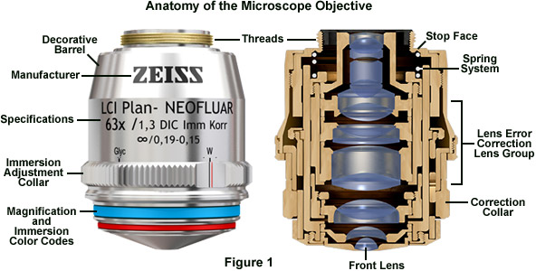
Microscope objectives are by far the most complex assemblies in the optical train. In contrast to the condenser and eyepieces, which contain between two and eight lenses, highly corrected objectives with numerical apertures above 1.0 can feature up to 15 or more lens elements and groups (see Figure 1). Objectives are fabricated with differing degrees of optical correction for both monochromatic (spherical, astigmatism, coma, distortion) and polychromatic aberrations, as well as field size and flatness, wavelength transmission band, birefringence, freedom from fluorescence and a variety of other factors that contribute to background noise. The two main criteria in objective manufacture are the elimination of chromatic errors and the flatness of the intermediate image that when perfectly corrected, provide an image with edge-to-edge sharpness, even with large fields of view. Depending upon the degree of correction, objectives are generally classified as achromats, fluorites, and apochromats, with a plan designation added to lenses with low curvature of field. Furthermore, objectives can be specifically classified into transmitted light and reflected light versions. The transmitted light versions popular in biological applications are usually designed for use with coverslips (in most cases, 170 micrometers in thickness). Reflected light (often termed Epi) objectives feature specially coated glass surfaces (antireflective coating) to avoid reflections in the optics when examining specimens lacking a coverslip. In fact, these objectives are specifically designed to be used on specimens without a coverslip.
Polarization oflight

Therefore, signals can represent an intriguing inversion of Wehner’s (1987) “matched filter”, where the features of a sensory system are shaped to detect a particular stimulus with high efficiency (see also von der Emde and Warrant [2016] for more about matched filters). In sensory drive, the stimulus is structured to be perceived by a sensory system. Nevertheless, there are a few cases in which sensory systems have converged onto signals which themselves have probably been shaped by environmental features. Perhaps the best example is found in the bioluminescent flashes of signaling fireflies. Here, it appears that the bioluminescent emission spectra of fireflies that are active at different times of the evening (early twilight, late twilight, or night) vary so as to be most effective in the illumination, and against the backgrounds, visible at that particular time and place (Seliger et al. 1982a, 1982b). In turn, the sensitivity spectra of receptors in their compound eyes are tuned to match almost perfectly these emission spectra across many species—perfect examples of matched filters (Lall et al. 1980, 1988; Cronin et al. 2000). This paper is not concerned with bioluminescence, but diverse bioluminescent signaling systems illustrate how sensory systems and signals coevolve in unexpected ways (see Haddock et al. 2010; Cronin et al. 2014).
The more highly corrected fluorite and plan-fluorite objectives have better color correction (at least three wavelengths) and feature flat fields (plan versions) in viewfields up to 26 millimeters in diameter. Due to the use of more advanced specialized glasses, fluorites are able to transmit ultraviolet wavelengths with high efficiency. Fluorite objectives are available for all contrast-enhancing modes, and special high-quality versions are available for polarized light and DIC. The apochromat objectives are the best performers and so are produced at the highest numerical aperture with color correction for at least four wavelengths. Plan versions reduce transmission efficiency, but produce spectacular images with a high degree of field flatness over the entire viewfield. As the need for specialized objectives grows with advances in technology, new apochromats are being designed to push the envelope with regards to color correction (360 to 700 nanometers or more), numerical aperture (up to 1.49), working distance, and suitability for various immersion media.
Crookes WJ, Ding LL, Huan QL, Kimbell JR, Horwitz J et al. , 2004. Reflectins: the unusual proteins of squid reflective tissues. Science 303:235–238.
Electric polarization
Endler’s presentation seemed to invert the usual way in which signal and sensory evolution were assumed to evolve. But on thinking about the idea further (and as he discussed at some length), sensory systems must in some way be tuned to the environment for many reasons fundamentally associated with survival and successful reproduction. In fact, the ideas behind sensory drive were first articulated soon after the publication of “The Origin of Species”. In 1879, Grant Allen wrote “… almost all the colours of vegetal life, except the uniform green of the foliage, are … due to the colour-sense of one or other of the great seeing classes, the vertebrate and the articulate”, adding “[Color vision has played a major role] in the moulding of organic forms (Allen 1879).” Effective signals must reach an intended receiver efficiently while being as inconspicuous as possible to potential predators. Obviously, while there are many constraints on signal evolution (avoiding the presence of predators when possible, reducing interception by competitors or unintended receivers, and so forth), the signal must be perceptible to the receiver and clearly different from confounding stimuli. It must be structured to excite the appropriate sensory system of the receiver, and thus under the evolutionary influence of properties of this sensory system. Often, the reduction of predation and unintended interception are managed by the timing of signal production or by signaling mechanisms that direct it most effectively at the proper receiver.
202263 — Focal length is the distance measured in millimeters, between the optical center of the lens and the camera sensor, where the light information is recorded.
If you look up at a clear blue sky, you would never know that your eyes are being flooded with polarized light. Humans are essentially blind to this electromagnetic property, but many animals are as capable of seeing and analyzing light’s polarization as we are of seeing and analyzing color. Here, we discuss the properties, sources, and distributions of polarized light in nature.
Apparent polarization signaling in stomatopod crustaceans. (A) Single frame from a video taken in the field showing 2 individuals of the Caribbean species Odontodactylus havanensis as they encounter each other at the burrow entrance of the animal to the right. This animal poses vertically above its burrow, extending the strongly polarizing antennal scales (black arrow) in a prominent lateral display. The animal on the left subsequently turns away and retreats (not shown). (Video recorded by A Cheroske; the background has been somewhat retouched to emphasize the 2 stomatopods.) (B) Two individuals of the Indopacific species O. scyllarus mating in the laboratory (the male is to the left, approaching and grasping the female from behind). The female angles her polarized antennal scales backward and waves them toward the male (credit: R.L. Caldwell).
Animals use 2 other optical mechanisms to generate linearly polarized light. Some species of stomatopod crustaceans employ a special type of scattering to do this (Figure 4C; Chiou et al. 2008a; Jordan et al. 2016). Unlike the broad expanses of the sky or the surrounding light field in water, the polarization from some mantis shrimps’ scattering structures has a constant e-vector angle, invariant with the angle of illumination, so clearly it is not due to symmetrical scattering as from a molecule. Instead, the mechanism is unique to stomatopods, being based on a rather exotic structure constructed from stacked, elongated, and precisely dimensioned and spaced photonic features that act as tuned scatterers favoring one axis of polarization (Jordan et al. 2016). Other stomatopods possess polarizing structures that use dichroic filters to polarize light, a mechanism thought to be unique to these animals. These filters, functionally analogous to polaroid film (basically, the same material used in polarized sunglasses), have long, aligned molecules that preferentially transmit polarized light having its e-vector plane perpendicular to the long axes of the molecules. The filters work because the long molecules absorb light polarized parallel to them, so light polarized in the orthogonal plane passes through. Stomatopods use the long keto-carotenoid astaxanthin to polarize via dichroism (Figure 4D), so the light that is transmitted is red (because astaxanthin absorbs shorter wavelengths of light). Species using this mechanism take advantage of the fact that astaxanthin’s length is almost exactly equal to the distance between the layers of the plasma membrane’s lipid bilayer. Consequently, the molecule spans the membranes of cells used in the polarizer, with its polarization axes perpendicular to these membranes. The transmitted horizontally polarized light is parallel to the plane of the plasma membrane, so such a polarizer is best seen when viewed at an angle. Stomatopods enhance the effect by placing membrane sheets containing the astaxanthin over a reflecting layer of cuticle (Chiou et al. 2012).
Cronin TW, Järvilehto M, Weckström M, Lall AB, 2000. Tuning of photoreceptor spectral sensitivity in fireflies (Coleoptera: Lampyridae). J Comp Physiol A 186:1–12.
Engravings found on the condenser housing include its type (achromatic, aplanatic, etc.), the numerical aperture, and a graded scale that indicates the approximate adjustment (size) of the aperture diaphragm. Condensers with numerical apertures above 0.95 perform best when a drop of oil is applied to their upper lens in contact with the undersurface of the specimen slide. This ensures that oblique light rays emanating from the condenser are not reflected from underneath the slide, but are directed into the specimen. In practice, this can become tedious and is not commonly done in routine microscopy, but is essential when working at high resolutions and for accurate imaging using high-power (and numerical aperture) objectives.
Probably the strongest case for the use of polarization signals is in the nymphalid butterfly Heliconius cydno. Sweeney et al. (2003) showed that this species reflects polarization patterns and that males of the species are far more likely to approach females viewed through windows that transmit polarized light than those viewed through depolarizing windows. Encouragingly, the congener Heliconius melpomene, which does not reflect such patterns, showed no differences in approach between the 2 types of windows. Overall, these results clearly suggest that H. cydno males indeed recognize the polarized-light reflections from females as signals. Taking these results further, Douglas et al. (2007) measured the polarization reflections from specimens of 144 nymphalid species, and found that about half (75) reflect polarized patterns (e.g., Figure 4A). Species inhabiting dimly lit forest habitats were very likely to reflect these patterns (68% of these species), while species from bright, open habitats rarely used them (only 10% of species). Douglas et al. (2007) argued that polarization patterns are more likely than color patterns to stand out in a dim, achromatic world, and thus should make effective signals. Many other families of butterflies have iridescent, polarized reflections which could be important for signaling (see Marshall et al. [2014] for a review).
Linearpolarization
An excellent and approachable discussion of actual signals, including definitions, can be found in Maynard Smith and Harper’s (2003) review, although they do not consider the sensory aspects of signaling. Still, defining (or even recognizing) a “signal” is sometimes problematic (Bradbury and Vehrencamp 1998). Just demonstrating that a particular action or marking is a signal can be challenging, since the generation of the signal, the behavior of both sender and receiver, and the benefits of such behavior are observed rarely or not at all under natural circumstances. This is particularly true of polarization markings and patterns, which certainly are strong candidates for signals. Most of these have never been observed in use in nature, and even under experimental conditions their validity as signals is only inferred, as will be noted later, when candidate signals are discussed. Nevertheless, there is compelling evidence that at least some polarization features are signals and that they are shaped by sensory drive. It is now time to turn to polarized light, its generation in nature, and its perception by animals.
Perception of circularly polarized light seems to be very rare, and in fact is only described in a few species of stomatopod crustaceans (mantis shrimps). The mechanism used is the reverse of that used to create circularly polarized light in the laboratory (Figure 1), with the light entering the diagrams in Figure 1A,B from the left. Some mantis shrimp eyes contain structures that function as quarter-wave plates, and these recombine the out-of-phase circular components into a linearly polarized output (Chiou et al. 2008b; Roberts et al. 2009). This is then directed to underlying linear polarization receptors built from aligned microvilli, like those just described. The overall result is that these receptors respond differentially to circular polarization with clockwise or counterclockwise rotation.
While some of the microscope optical components act as image-forming elements, others serve to produce various modifications to illumination of the specimen and also have filtering or transforming functions. Components involved in formation of images by the microscope optical train are the collector lens (positioned within or near the illuminator), condenser, objective, eyepiece (or ocular), and the refractive elements of the human eye or the camera lens. Although some of these components are not typically thought of as imaging components, their imaging properties are paramount in determining the final quality of the microscope image.
How MJ, Porter ML, Radford A, Feller KD, Temple S et al. , 2014. Out of the blue: the evolution of horizontally polarized signals in Haptosquilla (Crustacea, Stomatopoda, Protosquillidae). J Exp Biol 217:3425–3431.
When the objective is changed, for example from a 10x to 20x, the aperture diaphragm of the condenser must also be adjusted to provide a new light cone that matches the numerical aperture of the new objective. This is done by turning the knurled knob or lever that controls the condenser aperture diaphragm. There is a small yellow or white dot, arrow, or index mark located on the condenser that indicates the relative size of the aperture when compared to the linear gradation on the condenser housing. Many manufacturers will synchronize this gradation to correspond to the approximate numerical aperture of the condenser. For example, if the microscopist has selected a 10x objective of numerical aperture 0.25, then the arrow would be placed next the value 0.18-0.20 (about 80 percent of the objective numerical aperture) on the gradation inscribed on the condenser housing.
Lall AB, Strogher GK, Cronin TW, Seliger HH, 1988. Modification of spectral sensitivities by screening pigments in the compound eyes of twilight-active fireflies (Coleoptera: Lampyridae). J Comp Physiol A 162:23–33.
Cronin TW, Shashar N, 2001. The linearly polarized light field in clear, tropical marine waters: spatial and temporal variation of light intensity, degree of polarization, and e-vector angle. J Exp Biol 204:2461–2467.
Roberts NW, Chiou TH, Marshall NJ, Cronin TW, 2009. A biological quarter-wave retarder with excellent achromaticity in the visible wavelength region. Nat Photonics 3:641–644.
Circular polarization reflections from animals. (A) Scarab beetle Chrysina beyeri. Left panel: left circular polarization. Right panel: right circular polarization. Like many scarabs, the animal reflects left-handed highly polarized light, both by the green dorsal surface of the body (green) and by the purplish legs (credit: A. Harryman). (B) A male of the stomatopod Odontodactylus cultrifer. The white arrow shows the tall median keel of the animal’s telson (credit: R.L. Caldwell). (C) Images of the keel in circularly polarized light. As in panel A, in each pair the image to the left is taken in left circular polarization, and the right image is taken in right circular polarization. From the left side (top pair), the keel preferentially reflects left circular polarization; from the right side (bottom pair), right circular polarization dominates (credit: T.-H. Chiou). (D) Circular polarization reflections from an individual of the Indopacific stomatopod Gonodactylaceus falcatus in a defensive pose at its burrow entrance. The bright red areas (on the legs and other appendages) are false-colored to show strong left circularly polarized reflections (from Gagnon et al. [2015]; used with permission).
Linear Polarization is also known as Linear Sweep Voltammetry (LSV). It is an electrochemical technique, where the potential is increased or decreased with time ...
Foster JJ, Temple SE, How MJ, Daly IM, Sharkey CR et al. , 2018. Polarisation vision: overcoming challenges of working with a property of light we barely see. Sci Nat 105:27.
Circularly polarized signals could also be useful in aggressive encounters. A different mantis shrimp species, Gonodactylaceus falcatus, displays highly circularly polarized markings on its legs and anterior body parts when it poses in its burrow entrance (Gagnon et al. 2015; Figure 6D). The polarization is left-handed, and G. falcatus can learn to discriminate circular polarization from depolarized light, although (unlike Odontodactylus) it seems not to discriminate right from left circular polarization. The circularly polarized markings displayed at the burrow entrance (as in Figure 6D) apparently act defensively, as naive individuals of this species avoid burrows with entrances marked with circularly polarized filters. Gagnon et al. (2015) suggest that the circular polarization can be considered a true “covert” signal, in that no animals besides stomatopod crustaceans would be able to recognize it. Gonodactylaceus falcatus is often sandy colored and thus well camouflaged against its coral sand or rock environment, but the circularly polarized reflections could be obvious to another mantis shrimp.
Polarization
Chiou TH, Calwell RL, Hanlon R, Cronin TW, 2008a. Fine structure and optical properties of biological polarizers in crustaceans and cephalopods. In: Chenault DB, Goldstein DL, editors. Proceedings of SPIE 6972. Polarization: Measurement, Analysis, and Remote Sensing VIII. Bellingham (WA): SPIE Press.
Extra Large LED Handheld Magnifying Glass with Light - 2X 4X 10X Lens - Best Jumbo Size Illuminated Reading Magnifier for Books · With Light Or NotYes · Material ...
Get Sugared! Book Online Now! My account.
The animals that have the widest known diversity of intriguing polarization patterns are the mantis shrimps—stomatopod crustaceans. Mantis shrimp patterns are not produced by surface reflection (so far as is known), but by photonic, reflective scattering (Jordan et al. 2016) or by oriented, dichroic molecules (Chiou et al. 2012). To us, the photonic structures appear to be shiny blue (Figure 4C), so it is difficult to disentangle their signaling value as a color signal from their polarization features. Scattering polarizers are found on the anterior appendages of several stomatopod species (Cronin et al. 2003; Chiou et al. 2008a; How et al. 2014; Marshall et al. 2014), where they may be moved rapidly in a characteristic mating display (Chiou et al. 2011). In contrast to the relatively restricted locations of scattering polarizers, dichroic polarizers in mantis shrimps (Figure 4D) are found in many body structures, including antennal scales (the flaps that project laterally out from the base of the antennae), legs, carapace, telson, and uropods (Cronin et al. 2003, 2014; Chiou et al. 2008a, 2008b, 2012; Marshall et al. 2014). Because of the use of astaxanthin to produce the polarization (Chiou et al. 2012), this type of polarizer generally appears bright red, so again it is possible that polarized and colored signals are combined in such structures.
When John Endler introduced the term “sensory drive” (Endler 1992), the idea encouraged the view that signals and the sensory systems of animals work together. The scheme he discussed was that sensory system evolution is heavily influenced by local environmental conditions, such as the sources of relevant stimuli, the characteristics of the transmission of stimuli in a particular environment, and the background features or ongoing irrelevant stimuli (“noise”) from which significant stimuli must be discriminated. Animal “signals,” stimuli significant for communication that originate from other animals, are in turn shaped—at least in part—by the properties of the sensory systems to which they are directed. Thus, environment shapes sensation, and sensation shapes signals.
Research-level microscopes also contain one of several light-conditioning devices that are often positioned between the illuminator and condenser, and a complementary detector or filtering device that is inserted between the objective and the eyepiece or camera. The conditioning device(s) and detector work together to modify image contrast as a function of spatial frequency, phase, polarization, absorption, fluorescence, off-axis illumination, and/or other properties of the specimen and illumination technique. Even without the addition of specific devices to condition illumination and filter image-forming waves, some degree of natural filtering occurs with even the most basic microscope configuration.
SOLYX® Light Diffusing Window Films feature a collection of light diffusing films designed to allow light through while cutting glare and protecting ...
Chiou TH, Mäthger LM, Hanlon RT, Cronin TW, 2007. Spectral and spatial properties of polarized light reflections from the arms of squid Loligo pealeii and cuttlefish (Sepia officinalis L.). J Exp Biol 210:3624–3635.
Tuthill JC, Johnsen S, 2006. Polarization sensitivity in the red swamp crayfish Procambarus clarkii enhances the detection of moving transparent objects. J Exp Biol 209:1612–1616.
With our very limited polarized-light sensitivity (Temple et al. 2015), humans probably have no natural behavior that involves using the polarization of light. Thus, we tend to discount the significance of polarization vision, but the huge majority of animals would disagree with us. In fact, linear polarization sensitivity is widespread among invertebrate phyla and is likely to be important in some vertebrates as well (Cronin et al. 2003, 2014; Marshall and Cronin 2011; Marshall et al. 2011). Detection of linearly polarized light is inherent to visual pigments of all animals, because the chromophores of these molecules absorb photons most effectively when the e-vector of a given photon is parallel to the chromophore’s long axis. To give an entire receptor cell linear polarization sensitivity, chromophores must therefore be aligned roughly in parallel throughout the receptive membranes of the cell (where the visual pigments are contained).
Linearpolarization example
Izumi M, Sweeney AM, DeMartini D, Weaver JC, Powers ML et al. , 2009. Changes in reflectin protein phosphorylation are associated with dynamic iridescence in squid. J R Soc Interface 10:1098.
Shashar N, Rutledge PS, Cronin TW, 1996. Polarization vision in cuttlefish: a concealed communication channel? J Exp Biol 199:2077–2084.
All signals discussed so far are linearly polarized, but a few animals reflect circularly polarized light. As mentioned before, most circularly polarized light includes a constantly oriented, linearly polarized component and is properly called “elliptically polarized light,” but here I’ll focus specifically on the circularly polarized component.
Because the overhead light field is bright during the day (especially on sunny days), the terrestrial environment provides an abundance of horizontally polarized features (Horváth and Varjú 2003; Cronin and Marshall 2011; Marshall and Cronin 2011; Cronin et al. 2014; Horváth 2014). Insects, in particular, use horizontally polarized reflections from water surfaces (e.g., Figure 2B) to locate natural ponds and pools (Schwind 1984a, 1984b; Horváth and Varjú 2003; Horváth and Csabai 2014). In fact, the response is so widespread and invariant among aquatic insects that it has become a problem. These animals are fatally attracted to shiny artificial surfaces (Horváth et al. 2014b), and many human structures lure them in large numbers, where they perish. The sensory impact of horizontal polarization is illustrated in the oviposition behavior of an Australian swallowtail butterfly, Papilio aegeus (Kelber 1999). Females of this species select horizontally polarized surfaces on which to ovoposit. The choice is driven in part by the color of the substrate; green is preferred over both yellow and blue, but the butterflies will choose a horizontally polarized target of any color over a vertically polarized one of the same color. Furthermore, females prefer a horizontally polarized yellow surface over a vertically polarized green one, emphasizing the role of polarization in making a choice. Innate preferences like this easily could guide the evolution of horizontally polarized signals (like the patterns on butterfly wings; Figure 4A).
Temple SE, Pignatelli V, Cook T, How MJ, Chiou TH et al. , 2012. High-resolution polarisation vision in a cuttlefish. Curr Biol 22:R121–R122.
Unpolarizedlight
I thank Rickesh Patel for critically reading and commenting on an earlier draft of the manuscript, Eleanor Caves for assistance with defining signals and communication, and an anonymous reviewer for introducing me to Allen’s book.
Oxford University Press is a department of the University of Oxford. It furthers the University's objective of excellence in research, scholarship, and education by publishing worldwide
Cronin TW, Marshall J, 2011. Patterns and properties of polarized light in air and water. Phil Trans R Soc B 366:619–626.
As it travels through space, each vibrating photon in a beam of light creates a wave of electromagnetic energy with 2 properties: wavelength and polarization. The wavelength is the distance in space between identical points, or phases, of the wave; in vision it ranges from ∼300 to ∼725 nm, varying with species. Polarization is a different property; it refers to the orientation of the wave. Single photons have a characteristic plane of vibration; the orientation of the electrical component of this vibration relative to the direction of travel is called the e-vector axis (or plane of polarization; measured clockwise relative to the detector’s view, with 0°/180° being the vertical plane). By definition, each individual photon is polarized, but beams of light—such as light coming from a patch of the sky—contain countless photons, each vibrating with its individual wavelength on its individual e-vector axis. The mix of wavelengths determines the spectrum of the beam; to us and many other animals, it determines its color. The polarization of the beam as a whole similarly depends on the mix of e-vector axes of all the photons making it up. If the e-vectors are completely random, the beam is said to be unpolarized (0% polarized); if all are completely parallel, the beam is fully polarized (100% polarized). Much light contains photons with mixed axes and is partially polarized (see Johnsen 2012).
The next level of condenser sophistication is split between the aplanatic and achromatic condensers that are corrected exclusively for either spherical (aplanatic) or chromatic (achromatic) optical aberrations. Achromatic condensers usually contain three to four lens elements and are corrected in two wavelengths (red and blue) for chromatic aberration. The achromatic condenser usually contains four lens elements and has a numerical aperture ranging from 0.9 to 1.4. This condenser design is useful for both routine and critical laboratory analysis with "dry" or oil immersion objectives and also for black and white or color photomicrography and digital imaging. The highest level of correction for optical aberration is incorporated in the aplanatic-achromatic condenser. This condenser is well corrected for both chromatic and spherical aberrations and is the condenser of choice for use in critical color imaging with white light. A typical aplanatic-achromatic condenser features eight internal lens elements cemented into two doublets and four single lenses.
Modern microscope objectives belong to a broad family known as infinity color corrected optics that produce a parallel bundle of wavefronts (leaving the rear focal plane), which are then focused onto the intermediate image plane using a tube lens. Because the light rays in these infinity optics are projected in parallel between the objective and the tube lens, filters, prisms, beamsplitters, reflectors, and other plane-parallel components can be inserted into the optical train without the need for additional optical components. Also, infinity corrected objectives are specifically matched in terms of optical factors to a tube lens to produce the final, fully-corrected intermediate image. Classical microscopes with finite optical systems require the eyepiece lenses to perform a portion of the aberration compensation work. The parfocal length of infinity-corrected optical systems (in effect, the distance from the objective mount to the specimen) is in most cases 45 millimeters so that individual objectives are optically and mechanically parfocalized in such a manner that the focal plane is maintained after an objective change without significant re-focusing.
Giraldo MA, Stavenga DG, 2016. Brilliant iridescence of Morpho butterfly wings due to both a thin film lower lamina and a multilayered upper lamina. J Comp Physiol A 202:381–388.
Examples of polarization reflections from animals thought to act as signals. (A) An individual of the Neotropical nymphalid butterfly, Prepona pylene gnorima, photographed in white light (top) and in a false-color polarization image (bottom). The color scale in the lower panel shows the percent of polarization from 0 to 100. The blue, iridescent patches act as reflective polarizers (credit: J. Douglas). (B) Intensity (top) and false-color polarization images (bottom) of the tentacle of a squid, Doryteuthis pealeii. As in A, the color scale shows the degree of polarization. The reflective iridophores in the skin strongly polarize light, especially on the upper curved surface of the tentacle (credit: T.-H. Chiou). (C) The blue maxilliped polarizer (arrow) of the mantis shrimp, Haptosquilla trispinosa, as the animal displays at the burrow entrance. This pair of photographs is taken through vertical or horizontal polarizers, as indicated by the 2-headed arrows. Scattering reflections from this structure are strongly horizontally polarized (credit: R.L. Caldwell). (D) Uropod of the mantis shrimp Busquilla plantei, illustrating a dichroic polarizer. The 2-headed arrows indicate the polarization axis of each image. The bright color seen when the white horizontal polarization is extinguished (top panel) is the red color typical of astaxanthin (credit: R.L. Caldwell).
Simple eyepieces such as the Huygenian and Ramsden and their achromatized counterparts will not correct for residual chromatic difference of magnification in the intermediate image, especially when used in combination with high magnification achromatic objectives as well as any fluorite or apochromatic objectives. To remedy this, manufacturers produce compensating eyepieces that introduce an equal, but opposite, chromatic error in the lens elements. Compensating eyepieces may be either of the positive or negative type, and must be used at all magnifications with fluorite, apochromatic and all variations of plan objectives (they can also be used to advantage with achromatic objectives of 40x and higher). In recent years, modern microscope objectives have their correction for chromatic difference of magnification either built into the objectives themselves or corrected in the tube lens.
Animals convert depolarized environmental light to polarized light by a surprising diversity of optical mechanisms. Polarization patterns and polarized-light-producing structures are widespread and diverse among animals; many of these are likely to be used for signaling. Most such structures provide horizontal polarization, which is consistent with the hypothesis of evolution by sensory drive since many polarization-related behaviors similarly involve horizontally polarized light. At this point, all examples of possible polarization signals are drawn from arthropods (butterflies and mantis shrimp) and cephalopods (primarily cuttlefish), and it will be important to extend the field to other taxa to know more about the biological extent of the use of polarization in communication. In addition, the huge majority of examples come from marine animals, specifically cephalopods and mantis shrimps. This suggests that polarization signaling is particularly useful in an environment that varies enormously in illumination and spectrum, but that has a fairly constant polarization content (Cronin et al. 2003). The connection to marine animals is particularly interesting because they do not normally reflect polarized light without specializations because of the weak specular reflectivity of surfaces in water, yet they have evolved exotic mechanisms to produce polarizations. The disproportionate expression of polarization reflective patterns in forest-dwelling nymphalid butterflies (Douglas et al. 2007) is consistent with the idea that polarization signaling is favored in dim environments.
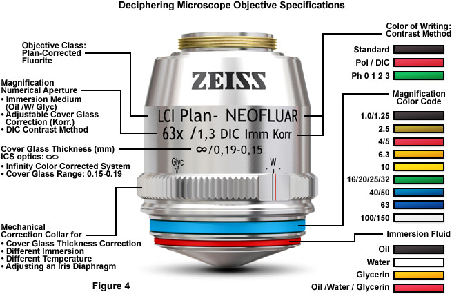
Most of the high-performance objectives feature spring mounts (see Figure 1) to protect the specimen, and many of the immersion objectives have nosepieces that can be snapped into the top position of their spring mount to enable the easy application of immersion fluids. The glass selected for objective fabrication must have suitable refractive index and dispersion, homogeneity, absence of strain, high chemical resistance, low thermal expansion, resistance to climatic changes, and high light transmission through the near-ultraviolet, visible, and near-infrared portions of the spectrum. In terms of how the various correction factors for objectives are categorized, the achromats have good color correction in two wavelengths, feature relatively flat fields in the center of the image, but require refocusing to observe details in the peripheral areas. Achromats are produced in versions designed for polarized light and phase contrast, but not fluorescence or differential interference contrast (DIC). Plan and epi-plan objectives are improved achromat versions with excellent flatness of field up to diameters of 24 millimeters or more. In addition, reflected light achromat objectives exhibit excellent contrast and a variety of working distances. The specifications required to identify objectives (see Figure 4) are usually inscribed on the decorative barrel protecting the internal lens elements.
Polarized light fields in nature. In each pair of panels, the top view is photographed through a linear polarizer with its e-vector oriented horizontally and the bottom view through a vertical polarizer (also indicated by the 2-headed arrows). (A) Full-sky images taken using a fisheye lens viewing the entire celestial hemisphere, showing scattering in a partly cloudy sky at sunrise; North is to the top and East to the left. A band of polarization passing overhead with the e-vector polarized North: South is obvious. Even though the sky is partly covered with clouds, the pattern persists in open blue sky areas. (B) Reflective polarization from the surface of a pond. Note the strong horizontal polarization, especially in the lower part of the image which is viewed near Brewster’s angle. (C) Scattering-induced polarization in the clear blue water over a coral reef. The light is ∼50% polarized with a horizontal e-vector. When the horizontal polarization is removed there is greater contrast (lower panel), especially at longer viewing distances.
Circular polarization is rare in nature; in fact, the only described case on earth is for linearly polarized light in aquatic habitats that becomes internally reflected from the air:water interface (Ivanoff and Waterman 1958). A few animals, however, create circularly polarized light by a variety of optical mechanisms that will be discussed in the section on circularly polarized signals (see also Johnsen 2012; Cronin et al. 2014). Because circularly polarized light is not common, most of the discussion here considers linear polarization. When writing about circular polarization, I note this specifically.
Before proceeding further, I want to go into a bit of semantics. Sensory drive acts on signals, which are used in intraspecific or interspecific communication. When communicating with conspecifics, such signals may be directed at males, females, or both, and may be involved in mate selection, behavioral intent, competition, or agonistic behaviors. Signals directed at heterospecifics are often concerned with predator deterrence (agonistic or aposematic signals). “Biological communication” is very difficult to define, and definitions tend to center either on adaptiveness or on function (information content); see Scott-Phillips (2008) for a discussion of this dilemma. Bradbury and Vehrencamp (1998, p. 357) define communication as “an exchange of a signal between a sender and a receiver to the benefit of both parties.” This definition rules out camouflage or deceptive signals, such as bluffs or exaggerations (where only one party, the sender, benefits), even though such signals are obviously amenable to evolution via sensory drive. Owren et al.’s (2010) definition of communication focuses on a signal’s influence on a receiver rather than on the information content of a signal; since the function of crypsis is to avoid detection by a receiver, it is not a signal under this definition, and we will not consider it here.
While many surfaces of animals have the ability to reflect polarized light, the structures just described appear to be specialized to form visual signals. Understanding the nature (or even the presence) of polarized-light signals, not to mention their evolution, is very challenging. Marshall et al. (2014) strongly argued that the question has received “overenthusiastic optimism” in generating hypotheses and interpreting results. The problem stems from the simple fact that animal surfaces naturally reflect all sorts of polarization patterns. We see none of these, so when careful polarimetry reveals a striking pattern in the polarization domain, it is natural to jump to the conclusion that it means something to a viewer with polarization vision. That these patterns are hidden to us then suggests that the signaling is “secret,” carrying privileged, encrypted intraspecific information. (Similar arguments were once advanced for patterns visible in ultraviolet light.) There are 2 types of problems with this kind of thinking. In the first place, knowing that an animal’s visual system responds to polarized light is no demonstration that it even is interested in polarized objects, much less that it is used in communication (see Cronin et al. 2014; Marshall et al. 2014). Also, impressive patterns of polarization reflection are likely to be a consequence of unusually shaped body features or of underlying structural elements, and thus may be unrelated to signal production. A final set of issues arises from the experimental challenges of polarization vision research in general, where it is very easy to misinterpret experimental results (Foster et al. 2018).
Owren MJ, Rendall D, Ryan MJ, 2010. Redefining animal signaling: influence versus information in communication. Biol Philos 25:755–780.
by RB SAPER · 2009 · Cited by 419 — Commercial zinc supplements contain 7 to 80 mg of elemental zinc, and are commonly formulated as zinc oxide or salts with acetate, gluconate, and sulfate. In ...
Horváth G, Blahó M, Egri Á, Hegedüs R, Szél G, 2014a. Circular polarization vision of scarab beetles. In: G Horváth, editor. Polarized Light and Polarization Vision in Animal Sciences. Berlin: Springer, 147–170.
The optical components contained within modern microscopes are mounted on a stable, ergonomically designed base that allows rapid exchange, precision centering, and careful alignment between those assemblies that are optically interdependent. Together, the optical and mechanical components of the microscope, including the mounted specimen on a glass microslide and coverslip, form an optical train with a central axis that traverses the microscope base and stand. The microscope optical train typically consists of an illuminator (including the light source and collector lens), a substage condenser that serves to prepare illumination for imaging, specimen, objective, eyepiece, and detector, which is either some form of camera or the observer's eye.
Ivanoff A, Waterman TH, 1958. Factors, mainly depth and wavelength, affecting the degree of underwater light polarization. J Mar Res 16:283–307.
Endler (1992) focused on sensory drive and visual signals in his presentation of the topic, and since then it’s likely that a strong majority of studies of this process have concerned visual systems and visual signals (although there is no scarcity of other systems represented). Most of this work centers on spectral sensitivity tuning and color signals and patterns. However, a diverse assortment of animals uses an entirely different property of light for signals: its polarization.
It has long been recognized that many scarab beetles reflect circularly polarized light (Figure 6A). Michelson (1911) first noted this, and the phenomenon has recently been described in some detail by Goldstein (2006). Brady and Cummings (2010) published results of spontaneous phototaxis experiments that suggested that one species of scarab beetles Chrysina gloriosa could discriminate vertical linearly polarized from left circularly polarized light, while another C. woodi, could not. Exhaustive tests with hundreds of beetles of 4 other scarab species by Horváth’s laboratory, however, found no evidence that any of these beetles respond differentially to circularly polarized light of either handedness during feeding or when presented with individuals of their own species, whether walking or flying (Blahó et al. 2012; Horváth et al. 2014a; see also Foster et al. 2018). Since eyes of scarab beetles do not appear to have any optical specializations that would permit them to detect circular polarization, it now appears that their circularly polarized reflections are not involved in signaling behavior.
Unlike the evolution of putative linearly polarized signals, these circularly polarized ones seem not to have originated with a push from sensory drive. Except for rare and transient optical phenomena, such as internal reflection from the water’s surface (Ivanoff and Waterman 1958), the marine environment is thought to be devoid of circular polarization. Indeed, it is mystifying that mantis shrimps even are able to detect it, given the optical demands of constructing a circular polarization analyzer and the apparent inutility of sensing it in the first place. It now appears that circular polarization sensitivity exists because of an odd arrangement of receptor cells in a subset of receptors in the midband, a specialized region of mantis shrimp compound eyes. In 2 rows of ommatidia, excellent orthogonally arranged polarization receptors exist (similar in principle to Figure 3C). However, they are placed under a different receptor that absorbs ultraviolet light, but transmits longer-wavelength light that the polarization receptors absorb. Uniquely, this ultraviolet receptor is an optical, achromatic retarder, converting incoming circularly polarized light to linearly or elliptically polarized light that passes through to the underlying polarization receptors (Chiou et al. 2008b; Roberts et al. 2009). Simply put, it converts light that the polarization receptors would not normally discriminate from unpolarized light into discriminable light. The efficiency of the conversion depends on the length of the ultraviolet receptor, but at typical lengths the efficiency is fairly high (Roberts et al. 2009). The end result is a circular polarization analyzing system without a circularly polarized stimulus to analyze. Due to the often incomplete conversion of circular to linear polarization, the ellipticity of the light entering the linear polarization receptors is high enough to permit them to continue to detect its linear polarization content, while also responding to occasional circular polarization.
Erin E. Wilson and Michael W. Davidson - National High Magnetic Field Laboratory, 1800 East Paul Dirac Dr., The Florida State University, Tallahassee, Florida, 32310.


 Ms.Cici
 8618319014500