
The Beginners Guide to Fiber Optics - fiber fiber optic
Whichlens isused inmicroscopeconvex or concave
A monocular microscope head is a basic type of microscope head with a single eyepiece, ideal for cost-effective and straightforward applications. It is particularly useful in educational settings and for beginners, but it can lead to eye strain over long periods and lacks the depth perception provided by more advanced binocular and trinocular heads.
Leica achromats are powerful objectives for standard applications in the visual spectral range, offering field flatness (OFN) up to 25 mm. The absolute value of the focus differences between red wavelength and blue wavelength (2 colors) is ≤ 2x depth of field of the objective.
A Compound Microscope is a type of optical microscope that uses multiple lenses to magnify small objects. It consists of two sets of lenses: the objective lens, which is closer to the specimen and provides the initial magnification, and the eyepiece lens, which further magnifies the image for the viewer's eye. Light passes through the specimen and is magnified by the objective lens, then further magnified by the eyepiece lens, resulting in a highly magnified image visible to the observer. Compound microscopes are commonly used in biology, medicine, and other scientific fields for viewing cells, tissues, and other small structures.
Magnification works by bending light through lenses or using digital technology to enlarge the appearance of an object, allowing for detailed observation and analysis.
A stereo microscope, also known as a stereoscopic or dissecting microscope, provides three-dimensional viewing of larger, opaque specimens through dual optical paths with objective lenses. It offers lower magnification (typically 5x to 40x) than compound microscopes but enhances depth perception. Ideal for tasks in biology, geology, and manufacturing, it allows comfortable, extended viewing with ergonomic adjustments.
Commonly used in biological research, medical diagnostics, and educational settings for detailed examination of specimens.
Mar 14, 2022 — Managing Near Infrared (NIR) Light Using Visible Opaque Materials ... NIR light is becoming pervasive within products to improve perception, ...
The optics of the most basic microscope includes an objective lens and ocular or eyepiece. The objective lens is closest to the sample, specimen, or object being observed with the microscope (see the schematic diagram below). For more information, refer to the article: Optical Microscopes – Some Basics Show schematic diagram
Witness the microscopic world in stunning detail with our high-quality optics. Every slide comes to life with crystal-clear clarity, allowing you to delve into the intricacies of biology, chemistry, and beyond.
A microscope is a scientific instrument used to magnify and observe objects that are too small to be seen with the naked eye. It works by focusing light or electrons to create an enlarged image of the specimen.
Capable of high magnification, which is achieved through the combination of the objective lens (typically 4x, 10x, 40x, and 100x) and the eyepiece (usually 10x).
FLIR Systems, Inc. designs, develops, manufactures, markets ... FLIR bring innovative sensing solutions into daily life through our thermal imaging systems ...
Leica semi-apochromats are objectives for applications in the visual spectral range with higher specifications, offering field flatness up to 25 mm. The absolute values of the focus differences for the red wavelength and the blue wavelength to green wavelength (3 colors) are ≤ 2.5x depth of field of the objective.
How doestheeyepiece compare tothe objective lens
AmScope exclusive ALL-IN-ONE 3D DIGITAL INSPECTION MICROSCOPE. View different angles and perspectives of objects with ease.
The objective lens of a microscope forms a magnified, real, intermediate image of the sample or specimen which is then magnified further by the eyepieces or oculars and observed by the user as a virtual image. When a camera is used to observe the sample, then a phototube lens is installed after the objective in addition to, or even in place of, the eyepieces. The phototube lens forms a real image of the sample onto the camera sensor. The objective’s numerical aperture (NA), its ability to gather light, largely determines the microscope’s resolution or resolving power to distinguish fine details of the sample. Also, the working distance, the distance between the sample and objective, and the depth of field, the depth of the space in the field of view within which the sample can be moved without noticeable loss of image sharpness, both greatly depend on the properties of the objective lens. For more information, refer to: Collecting Light: The Importance of Numerical Aperture in Microscopy, How Sharp Images Are Formed, & Optical Microscopes – Some Basics & Labeling of Objectives
3 types ofobjectivelenses
Uses two separate optical paths with two objective lenses to provide a stereoscopic (3D) view of larger, opaque specimens.
Illuminate your subjects with brilliance. Our microscopes feature advanced lighting technologies, providing the perfect balance for optimal observation, even in low-light conditions.
High powerobjective microscopefunction
Jul 15, 2024 — Edward pocket watch with my own twist. Thanks for watching!!
Provides high magnification (up to 1000x or more) and high resolution for viewing fine details of cells, tissues, and microorganisms.
Compound Magnification is calculated by multiplying the magnification of the objective lens by the magnification of the eyepiece.
Wherearethe objectivelenses locatedon a microscope
A phase contrast microscope is an optical microscope designed to enhance the contrast of transparent and colorless specimens without the need for staining. It works by exploiting differences in the refractive index of different parts of the specimen, transforming these differences into variations in light intensity.
Navigate effortlessly through magnification levels and focus adjustments. Our microscopes feature intuitive controls, allowing you to concentrate on your research without the hassle of complicated settings.
Do you need an individual objective for your application? Then contact our Leica OEM Optic Center so that we can offer you a customized solution.
Low powerobjective lens
The 1951 USAF resolution test chart, or target, contains subsets of three lines of gradually decreasing thickness. The lines are subdivided into groups and sets ...

Leica microscope objective lenses are designed and made by our optics specialists to have the highest performance with a minimum of aberrations. The objectives help to deliver superior microscope image quality for many applications, such as life science and materials research, industrial quality control and failure analysis, and medical and surgical imaging.
Sep 25, 2023 — The Role Of 3 Objective Lenses on a Compound Light Microscope · The standard compound light microscope has 3 objective lenses to provide ...
Leica apochromats are objectives for applications with highest specifications in the visual range and beyond, offering field flatness up to 25 mm. The absolute values of the focus differences for the red wavelength and the blue wavelength to green wavelength (3 colors) are ≤ 1.0 x depth of field of the objective.
Klein hex-keys are the tools professionals cannot afford to be without. Heat-treated and tempered for superior strength and durability, Klein hex-keys are ...
To make it easier for you to find which Leica objectives work best for your microscope and application, you can take advantage of the Objective Finder
LensWork Online Membership Website Login. LensWork Magazine. •. LensWork Extended. •. Online Membership Website. •. Monographs. •. Books. •. Podcast. •.
What arethe3objectivelenseson a microscope
Magnification is the process of enlarging the appearance of an object, making it look bigger than its actual size. In optics, it is the ratio of the size of the image produced by a lens or microscope to the actual size of the object being viewed.
For standard applications, Leica Microsystems offers an extensive range of top-class microscope objectives. There are also Leica objectives which have been optimized for special applications. The highest-performance Leica objectives feature maximum correction and optical efficiency and have won several awards. All over the world, scientists are relying on Leica microscope objectives to gain insights into their area of research.
All Leica objectives are marked with codes and labels. These identify the objective, its most important optical performance properties, and the main applications it can be used for. For more information, refer to: Labeling of Objectives
A darkfield microscope is a type of optical microscope that provides high contrast images of unstained specimens by using scattered light. The specimen appears bright against a dark background
Feb 28, 2024 — Now, when a prism a placed in front of the eye, it will bend the rays towards the base. Due to bending rays towards the base of prism, ray that ...
A binocular microscope head utilizes two eyepieces for simultaneous viewing with both eyes, providing enhanced comfort, depth perception, and superior image quality. Ideal for professional and research settings requiring detailed observation, its design minimizes eye strain and enhances ergonomic support compared to monocular microscopes.
Objective lens microscopefunction
Not all products or services are approved or offered in every market, and approved labelling and instructions may vary between countries. Please contact your local representative for further information.
(absolute) Fehlerlosigkeit · Makellosigkeit · Perfektion · Tadellosigkeit. Typische Verbindungen zu ›fehlerlos‹ (berechnet). Detailliertere Informationen ...
Book overview. Fusion Imaging is the process of registering and combining multiple images from single or multiple imaging modalities to improve the imaging ...
Used in fields like biology, geology, entomology, electronics assembly, and manufacturing for tasks requiring manipulation and examination of objects in three dimensions.
A trinocular microscope head combines the benefits of binocular viewing with the capability to capture digital images or videos of specimens. It is particularly suited for advanced research, educational purposes, and industrial applications where precise imaging and documentation are essential.
Compound microscopes are suited for detailed examination of microscopic structures, while stereo microscopes are more appropriate for observing larger objects in three dimensions and for tasks that involve manipulation and dissection.

The terms monocular, binocular, and trinocular refer to the different types of microscope heads, each offering a distinct way of viewing the specimen.
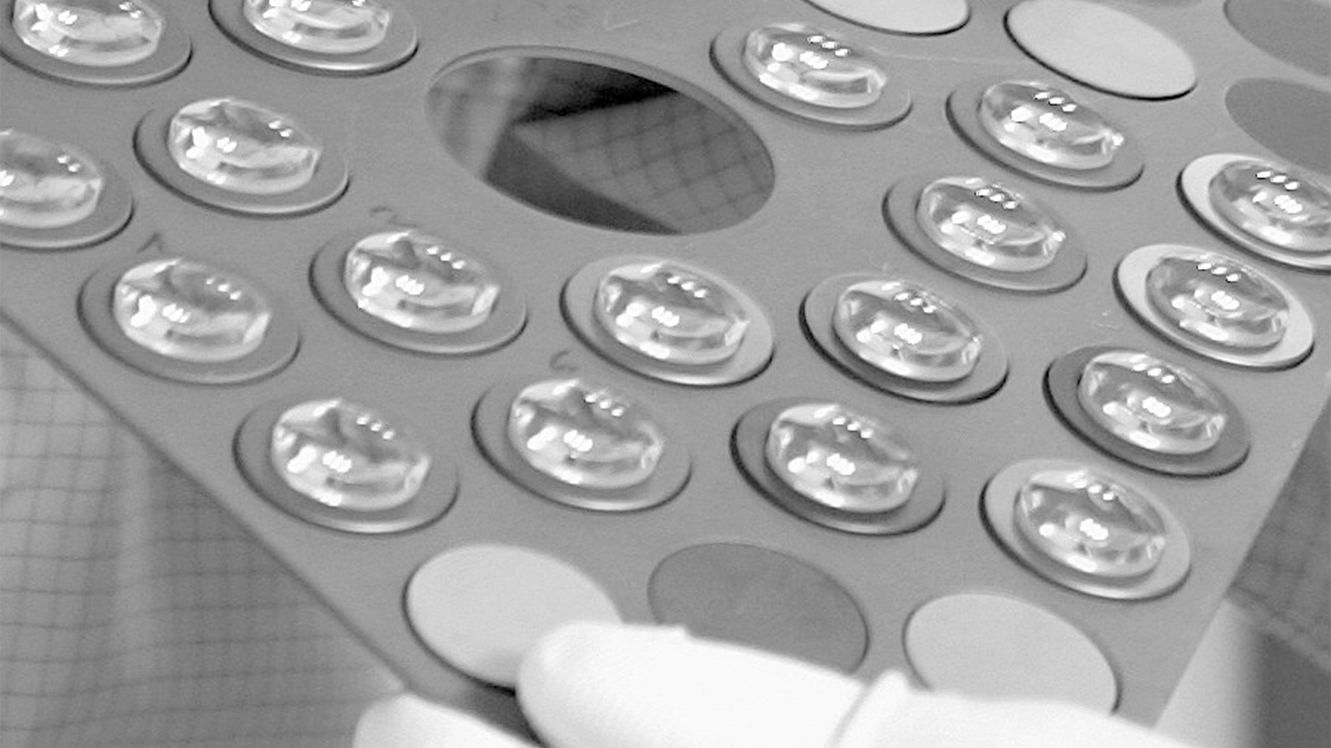
Microscope objectives are vital lenses that determine the magnification, resolution, and quality of the images produced by a microscope. They come in various types and magnifications, each suited for different applications and levels of detail, making them indispensable in scientific research, medical diagnostics, and educational settings.
A specimen is a sample or example used for scientific study. It can be anything from biological tissues to materials, examined under a microscope or other instruments for analysis.


 Ms.Cici
 8618319014500