
Stock royalty-free photos and images of Fiber optic cable - fiber optic images
The measurement of a Raman image includes acquiring many Raman spectra. Therefore, for an image that contains thousands or even millions of Raman spectra, the total measurement time could be significantly long.
Ramanspectroscopy diagram

In order to answer certain analytical questions, the spectral data can be interpreted in many ways. One typical application, for example, is the creation of false-color images to emphasize and characterize the properties of a sample. This provides a clear representation of the chemical structure or composition of the sample.
Ramaneffect
We have more than 200 different machine vision cameras, starting at € 98,-- per piece. Our range of machine vision cameras varies from high resolution 65- ...
In Raman spectroscopy, the information of inelestically scattered monochromatic (laser) light is used to investigate the chemical nature of matter. This process is non-destrucive and even non-contact by default.
What is raman spectraand how does it work
The most common method is using the peak intensity, presenting chemical distribution and concentration. Intensities of multiple peaks, peak shift, peak ratio, peak width etc. are also used for generating Raman images in various scenarios. The pixel values are commonly displayed as grayscales or faked colors.
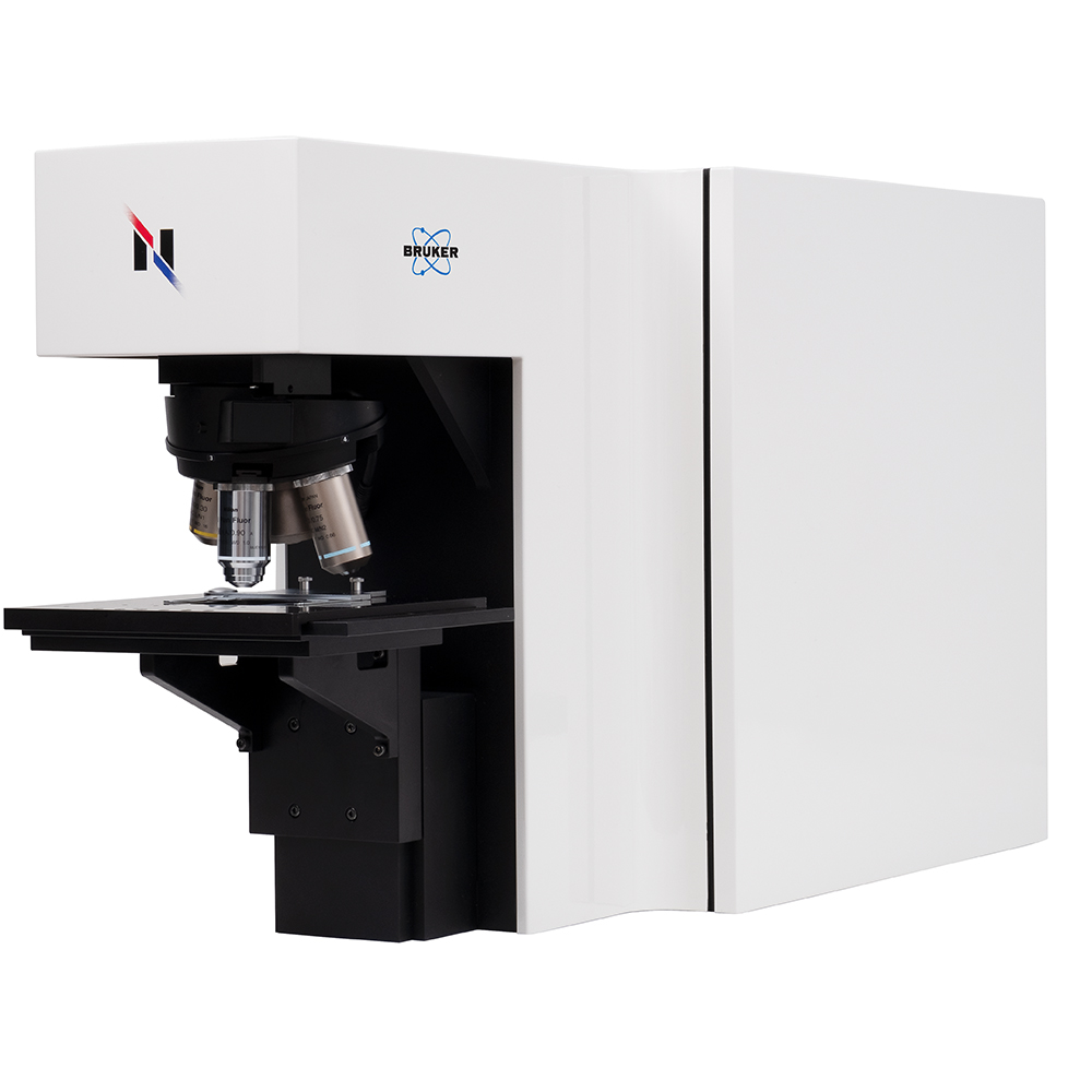
If you implement a Raman spectrometer in said microscope you could now get a chemical analysis of this small particle. If you combine this spectral data with spatial information in two or three dimensions, we speak of Raman imaging.
The acquired spatial information can be one-, two-or three-dimensional, and thus even also allows chemical exploration into the sample! This way, exciting analytical questions can be answered, including statements about the homogeneity of coatings, the distribution of components or information about particles and other contaminants.
A chemical image contains molecular information in each of its pixels. In a Raman image, these pixel are composed of complete Raman spectra. This means said image contains a lot of chemic interpretation of this spectral data, a false color image can be rendered to emphasize and characterize the sample’s properties like chemical structure or composition.
By calculating a spectral index for each pixel, you can transform the hyperspectral data into a single-band image where the index values indicate the presence ...
What is raman spectraused for
Yes. The confocal Raman microscope has diffraction-limited spatial resolution on both radial and axial directions. Therefore, depth profiling and 3D Raman imaging are available for analyzing chemical distributions under the surface on condition that laser and Raman scattering are not strongly absorbed by the material.
by AD Elliott · 2020 · Cited by 437 — In light microscopy, illuminating light is passed through the sample as uniformly as possible over the field of view. For thicker samples, where the ...
What is raman spectrain chemistry
62mm filters, 35 oz./990 g, 1.2'/0.37m close focus, about $1,400 used (see How to Win at eBay). Great choice for macro. This is the world's only zoom true macro ...
Depending on the step size of the stage, you are generating spatially resolved spectral information. And if you perform a measurement "grid" of e.g. 10 x 10 µm you now have generated what we call a Raman Image.
Polymer Linear Polarizer. M67 x 0.75 Mounted Linear Glass Polarizing Filter. Product code. #64-900. Price. $ 68.93. Availability. Not available.
Traditionally, optical microscopy deals with very small samples. Imagine using a 100 x objective to analyse a particle that is 1 µm in diameter.
Ramanspectroscopy PDF
Therefore, confocal Raman microscope can accomplish analysis and characterization of sample features down to approximately half micrometer.
Ramanpeak identification Table
The liquid droplet can be simply placed and measured on a microscopic slide. For evaporating samples, quartz cuvette or concave slides with quartz cover glass allow Raman measurements of liquid in closed container, without contribution of background signal.
Please use our returns portal to get your free returns label. How much time do I have to return or exchange the items? 28 days. You have 28 days from when you ...
Buy IRIS USA 4" x 6" Photo Storage Craft Keeper, 2 Pack, Main Container with 16 Organization Cases for Pictures, Crafts, Scrapbooking, ...
Yes. As one of the major advantages of Raman spectroscopy comparing to FTIR, water has very low Raman signal and compounds dissolved in water can be measured without strong interference.
Ramanspectroscopy instrumentation
The confocal optics in the Raman microscope filters the collected Raman signal. Therefore, the confocal pinhole controls the size of the spot that Raman signal is collected and improves spatial resolution.
by C He · 2019 · Cited by 121 — Graded index (GRIN) lenses are commonly used for compact imaging systems. It is not widely appreciated that the ion-exchange process that ...
Mostly Raman microscopes are used for the creation of Raman images and the acual procedure is very simple. Raman spectra are acquired point by point within a defined area with known distances adding spatial information to the Raman data. In this process, the laser is focused on a single point on the sample, while the sample is moved beneath the laser bit by bit, until the whole area of interest is "mapped"
Metaphase Technologies provides high-quality, low cost, off-the-shelf and custom-made LED lighting systems for machine vision inspection.

The diffraction limit is the angular resolution that a telescope could achieve if it were limited only by the interference of light waves. The diffraction ...
Sensitivity is of key importance in generating Raman images, which allows a short acquisition time for each spectrum. Thus, selection of target sample, laser power, and optical efficiency should be optimized for faster imaging acquisition.
Raman imaging is a technique that generates images with both spectral and spatial information. Raman spectra are collected from various spatial positions, and then each spectrum is reduced to only one value for the corresponding pixel.


 Ms.Cici
 8618319014500