
Silicon Nitride X-Ray Windows for TEM - silicon windows
by M Baillergeau · 2016 · Cited by 30 — Diffraction is the ultimate limit at which details of objects can be resolved in conventional optical spectroscopy and imaging systems.
Once each fluorescent probe in a sample can be imaged individually, its positions can be determined to high precision by finding the center of the single-molecule image (Thompson et al., 2002; Yildiz et al., 2003). The uncertainty in determining the molecule's position (i.e., the localization precision) scales approximately with the inverse square root of the number of photons detected from the molecule. It can be approximated by a‒≈Δ∕N, wherein Δ denotes the width of the diffraction-limited point spread function and N is the number of photons detected (Thompson et al., 2002). For bright fluorescent dyes, about one million photons can be detected from a single molecule, leading to a localization precision of ≤ 1 nm (Pertsinidis et al., 2010; Yildiz et al., 2003). The relative position of multiple fluorophores can also be determined by taking advantage of different emission colors (Churchman et al., 2005; Lacoste et al., 2000; van Oijen et al., 1998), the sequential photobleaching of individual fluorophores (Gordon et al., 2004; Qu et al., 2004), or the stochastic blinking of quantum dots (Lagerholm et al., 2006; Lidke et al., 2005).
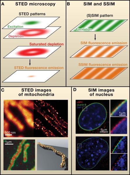
In STED microscopy, the patterned illumination prevents fluorophores from emitting light (Hell, 2007; Hell and Wichmann, 1994; Klar and Hell, 1999). This suppression is achieved by stimulated emission, a process in which a light source, called the depletion light, brings an excited fluorophore down to the lowest energy state (i.e., the ground state) before it can emit fluorescence signal. In practice, the depletion light is applied as a pattern surrounding the focal spot of the excitation laser. This reduces the size of the region of molecules that fluoresce, as if the “focal spot” of the microscope is sharpened (Figure 2Afig2). Scanning this sharpened spot across a sample then allows a super-resolution image to be recorded. Thus, this negative patterning approach elegantly generates a positive image without the need of any postacquisition processing.
In these cabins, the heating elements are typically heated to about 300– 400° C and the emission is in the FIR range, that is, the heat exchange between the body and the environment is almost purely radiative (radiant heating) with cabin air temperature being at around 40°C or less (Figure 4). Heating of the skin with FIR warming cabins is faster (in comparison with the conventional saunas) but higher irradiance of the skin must be applied in order to produce noticeable sweating. These cabins are frequently used in Japan where the practice is called “Waon therapy” [20, 21]. Waon therapy has been used extensively in Japan [22] and Korea [23] for cardiovascular conditions and diseases, particularly chronic heart failure [24, 25] and peripheral arterial disease [26, 27]. FIR sauna therapy has been used to improve cardiac and vascular function and reduce oxidative stress in patients with chronic heart failure [28]. Beever [29] asked whether FIR saunas could have a beneficial effect on quality of life in those patients with type II diabetes. The study consisted of 20 min, three times weekly infrared sauna sessions, over a period of 3 months. Physical health, general health, social functioning indices, and visual analogue scales (VAS) measurements for stress and fatigue all improved in the treatment group. A study of patients with rheumatoid arthritis and ankylosing spondylitis showed a reduction in pain, stiffness, and fatigue during infrared sauna therapy [30].
The basic form of SIM (i.e., the linear form) does not rely on any special photophysics of the fluorophores but purely on the optics of the microscope. Therefore, any fluorescent probe that is compatible with conventional fluorescence imaging is compatible with SIM. In addition, multicolor imaging can be achieved with SIM as it is with conventional fluorescence microscopy. Up to four colors can be imaged in the visible to near infrared (IR) spectrum range without incurring much crosstalk between the different color channels. As an example, Figure 2D shows a three-color SIM image of DNA, lamin B, and nuclear pore complexes in the nucleus (Schermelleh et al., 2008). In contrast, SSIM requires special fluorophores that have high photostability because the fluorophores are maintained in the highly reactive excited state most of the time in this imaging scheme.
Anyone who has used a light microscope has wished that its resolution could be a little better. Now, after centuries of gradual improvements, fluorescence microscopy has made a quantum leap in its resolving power due, in large part, to advancements over the past several years in a new area of research called super-resolution fluorescence microscopy. In this Primer, we explain the principles of various super-resolution approaches, such as STED, (S)SIM, and STORM/(F)PALM. Then, we describe recent applications of super-resolution microscopy in cells, which demonstrate how these approaches are beginning to provide new insights into cell biology, microbiology, and neurobiology.
In the literature, the localization precision is often reported as the standard deviation of multiple localization measurements of a single object, whereas both standard deviation and full width at half maximum (=2.35 × standard deviation for a Gaussian distribution) have been used as measures of the resolution. In this Primer, we use full width at half maximum to describe resolution because it illustrates better the closest resolvable separation between objects.
Diffractiongrating
For FIR used as a therapeutic modality the alternative terms “biogenetic radiation” and “biogenetic rays” have been coined and widely used in the popular literature. FIR wavelength is too long to be perceived by the eyes, however, the body experiences its energy as a gentle radiant heat which can penetrate up to 1.5 inches (almost 4 cm) beneath the skin. FIR energy is sufficient to exert rotational and vibrational modes of motion in bonds forming the molecules (including the water molecules) as well as resonate with cellular frequencies. Resulting epidermal temperature is higher when the skin is irradiated with FIR than if similar thermal loads from shorter wavelengths are used. The prolonged erythermal response due to FIR exposure has been proposed to be due to increased epidermal temperatures associated with it, but levels of FIR that do not produce any detectable skin heating can also have biological effects.
Although photoswitching offers the most versatile strategy for achieving the temporal control needed for super-resolution imaging, other strategies can also achieve similar control in certain biological samples, such as through the binding and dissociation of fluorescent molecules (Sharonov and Hochstrasser, 2006). It is possible to obtain subdiffraction limit image resolution even when the single-molecule imaging condition is not rigorously satisfied. When the density of fluorophores is not too high, such that temporal fluctuations of neighboring pixels are distinct, higher-order correlation analysis of the temporal fluctuations of individual pixels can be used to substantially improve the image resolution (Dertinger et al., 2009).
If it can be proved that non-heating FIR has real and significant biological effects, then the possible future applications are wide ranging. Not only could bandages and dressings made out of NIR emitting fabrics be applied for many medical conditions and injuries that require healing, but there is a large potential market in lifestyle enhancing applications. Garments may be manufactured for performance enhancing apparel in both leisure activities and competitive sports areas. Cold weather apparel would perform better by incorporating FIR emitting capability and sleeping environments could be improved by mattresses and bedding emitting FIR.
In this regard, the dynamics of water-clusters has attracted considerable interest since there is a noticeable difference with respect to the dynamics of bulk-liquid-water, and this may have significant implications in biological environments. Local changes in the molecular environment (caused by solvation or confinement) are shown to affect substantially the translational and vibrational modes in FIR frequency range. It is found that water cluster size and temperature affect the FIR absorption spectrum significantly [3].
As is true for the depletion light pattern used in STED, the illumination pattern created by interference is also limited by diffraction. Therefore, when the fluorescence signal scales linearly with the intensity of the excitation light, SIM results only in a doubling of spatial resolution (Figure 2B), which is ~100 nm in the lateral dimensions(Gustafsson, 2000).
Celliant® (Hologenix, Santa Monica, CA, USA) is a polyethylene terephthalate (PET) fiber that incorporates FIR emitting ceramic nanoparticles. York and Gordon [42] studied socks manufactured from Celliant® fiber material in patients with chronic foot pain resulting from diabetic neuropathy or other disorders. A double-blind, randomized trial with 55 subjects (38 men, 17 women, average age 59.7 ± 11.9 years) enrolled 26 patients with diabetic neuropathy and 29 with other pain etiologies. Subjects twice completed the VAS, brief pain inventory (BPI), McGill pain questionnaire (MPQ), and a multipurpose, short form health survey with 36 questions (SF-36) a week apart [W(1+2)] before receiving either control or Celliant® socks. The same questionnaires were answered again 1 and 2 weeks later [W(3+4)]. The questionnaires provided nine scores for analyzing pain reduction: one VAS score, two BPI scores, five MPQ scores, and the bodily pain score on the SF-36. Mean W(1+2) and W(3+4) scores were compared to measure pain reduction. More pain reduction was reported by Celliant® subjects for eight of the nine pain questions employed, with a significant (p = 0.043) difference between controls and Celliant® for McGill question III. In neuropathic subjects, Celliant®; caused greater pain reduction in six of the nine questions, but not significantly. In non-neuropathic subjects eight of nine questions showed better pain reduction with the Celliant® socks.
The resolution for optical microscopy is limited by the diffraction, or the “spreading out,” of the light wave when it passes through a small aperture or is focused to a tiny spot. Because this property is intrinsic to all waves, breaking the diffraction barrier of light microscopy has been deemed impossible for a long time. However, such limitations have not deterred a small group of scientists from pursuing “super-resolution” fluorescence microscopy that breaks through this seemingly impenetrable barrier.
In the IR radiation bands, only FIR transfers energy purely in the form of heat which can be perceived by the thermoreceptors in human skin as radiant heat [1]. Not only is FIR absorbed by the human body but it is also emitted by the body in the form of black body radiation (3–50 μm with an output peak at 9.4 μm).
Corresponding author: Michael R. Hamblin, Wellman Center for Photomedicine, Massachusetts General Hospital, Boston, MA, USA, hamblin@helix.mgh.harvard.edu
(B) Structured illumination microscopy (SIM) and saturated SIM (SSIM) use pattered illumination to excite the sample and generate fluorescence. This patterned excitation typically has a sinusoidal shape (green, top layer). Such illumination generates a similarly shaped fluorescence emission pattern when the fluorescence responds in a linear manner (orange, middle layer). With strong excitation, fluorescence saturates, generating a saturated emission profile with narrow dark regions (orange, bottom layer) that provide spatial information substantially beyond the diffraction limit.
The classification of the International Commission on Illumination (CIE) has three sub-divisions for the IR radiation as given in Table 1. An alternative classification provided in ISO 20473 standard for the sub-division of the IR ranges is given in Table 2.
For imaging dynamic events in living systems, time resolution is critical. In all imaging methods, there is an intrinsic trade-off between spatial and temporal resolution. This trade-off can be best understood from the Nyquist sampling theory. In the case of STED microscopy, which acquires an image by scanning the focal spot across the sample, the Nyquist criterion requires the scanning step size to be smaller than half of the desired resolution. The time resolution is then determined by the integration time per scanning step, the scanning step size, and the size of the imaging field. Better spatial resolution requires a smaller scanning step size and, consequently, a longer time to image the same field of view.
For many years, several imaging techniques have pushed the boundary of the diffraction limit of light microscopy. Among these methods, confocal microscopy and multiphoton fluorescence microscopy not only enhance the image resolution, but also reduce the out-of-focus fluorescence background, allowing optical sectioning and thus three-dimensional imaging. In addition, infrared light experiences a lower amount of scattering from tissues, allowing deep tissue imaging with two-photon microscopy (Zipfel et al., 2003). 4Pi microscopy and I5M use two opposing objective lenses to increase the effective numerical aperture of the microscope and thereby improve the image resolution (Gustafsson et al., 1995; Hell and Stelzer, 1992; Hell, 2003). Although these methods significantly improve the resolution, they are still fundamentally limited by diffraction and, in practice, achieve resolutions of ~100 nm in all three dimensions (Hell, 2003).
In SIM, a 3D illumination pattern can be created by the interference of three excitation light beams. Similar to the 2D counterpart, 3D SIM can double the resolution in all three dimensions, resulting in ~100 nm resolution in the lateral directions and ~300 nm in the axial direction (Gustafsson et al., 2008; Schermelleh et al., 2008). Figure 2D shows such a 3D SIM image of the nucleus. This 3D illumination pattern can be further combined with I5M using two-opposing objectives to achieve an isotropic resolution of ~100 nm in all three dimensions (Gustafsson et al., 2008). 3D SSIM has not yet been implemented.
Although these two categories of methods use different approaches to accomplish subdiffraction resolution, these techniques also share important commonalities. In both cases, a physical or chemical property of the fluorophore is used to maintain neighboring molecules in different states (i.e., “on” and “off”), enabling them to be resolved from each other (Hell, 2007).
(A) In stimulated emission depletion (STED) microscopy, fluorophores are excited by a focused light beam (green, top layer), and an additional depletion light beam (red, second layer) is used to bring molecules back to the ground state by a process called stimulated emission. The intensity profile of this additional beam at the focal plane typically has a ring shape, depleting the population of molecules that can generate fluorescence, especially near the edge of the focal spot. The depletion efficiency can be described by the red pattern shown in the third layer. This depletion effect substantially reduces the size of the fluorescent spot (orange, bottom layer), thereby improving the image resolution.
SIM acquires an image by scanning an illumination pattern that covers the entire image field. Thus, the image acquisition time of SIM is limited by how fast the illumination pattern can be modulated and by how fast the camera can read out a snapshot. For 2D SIM, which requires nine different illumination patterns to reconstruct a resolution-enhanced image, a time resolution of ~0.1 s has been demonstrated for live-cell imaging (Kner et al., 2009). Therefore, SIM is excellent for live-cell applications that require a large view field but not very high spatial resolution. Although live-cell imaging has not yet been demonstrated with SSIM, one would expect it to have higher spatial resolution at the cost of lower time resolution, as compared to SIM, because more illumination patterns are required to reconstruct an SSIM image (Gustafsson, 2005).
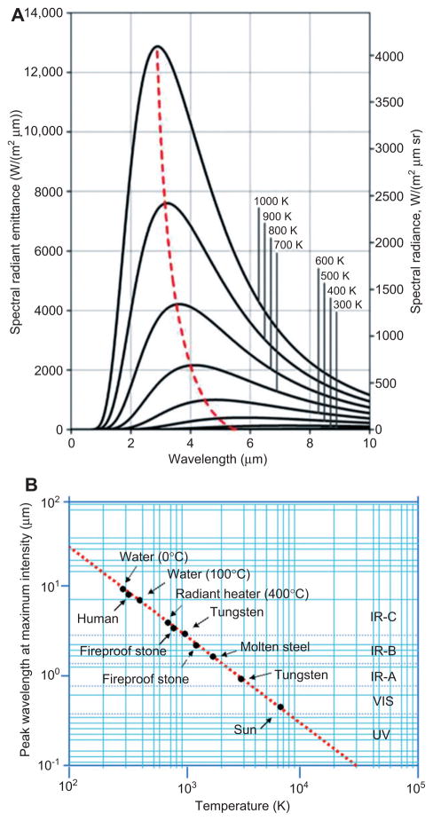
The term “black body” was first used by Gustav Kirchoff in 1860. In essence, all matter absorbs electromagnetic radiation to some degree and an object that absorbs all radiation falling on it (at all wavelengths and frequencies) is called a black body, i.e., a perfect absorber. When a black body is at a uniform temperature state, it emits back this absorbed energy, and it is termed as “ black body radiation ”. This is a type of radiation and has continuous frequency/intensity which depends only on the black body’s temperature, and the type of spectrum it generates is called the Planck spectrum. In this type of spectrum, spectral peaks at characteristic frequencies are shifted to higher values (shorter wavelengths) with increasing temperature values. For instance, at room temperature most of the emission of the black body is in the infrared region of the electromagnetic spectrum. At a typical environmental background temperature, which is around 300 K, the peak emission is at about 9.7 μm (and the curve covers the FIR region as well); at around 1800 K (temperature of molten steel), the peak is shifted to 1.6 μm; at around 6000 K (surface temperature of the sun), the peak is shifting even further, 0.48 μm, which now is in the visible (blue) region of the spectrum. Peak shifts of some representative black body temperatures and the range of electromagnetic radiation they fall into are given in Figure 2A, B. This type of shift in the emission peaks of the black bodies (to shorter wavelengths at higher temperatures) is governed by Wien’s displacement law.
In their most recent study the Leung group studied the repair effect of cFIR in human breast epithelial cells (MCF-10A) after H2O2 and after ionizing radiation from an X-ray source [19]. Their results show that in both, H2O2 toxicity and radiation exposure models, the cFIR treated cells demonstrated significantly higher cell survival rates than the control groups. In view of the experimental results and taking into account the relationship between indirect ionizing radiation and the oxidative stress-induced cell damage, and accumulation of free radicals, they proposed that the ionizing radiation protective ability of cFIR occurs predominantly through an antioxidant mechanism. They are suggesting that cFIR provides cells with a defensive mechanism during the irradiation process and promotes cell repair during post exposure period through hydrogen peroxide scavenging and COX-2 inhibiting activities.
Ishibashi et al. [8] did an in vitro study with five human cancer cell lines (A431, vulva; HSC3, tongue; Sa3, gingival; A549, lung; and MCF7, breast) to assess the effects of FIR irradiation. For that purpose, they used a tissue culture incubator with an imbedded FIR lamp that could continuously irradiate cells with FIR (lamp operating wavelength range being 4–20 μm with an emission peak height at 7 –12 μm). The overall observation was that the FIR effect varied in these five cancer cell line types, as can be expected. The study results showed that basal expression level of heat shock protein (HSP) 70A mRNA was higher in A431 and MCF7 cell lines in comparison with the FIR-sensitive HSC3, Sa3, and A549 cell lines. The study showed that the over expression of HSP70 inhibited FIR-induced growth arrest in HSC3 cells, and that HSP70 siRNA inhibited the proliferation of A431 cells after FIR treatment. A summary of the results of this study indicated that the proliferation-suppressing effect of FIR, in some cancer cell lines, is controlled by the basal expression level of the HSP70A. These findings suggest that FIR irradiation may be used as an effective medical treatment avenue for some cancer cells which have low levels of HSP70.
Photoactivation has also been combined elegantly with single-particle tracking to visualize dynamic events of living cells (Hess et al., 2007; Manley et al., 2008). Unlike conventional single-molecule tracking experiments, which require low density of target molecules, the use of photoswitchable probes allows a high density of target molecules to be labeled and tracked. Although it may still take a substantial amount of time to accumulate the large number of localizations required to define a structure with high resolution, the motion and dynamics of the molecules inside of the structures can be obtained with millisecond temporal resolution from these single-molecule traces. This imaging mode greatly extends the power of dynamic single-molecule imaging.
Since the principle chromophore at FIR wavelengths is not CCO but rather water, we must ask ourselves how can the biological effects of red and NIR absorption be so similar to those seen with FIR ? Perhaps some clue can be obtained by considering the difference between the two types of FIR therapy (heating and non-heating). While heating FIR therapy is reported to increase blood flow, this result may be the simple response of increased thermoregulation that is known to occur when tissue is warmed. However, it is possible that the increase in blood flow, seen in non-heating FIR therapy, may be similar in nature to that seen in LLLT, in other words, a vasodilation due to NO release from stores in CCO [48] as well as from NO bound to hemoglobin and myoglobin [49]. How are we to explain cellular responses from low fluences of FIR that are insufficient to produce bulk heating of water in the tissue ? Perhaps the answer lies in the concept of nanostructured water layers [50]. These are thin (nano meters) layers of water that build up on hydrophobic surfaces such as cellular membranes, and they can be considered as “concentrated water ” [51]. If this description is correct, it is reasonable to assume that relatively small amounts of vibrational energy delivered by non-heating FIR could perturb the structure of the membrane underlying the nanoscopic water layer without bulk heating. Small perturbations in membrane structure could have big effects at the cell level if the membrane contains an ion channel. Ion channels (many kinds for both cations and anions [52]) are present in all cell membranes, but are particularly common in mitochondrial membranes (both inner and outer [53]). If mitochondrial ion channels (particularly calcium channels [54]) could be opened by non-heating FIR, thus increasing mitochondrial respiration, it would explain how the overall therapeutic outcomes of LLLT and non-heating FIR therapy are so similar.
All living organisms are subjected to the natural electromagnetic radiation reaching the earth from the sun. Living organisms experience the beneficial as well as adverse effects of it at all levels, starting from sub-cellular organelles and ending with the whole body. Thermal radiation (or infrared) is a band of energy in the complete electromagnetic spectrum and it has been used effectively for millennia to treat/ease certain maladies and discomforts. Heated saunas are only one of the avenues (and perhaps the oldest) to deliver the radiation in a controlled environment and within a convenient treatment time. With the development of better technology to deliver pure far infrared radiation (FIR), the benefits from its effects have widened. Nowadays, specialty FIR emitting heat lamps and garments made up of filaments (fibers) impregnated with FIR emitting nanoparticles are becoming used to deliver these thermal radiation effects. In this paper we explore the use of FIR as a promising treatment modality for certain medical conditions. We cover both traditional applications and novel applications, and survey the latest technological advancements and most recent scientific studies in the field.
Super-resolution microscopy ... Super-resolution microscopy is a series of techniques in optical microscopy that allow such images to have resolutions higher than ...
The unrivaled combination of molecule-specific contrast and live-cell imaging capability makes fluorescence microscopy the most popular imaging modality in cell biology. Browse through any cell biological journal, and the impact of fluorescent microscopy is obvious, with > 80% of the images of cells in the book usually acquired with a fluorescent microscope. However, the application of fluorescence microscopy to many areas of biology is still hindered by its moderate resolution of several hundred nanometers. This resolution is approximately the size of an organelle and thus is inadequate for dissecting the inner architecture of many subcellular structures.
A belt containing FIR-emitting sericite mineral (a fine grained mica) was used to study the relief of menstrual pain [38]. In this study, 104 patients with primary dysmenorrhea were randomized to wear a sericite or placebo belt during sleep for three menstrual cycles, and then followed up for two additional menstrual cycles with no belt. Hot packs were used to heat the ceramics and ensure slight pain relief in both groups. Although the severity of dysmenorrhea decreased during the treatment period in both groups, it was found that during the follow-up period, the decreased VAS (pain) score was maintained in the experimental group, whereas the VAS score gradually returned to baseline in the control group, which resulted in significant difference between the groups (p = 0.0017).
By determining the position of individual molecules in all three dimensions, super-resolution microscopy based on single-molecule switching can be extended to 3D. The first implementation of this approach uses a simple optical design that takes advantage of astigmatism in which light waves in perpendicular directions have different focal points. Specifically, a cylindrical lens is inserted in the imaging path, such that the shape of a single-molecule image becomes elliptical. This makes it possible to determine the axial position of the molecule from the ellipticity and the lateral position from the center position of the image (Huang et al., 2008).Figure 3B shows 3D images of clathrin-coated pits taken with this approach, resolving the nanomorphology of these structures (Huang et al., 2008). Other implementations have utilized a variety of 3D localization methods, such as capturing defocused images at two different focal planes (Juette et al., 2008), engineering a PSF with a double-helical shape (Pavani et al., 2009), and using a mirror to project the axial view to the lateral direction (Tang et al., 2010). Axial resolutions of 40–70 nm have been reported using these methods.
There have been a few laboratory studies that have reported the biological effects of FIR. A recent important paper describes the in vitro use of an FIR generator (WS TY-301R®; M/s WS Far Infrared Medical Technology Co., Ltd., Taipei, Taiwan; see Figure 3) as a radiation source to irradiate human umbilical vein endothelial cells (HUVECs) [4]. In the study, FIR exposure (a low non-thermal irradiance) of 0.13 mW/cm2 for 30 min inhibited proliferation and the vascular endothelial growth factor (VEGF)-induced phosphorylation of extracellular signal-regulated kinases in HUVECs. Furthermore, FIR exposure induced the phosphorylation of endothelial nitric oxide synthase (eNOS) and nitric oxide (NO) generation in VEGF-treated HUVECs. Both VEGF-induced NO and reactive oxygen species (ROS) generation was involved in the inhibitory effect of FIR. Nitrotyrosine formation increased significantly in HUVECs treated with VEGF and FIR together. Inhibition of phosphoinositide 3-kinase (PI3K) by wortmannin abolished both the FIR-induced phosphorylation of eNOS and serine/threonine-specific protein kinase in HUVECs. In addition to that, FIR exposure upregulated the expression of PI3K p85 at the transcriptional level. It was observed that FIR exposure induced the nuclear translocation of promyelocytic leukemia zinc finger protein in the cells. These data provide information on how FIR exposure could affect microcirculation, independent from thermal effects. The same group had previously shown that non-thermal FIR therapy increased skin blood flow in rats [5]. Toyokawa et al. [6] used home-made ceramic FIR emitters to stimulate full thickness excisional skin wound healing in rats. After constant exposure to FIR, wound healing was significantly quickened and transforming growth factor (TGF)-beta1 expressing myofibroblasts and collagen content were increased.
Small particles (nanoparticles and microparticles) of FIR-emitting ceramic material have been incorporated into fibers that are then woven into fabrics. These fabrics can be manufactured into various garments that can be worn on different parts of the body.
In their recent clinical study, Liau et al. [39] looked into the benefits of using an FIR emitting belt for managing the discomfort of primary dysmenorrhea in female patients. Taking into account several parameters, such as body temperature, abdominal blood flow, pain assessment, and heart rate variability, they showed that FIR belts used increased the local surface body temperature as well as the abdominal blood flow; in addition to reducing the pain and the discomfort from it. In this particular study, a THERMEDIC FIR belt (LinkWin Technology Co., Ltd., Taiwan) with the capability to generate 11.34 mW/ cm2 at 50°C was used.
(D) Comparison of STORM/(F)PALM images of clathrin-coated pits immunostained with the photoswitchable Alexa647 dye (green) or tagged with the mEos2 fluorescent protein (red).
Numerical aperture
by M Young · 1986 · Cited by 20 — In the context of the scratch-and-dig standard, such a specification means an absence of scratches and digs as seen by the naked eye from an ~25-cm distance and ...
FIR emitting ceramics have been known for some time [9, 10]. All ceramics have the property of emitting IR radiation depending on their temperature. In the age of gas lighting, ceramic mantles were heated by gas flames to emit both IR and visible radiation depending on the temperature attained. The exact chemical composition of the ceramic material governs the relationship between the temperature and the amount of IR radiation. The radiated energy follows the Stefan-Boltzmann law which says that the total energy radiated per unit of surface area per unit of time is directly proportional to the fourth power of the black body’s absolute temperature. The wavelength range also depends strictly on the temperature according to Wien’s displacement law [11].
The dynamic nature of membrane components requires live-cell imaging with high time resolution. Photoactivation-facilitated high-density single-particle tracking provides a powerful approach to study these dynamics on millisecond to second time scales. Studies of influenza hemagglutinin (Hess et al., 2007), HIV Gag protein, vesicular stomatitis virus G protein (VSVG) (Manley et al., 2008), and epidermal growth factor receptor (Subach et al., 2009, 2010) have uncovered heterogeneous clustering and diffusion behaviors of these membrane proteins. Complementary to this single-particle tracking approach, STED-FCS probes the diffusion behavior of molecules within a subdiffraction focal spot with microsecond time resolution. Studies with STED-FCS discovered that sphingolipids and GPI-anchored proteins can be briefly (10–20 ms) trapped in cholesterol-associated membrane domains of < 20 nm in size, whereas phosphoglycerolipids exhibit free diffusion (Eggeling et al., 2009).
It cannot be excluded that FIR could itself have effects on CCO activity. A recent study has elucidated the existence of weakly H-bonded water molecules in bovine CCO that might change during catalysis [55]. Fitting with Gaussian components indicated the involvement of up to eight waters in the photolysis transition. The fact that Fourier transform infrared (FTIR) spectroscopy is extensively employed to study the structure, function, and dynamics of CCO [56, 57] suggests that it is possible that the same wavelengths (FIR uses comparable wavelengths to FTIR) could produce changes in conformation affecting enzyme activity or binding of NO to the CuB site.
Abbediffraction limit
Despite these requirements, a large number of switchable fluorophores have been used for STORM/(F)PALM imaging (Huang et al., 2009; Patterson et al., 2010). These probes range from organic dyes to fluorescent proteins (Table S1 available online), allowing the labeling of biological samples with a variety of methods. To provide readers with general guidelines for choosing the appropriate fluorophore, we describe the advantages and disadvantages of several switchable fluorophores in the Supplemental Information.
Super-resolution imaging also allows the visualization of fine structures within membrane organelles. For example, biochemical measurements found that human voltage-dependent anion channels (hVDAC) in mitochondria membranes associate with the cytosolic hexokinase-1 (Neumann et al., 2010). In contrast, two-color STED images of mitochondria revealed that a substantial fraction of hVDAC does not colocalize with the pool of hexokinase-I bound to mitochondria. The STED images also reveal that the three hVDAC subtypes exists in distinct domains on the mitochondria outer membrane. IsoSTED has resolved the cristae in the mitochondria inner membrane (Schmidt et al., 2009), suggesting the potential to study protein interactions in the interior of mitochondria.
Super-resolution fluorescence microscopy may be the imaging tool for which microbiologists have long been waiting. In bacteria, life processes occur in a small, crowded volume of ~1 um3, in which thousands of different protein and RNA species reside. Our view of bacterial structure has undergone a major transformation in recent years. Instead of being viewed simply as a bag of randomly distributed molecules colliding with each other, we now realize that bacteria contain highly organized chromosomes, dynamic cytoskeletal structures, and specific subcellular regions involved in signaling and biosynthetic processes. However, our understanding of the molecular organization in bacterial cells is still primitive.
The diffraction-limited resolution occurs only to light that has propagated for a distance substantially larger than its wavelength (i.e., in the far field). Therefore, one route to bypass this constraint is to place the excitation source or detection probe (usually an optical fiber, a metal tip, or simply a small aperture) near the sample (i.e., in the near field) (Synge, 1928). Indeed, near-field microscopy has achieved resolution substantially below 100 nm (Betzig et al., 1986; Lewis et al., 1984; Novotny and Hecht, 2006; Pohl et al., 1984). However, the requirement that the excitation source or detection probe be physically close to the target object (often within tens of nanometers) has made it difficult to look “into” a cell or a piece of tissue with near field microscopy, limiting the applications of this technique in biology.
In addition to stimulated emission, other saturable optical transitions that send the molecule to dark states can also be used to shrink the area of molecules that fluoresce in a focal spot (Bretschneider et al., 2007; Hofmann et al., 2005). This extension of the STED approach, called reversible saturable optically linear fluorescence transitions (RESOLFT) microscopy, allows super resolution to be implemented with a substantially lower-depletion light intensity, causing less damage to delicate biological samples (Hell, 2007; Hofmann et al., 2005).
Keywords: far infrared radiation, radiant heat, blackbody radiation, biogenetic rays, FIR emitting ceramics and fibers, infrared sauna
Airy disk formula
FIR sauna. (A, B) Comparison of FIR sauna with conventional heated sauna. (C) Cabin incorporating FIR emitting “cold” unit(s) (Anhui Hi-Tech Electronic Commerce Co., Ltd., Hefei, China).
In living systems, in addition to the water molecules association with the electromagnetic field and effects of that, one has to consider the “meso-structure” effect where proteins and charged groups (located at specific sites on the proteins) are crucial for the overall biological activity. These specifically located charged groups associate with the water molecules and by doing this influence the dielectric behavior of the whole molecular-assembly, which in turn effects its biologic functioning. Thus, the dielectric properties of tissues (even at cellular level) depend on and vary with the water content. In addition, the relaxation of these molecular “meso-structures” can show variations with frequency. For these reasons, water content is a critical factor in the interaction between FIR and living organisms.
In this category of techniques, a patterned field of light is applied to the sample to manipulate the fluorescence signal emitting from its fluorophores. This spatial modulation can be implemented either in a “positive” or “negative” manner. In the positive case, the light field used to excite the sample and generate fluorescence is directly patterned. In contrast, the negative patterning approach enlists the help of an additional patterned light field to suppress the population of molecules that can fluoresce in the sample. In both of these approaches, the spatial information encoded into the illumination pattern allows neighboring fluorophores to be distinguished from each other, leading to enhanced spatial resolution.
The advantage of a convex LED or fluorescent desk magnifyng glass with light or table magnifying glass with light and tabletop magnifying glass lamp is that ...
The same research group went on to study a rabbit model of rheumatoid arthritis in which rabbits received intra-articular injections of LPS to induce inflammation that mimics the rheumatoid arthritis [18]. Fluorodeoxyglucose(18F) coupled with positron emission tomography (FDG-PET) scans were used to monitor the inflammation in 16 h and 7 days after the LPS injection. Rabbits to be treated with cFIR were placed in a cage surrounded by paper sheets impregnated with a thin layer of the ceramic powder, while the control group was surrounded by the same sheet without the material. Comparison of the final and initial uptakes of FDG isotopes in the LPS-injected left knee-joints of the rabbits indicated larger decreases in the cFIR exposed group than in the control group indicating that FIR reduced inflammation.
Publisher's Disclaimer: This is a PDF file of an unedited manuscript that has been accepted for publication. As a service to our customers we are providing this early version of the manuscript. The manuscript will undergo copyediting, typesetting, and review of the resulting proof before it is published in its final citable form. Please note that during the production process errors may be discovered which could affect the content, and all legal disclaimers that apply to the journal pertain.
(A) The focal spot of a typical objective with high numerical aperture, depicted by the cyan ellipsoid, has a width of ~250 nm in the lateral directions and ~550 nm in the axial direction. The image of a point emitter imaged through the objective, namely the point spread function, also has similar widths. These widths define the diffraction-limited resolution. Two objects separated by a distance larger than this resolution limit appear as two separate entities in the image. Otherwise, they appear as a single entity (i.e., unresolvable). These two cases are exemplified by the two cross sections of the microtubule image, cyan curves A and B in the right panel, at the corresponding positions indicated by the white lines in the middle panel.
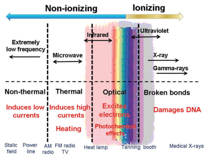
With respect to the complete electromagnetic radiation spectrum, the infrared radiation (IR) band covers the wavelength range of 750 nm–100 μm, frequency range of 400 THz–3 THz, and photon energy range of 12.4 meV– 1.7 eV. It lies between the long wavelength red edge of the visible and the short edge of the terahertz (starting at 3 THz) spectral bands (Figure 1).
Michael R. Hamblin, Department of Dermatology, Harvard Medical School, Boston, MA, USA; and Harvard-MIT Division of Health Sciences and Technology, Cambridge, MA, USA.
Rao et al. [40] used garments made out of bioceramic-coated neoprene in conjunction with a “topical cream” to treat cellulite of the legs. Each subject was randomized to receive occlusion by the garment on either the right or left leg, with the contralateral side serving as a control with no occlusion. Of the 17 subjects who completed the study, 76% noticed an overall improvement in their cellulite, with 54 % reporting greater improvement in the thigh that received garment occlusion. Further, the evaluators found the occluded thighs to show greater improvement than the non-occluded thighs in 65 % of subjects. Bioceramic-coated neoprene garment occlusion potentiated the effect of the topical agent in cellulite reduction. A follow up two-center, double-blinded, randomized trial found similar results [41].
Secure .gov websites use HTTPS A lock ( Lock Locked padlock icon ) or https:// means you've safely connected to the .gov website. Share sensitive information only on official, secure websites.
(B) The size scale of various biological structures in comparison with the diffraction-limited resolution. (Left to right) A mammalian cell, a bacterial cell, a mitochondrion, an influenza virus, a ribosome, the green fluorescent protein, and a small molecule (thymine).
These early applications of super-resolution imaging to bacterial cells have shown the great promise of the approach. Considering the primitive knowledge that we have of the organization of the chromosomes and proteins in bacteria, we expect super-resolution fluorescence microscopy to transform microbiology in the coming years.
(C) Examples of STED images. (Top) Comparison between confocal (left) and STED (right) images of the outer membrane of mitochondria that is immunolabeled against the protein TOM20. Shown in the STED panel is an xy cross section of the 3D isoSTED image. (Bottom-left) Two-color isoSTED image of TOM20 (green) and the matrix protein HSP70 (red). (Bottom-right) Three-dimensional rendering of an isoSTED image of TOM20. Reprinted by permission from Macmillan Publishers Ltd: Nature Methods Schmidt et al., 2008. Reprinted with permission from Schmidt et al., 2009, American Chemical Society.
Ever since Cajal observed Golgi-stained neurons under light microscopes more than a century ago, we have known that the brain functions by sending information from the axons to the dendrites of neurons. Therefore, a “wiring diagram” detailing how neurons are connected to each other will provide a structural foundation for understanding brain function, as well as its malfunction in neurological disorders. However, such a wiring diagram has not been obtained yet except for Caenorhabditis elegans (White et al., 1986). Why not? The small diameter of neurites (as small as tens of nanometers) and their high packing density require image resolution at the nanometer scale for wire tracing. Moreover, to annotate a wiring diagram, we need to identify the neuronal connections (i.e., the synapses) and characterize their properties. These properties depend on the molecular content of the synapses and thus require imaging with molecular specificity. In fact, synaptic function is orchestrated by an elaborate protein machinery with hundreds of protein species packed together in a structure of submicron size. Understanding synaptic function and plasticity requires a detailed characterization of the organization and dynamics of these molecules at specific synaptic sites. Thus, a method to map neural circuitry needs molecule-specific contrast, nanometer-scale resolution, and, ideally, live tissue imaging capability. Super-resolution fluorescence microscopy uniquely satisfies these requirements.
Medical FIR sources. (A) WS TY-301R® and (B, C) WS TY-101N® FIR lamps (both by WS Far Infrared Medical Technology Co., Ltd., Taipei, Taiwan).
As with STED and SIM, there is also a trade-off between spatial and temporal resolution with STORM/(F)PALM imaging. In this case, the image is reconstructed from single-molecule localizations. When specifying the spatial resolution, a Nyquist resolution (defined as twice the average distance between neighboring localizations) should be considered in addition to the localization precision (Shroff et al., 2008). This consideration effectively limits the temporal resolution to the time required to collect enough localizations to give the desired Nyquist resolution.
Belts made out of these fabrics have been used for weight reduction. In one study, Conrado and Munin [37] investigated whether the use of a garment made with synthetic fibers embedded with powdered ceramic led to a reduction in body measurements. The study population comprised 42 women divided into two groups: active and placebo. The volunteers used clothing either impregnated or not impregnated with ceramic powder for at least 8 h/day for 30 days. The experimental data showed a reduction in body measurements, which may be a consequence of an increment in microcirculation and peripheral blood flow, and these changes might promote improved general health.
It is important to note that the depletion light pattern itself is created by the same diffraction-limited optics. Therefore, if the fluorophores respond to the depletion light in a linear manner, the resolution enhancement would be rather limited. STED microscopy surpasses this limit by taking advantage of the saturated response of fluorophores: once the depletion laser intensity is above the saturation level, the number of fluorophores remaining in the excited state (and thus capable of generating fluorescence) approaches zero. Thus, when a ring-shaped depletion light with peak intensity significantly above the saturation level is applied to the sample, only the molecules within a small region near the center of the ring can generate fluorescence (Figure 2A). The size of this region, and thus the resolution of the microscope, scales approximately with the inverse square root of the intensity of the depletion light (or δ≈Δ∕1+Idep∕Isat, wherein δ is the resolution, Δ is the full width at half maximum of the diffraction-limited focal spot size, and Idep is the intensity of the depletion laser) (Hell, 2007).
For centuries, light microscopy has greatly facilitated our understanding of how cells function. In fact, entire fields of biology have emerged from images acquired under light microscopes. For instance, more than 300 hundred years ago, Antonie van Leeuwenhoek used his self-ground optical lenses to discover bacteria and commence the field of microbiology. Then, ~200 years later, Ramón y Cajal used light microscopes to visualize Golgi-stained brain sections and create beautiful drawings of neurons, which led to his ingenious vision of how information flows in the nervous systems and helped to form modern neurobiology.
We analyzed the peer-reviewed applications of therapeutic FIR delivery systems and realized that there are three main techniques for FIR radiation delivery: i) FIR saunas, ii) FIR ray devices and iii) FIR emitting ceramics and fabrics.
Official websites use .gov A .gov website belongs to an official government organization in the United States.
STED microscopy can also use a wide range of existing fluorescent probes because all fluorophores can undergo stimulated emission (http://www.mpibpc.mpg.de/groups/hell/STED_Dyes.html). In practice, dyes that are photostable under a strong depletion light, such as Atto 647N, Atto 590, and Atto 565, provide greater resolution enhancement. Fluorescent proteins such as enhanced yellow fluorescent protein (EYFP) and Citrine have also been used. However, multicolor imaging with STED is less flexible compared to conventional fluorescence microscopy. To image one fluorophore, STED microscopy requires two lasers at the opposite ends of the absorption and emission spectra of the fluorophore for excitation and depletion, respectively. Therefore, the number of colors that can fit into the visible to near IR spectrum range is limited, and a maximum of two colors has been imaged simultaneously thus far (Donnert et al., 2007; Schmidt et al., 2008). Figure 2C shows a two-color STED image of the mitochondrial outer-membrane protein TOM20 and matrix protein HSP70 obtained using the latter approach (Schmidt et al., 2008).
20131220 — Join George Maestri for an in-depth discussion in this video, Silhouetting and flow, part of Learning Character Animation.
Super-resolution fluorescence microscopy is expected to expand our understanding of the structures inside of the nucleus. The highly condensed DNA packaging inside of the nucleus calls for super-resolution imaging with protein and nucleic acid sequence specificity, which is difficult to achieve by any other means. Super-resolution fluorescence microscopy provides the opportunity to resolve the interactions between nucleic acids and proteins inside of the nucleus. For example, studies with 3D SIM revealed morphological changes of the chromosomes and the nuclear lamin during early mitosis (Schermelleh et al., 2008). In addition, STORM/(F)PALM has shown promise in resolving chromatin fibers (Matsuda et al., 2010). We anticipate that, with further improved image resolution and sequence-specific nucleic acid labeling by FISH, DNA/RNA-binding proteins, or aptamers, the regulation of gene expression may be directly visualized inside of the nucleus using super-resolution imaging.
However, determining the complete neuronal wiring diagram may require even higher resolution than what is currently feasible by super-resolution microscopy. Although resolution of a few tens of nanometers is now routinely obtainable using various super-resolution fluorescence microscopy techniques, tracing thin and densely packed axons in the brain may likely require a resolution of a few nanometers. Studying subneuronal structures, such as synapses, could also benefit from substantially higher resolution than the current state of the art. A resolution of a few tens of nanometers allows the organization of molecular assemblies and organelles to be determined, whereas resolution at a few nanometers (i.e., a true molecular scale) would permit the direct visualization of interactions between molecules.
Far infrared (FIR) radiation (λ = 3–100 μm) is a subdivision of the electromagnetic spectrum that has been investigated for biological effects. The goal of this review is to cover the use of a further sub-division (3– 12 μm) of this waveband, that has been observed in both in vitro and in vivo studies, to stimulate cells and tissue, and is considered a promising treatment modality for certain medical conditions. Technological advances have provided new techniques for delivering FIR radiation to the human body. Specialty lamps and saunas, delivering pure FIR radiation (eliminating completely the near and mid infrared bands), have became safe, effective, and widely used sources to generate therapeutic effects. Fibers impregnated with FIR emitting ceramic nanoparticles and woven into fabrics, are being used as garments and wraps to generate FIR radiation, and attain health benefits from its effects.
Light Sheet Fluorescence Microscopy. LSFM. Our research group specializes in Light Sheet Fluorescence Microscopy (LSFM), an optical imaging technique that ...
Diffractionresolution
Discs and garments manufactured of FIR emitting ceramic material have been applied to the human body (Figure 5). For instance, a blanket containing discs has been reported to improve quality of sleep [34] and single discs were applied to the breasts of women who encountered difficulty in producing sufficient breast milk during lactation [35]. Gloves have been made out of FIR emitting fabrics and there have been reports that these gloves can be used to treat arthritis of the hands and Raynaud’s syndrome [36].
Official websites use .gov A .gov website belongs to an official government organization in the United States.
It must be emphasized that the above remains a hypothetical explanation at present, but is clearly a testable hypothesis. One could ask whether exposure of cells to non-heating FIR can affect mitochondria by for instance increasing ATP, increasing oxygen consumption, producing NO and ROS, affecting MMP and calcium levels. One could also ask whether cells that are rich in mitochondria respond well to non-heating FIR, in the same way as they do to LLLT.
When light is focused by the objective of a microscope, the notion of light “rays” converging to an infinitely sharp “focal point” does not happen. Instead, the light wave forms a blurry focal spot with a finite size due to diffraction (Figure 1Afig1). The size of the spot depends on the wavelength of the light and the angle at which the light wave converges; the latter is, in turn, determined by the numerical aperture of the objective. The width of the spot at half-maximum intensity (i.e., full width at half maximum) is ~0.6 λ/NA, wherein λ is the wavelength of the light and NA is the numerical aperture of the lens. Similarly, a point emitter, such as a single fluorescent molecule, also appears as a blurry spot with a finite size when imaged through a microscope. The intensity profile of this spot, which defines the point spread function (PSF) of the microscope, has approximately the same width as that of the focal spot described above. Consequently, two identical emitters separated by a distance less than the width of the PSF will appear as a single object, making them unresolvable from each other (Figure 1A).
These are only a few examples that illustrate the impact that super-resolution fluorescence microscopy is having on cell biology. We look forward to seeing more questions answered by the rapid adoption of super-resolution microscopy tools, especially given this technique's ability to resolve subcellular structures beyond the organelle level.
This resolution limit was originally recognized by Ernst Abbe ~150 years ago, and thus, it is also called the Abbe limit (Abbe, 1873). The diffraction-limited image resolution of objective lens with a high numerical aperture is ~250 nm perpendicular to the direction of light propagation (i.e., in the lateral dimensions) and 550 nm parallel to the direction of light propagation (i.e., in the axial dimension) (Figure 1A). Many subcellular structures are smaller than these resolution limits, and therefore, they are unresolvable by light microscopes (Figure 1B).
The depletion pattern in STED microscopy is created by inserting a spatial light modulator in the laser beam before it enters the microscope. As a ring-shaped pattern in the x-y plane improves the lateral resolution, a pattern having two maxima along the z axis improves the axial resolution (Hell, 2003). Overlaying these two patterns improves the resolution in both lateral and axial directions (Harke et al., 2008), allowing three-dimensional (3D) super-resolution imaging with a z resolution ~2.5 times the xy resolution. Subsequently, isoSTED has been realized with the z depletion pattern generated by two opposing objectives using the 4Pi configuration, reaching a resolution as high as ~30 nm in all three dimensions, as demonstrated by the 3D images of mitochondria as shown in Figure 2C (Schmidt et al., 2008, 2009). Recently, STED microscopy has also been demonstrated with two-photon excitation, allowing super-resolution imaging deep into tissue samples (Ding et al., 2009; Moneron and Hell, 2009).
To study very fast processes, STED microscopy can be combined with fluorescence correlation spectroscopy (FCS). FCS is traditionally used with confocal microscopy to probe the movement or dynamics of molecules by monitoring the fluctuation of the fluorescence signal. The smaller detection region in STED enables FCS to probe dynamics at a much smaller length scale. Indeed, STED-FCS has been used to monitor diffusion dynamics with microsecond time resolutions and at a length scale as small as 20 nm (Eggeling et al., 2009).
The high molecular specificity offered by various fluorescent labeling approaches and the live-cell compatibility of super-resolution fluorescence microscopy offer an ideal solution to the bacterial imaging problem. Indeed, this approach has been used to study several different protein organizations in bacterial cells. For example, STORM/(F)PALM was used to determined the distributions of the chemotaxis proteins Tar receptor, CheY, and CheW in fixed Escherichia coli cells (Greenfield et al., 2009). These proteins were found to form clusters with exponentially distributed sizes. The cluster locations were consistent with a stochastic self-assembly model that does not require active transport. In Calobactur crescentus, live-cell STORM/(F)PALM imaging of MreB, an actin analog, revealed a helical organization of the protein (Biteen et al., 2008). A similar approach was also used to study the partitioning (Par) apparatus in C. crescentus. These studies found that the ATPase ParA forms a narrow, linear polymer structure that runs through the center of the cell body, functioning in a way that is similar to the mitotic spindles observed in eukaryotic cells (Ptacin et al., 2010).
(D) Examples of 3D SIM images. (Top) Central cross-section of a confocal image of the nucleus stained for DNA, lamin B, and the nuclear pore complex. DNA (blue) is stained with DAPI. Lamin B (green) and the nuclear pore complex (red) are immunostained. The right panels show the magnified images of the boxed region in the left panel. (Bottom) Corresponding 3D SIM images. From Schermelleh et al., 2008. Reprinted with the permission from AAAS.
Like with the negative patterning approach, the saturating response of the fluorophore can also be exploited here to further enhance the resolution (Gustafsson, 2005; Heintzmann et al., 2002). With sufficiently strong excitation, the fluorescence emission from a fluorophore will saturate. Saturated SIM (SSIM) utilizes this phenomenon to create sharp dark regions where the excitation pattern has zero intensity, providing image resolution significantly beyond the diffraction limit (Figure 2B). With this approach, a resolution of 50 nm has been obtained for imaging fluorescence microspheres (Gustafsson, 2005).
Diffraction limitcalculator
(C) Three-dimensional images taken using an interferometry approach with apposing objectives. (Top) xy projection of the plasma membrane of a cell expressing photoactivatable Eos-fluorescent protein. The color of the localization points encodes their z coordinates. (Bottom) xz cross-section of the boxed region in the top panel. Images adapted from Shtengel et al., 2009.
In addition to illuminating subcellular structures inside of neurons, super-resolution fluorescence microscopy has also been used to study neuronal morphology. Beautiful STED images revealed the shape and dynamics of dendritic spines in live cells (Nägerl et al., 2008). Two-photon STED allowed the visualization of spine morphology in deep tissue samples (Ding et al., 2009). The application of super-resolution imaging to map neuronal connectivity is also currently under way.
Despite all these different uses of FIR in medical applications, the exact mechanisms of the hyperthermic effects and biological activities of FIR irradiation are still poorly understood. It is clear that two kinds of FIR therapy may exist. The first type (FIR saunas and some FIR generators powered by electricity) uses irradiances or power densities (tens of mW/cm2) that are sufficient to heat up the tissue, while others such as ceramic discs, powders, and fabrics (that use no external power but rely on energy from the body) have such low irradiances that they do not heat the tissue (0.1–5 mW/cm2). The question arises to what extent are the fundamental mechanisms of these two forms of FIR therapy the same, and to what extent are they different ? Furthermore, the question may be posed as to what degree of similarity that FIR therapy has with the reasonably well-established therapy called low level laser (light) therapy (LLLT) also known as photobiomodulation (PBM). Pertinent to this question is the fact that many devices used to deliver therapeutic visible or NIR light were approved by the US Food and Drug Administration as being equivalent to an “ infrared heat lamp ”. The cellular and molecular mechanisms of LLLT/PBM are to some extent understood and involve absorption of red or NIR light by mitochondrial chromophores such as cytochrome c oxidase (CCO, unit IV of the mitochondrial respiratory chain) [43]. This photon absorption activates the enzyme possibly by photo-dissociating the inhibitory molecule, NO, from the copper B (CuB) site [44]. This loss of NO allows electron transport, oxygen consumption, and adenosine triphosphate (ATP) to rapidly increase and results in a marked rise in mitochondrial membrane potential (MMP) that gives rise to a brief burst of ROS [45]. Signaling pathways are activated by ATP, NO, and ROS and these lead to activation of transcription factors (such as NF-κB) [46] that lead to the long-term effects on tissue (healing, anti-inflammatory and pain relief [47]) seen after relatively transient periods of illumination.
The requirements of STORM/(F)PALM put several constraints on the fluorescent probes. First, the probes should have a fluorescent state that emits light at a certain range of wavelengths and a “dark” state that does not emit light in this wavelength range. Second, to achieve high precision of localization, the probes should emit a large number of photons before going dark. Third, because only one fluorophore is activated within a diffraction-limited area at any time and the vast majority of fluorophores remain in the dark state, dark state emission should be minimal to ensure high-precision localization of the activated molecule. Finally, because all switchable fluorophores can be spontaneously activated by thermal energy or the imaging laser (as opposed to the activation laser), this spontaneous activation could cause more than one fluorophore in the diffraction limited area to be turned on even without activation light, preventing single-molecule detection. Therefore, a low spontaneous activation rate is also desired.
Supplemental Information Supplemental Information includes Extended Discussion and one table and can be found with this article online at *bxs.
It is worth noting that, in addition to the intrinsic optical resolution, which often depends on the brightness and photostability of the fluorescent probes, the effective image resolution is also limited by the labeling density and the size of the fluorescent labels. Therefore, in addition to improvements in the design of microscopes, developments in fluorescent probes and labeling chemistry are also critical for further improving the resolution of light microscopy. Indeed, recent advances in super-resolution fluorescence microscopy have already inspired a large amount of research activity in these areas. We foresee that, together, these efforts will allow live-cell imaging at a truly molecular-scale resolution, thereby enabling the direct visualization of molecular interactions and biochemical events in living cells.
(B) 3D images taken using an astigmatism approach with cylindrical lens. (Two far-left columns) Conventional image of clathrin-coated pits in a mammalian cell immunostained against clathrin, in comparison with the corresponding 3D super-resolution image showing an xy cross section near the plasma membrane. (Middle) Magnified super-resolution images of a single clathrin-coated pit in a cell-free reconstitution system with an xy projection (top), an xy cross-section at the lower portion of the pit (middle), and an xz cross section cutting through the middle of the pit (bottom). (Two far-right columns) Composite 3D image of clathrin (green), dynamin (cyan), and an F-BAR domain protein FBP17 (red) in the cell-free system. Shown here is the super-position of 59 images of clathrin and FBP17 aligned to the center of the clathrin-coated regions (left) and the super-position of 96 dynamin-FBP17 images aligned to the center of dynamin spot (right). Clathrin is directly labeled, whereas dynamin and FBP17 are immunolabeled. From Huang et al., 2008 and Wu et al., 2010. Reprinted with the permission from AAAS.
Along the same lines, Akasaki et al. [7] studied in vivo the effects of repeated FIR irradiation on angiogenesis in a mouse model of hindlimb ischemia. Following reports that FIR therapy upregulated the expression of arterial eNOS in hamsters (and it is known that NO constitutively produced by eNOS plays an important role in angiogenesis) they took a step further to investigate whether the FIR therapy increases angiogenesis in mice with the hindlimb ischemia. In their study, unilateral hindlimb ischemia was induced in apolipoprotein E-deficient mice and the group to receive the FIR irradiation was placed in a FIR dry sauna at 41°C for 15 min and then at 34°C for 20 min once daily, with total duration of the experiment of 5 weeks. Laser-Doppler perfusion imaging demonstrated that at the ischemic limb, blood perfusion ratio in the irradiated group increased significantly in comparison with the control group (0.79±0.04 vs. 0.54±0.08, p<0.001). Also, in the treated group, significantly greater capillary density was observed (757±123 per mm2 vs. 416±20 per mm2, p<0.01). Western blotting showed that thermal therapy has increased markedly the hindlimb eNOS expression. Furthermore, to study possible involvement of eNOS in thermally induced angiogenesis, the same FIR therapy was given to mice with hindlimb ischemia with or without N(G)-nitro-L-arginine methyl ester (L-NAME) administration for the duration of 5 weeks. It was observed that L-NAME treatment eliminated angiogenesis induced using the FIR thermal therapy and that the therapy did not increase angiogenesis in eNOS-deficient mice. The study led to the conclusion that angiogenesis can be induced via eNOS using FIR thermal therapy in mice with hindlimb ischemia.
The lenses focus the light onto film stored in the camera. The chemically coated film reacts to the light and records an image, or picture, of the object.
In this method, photoswitchable (or photoactivatable) fluorophores are used to achieve temporal control of the emission. These fluorophores can be converted between a fluorescent (or “on”) state and a dark (or “off”) state or states that fluoresce at different wavelengths. Therefore, when activation light of a sufficiently low intensity is applied to the sample, only a random, sparse subset of fluorophores is activated to the on state at any time, allowing these molecules to be imaged individually, precisely localized, and then deactivated by switching to a reversible dark state or permanent bleaching. Iterating this process—activation, imaging, and deactivation—then allows the locations of many fluorophores to be mapped and a super-resolution image constructed from these localizations (Figure 3A), either with synchronized activation (Betzig et al., 2006; Hess et al., 2006; Rust et al., 2006) or with asynchronous activation (Egner et al., 2007). The image resolution is then no longer limited by diffraction but instead by how precisely each fluorophore is localized. Using this approach, a lateral image resolution as high as ~20 nm has been achieved (Rust et al., 2006).
A time resolution of 25–60 s per frame was obtained when imaging focal adhesion complexes labeled with Eos fluorescent protein (EosFP) at a Nyquist resolution of 60–70 nm (Shroff et al., 2008). Similar spatial and temporal resolutions have been achieved when studying the cytoskeleton structure in bacteria using enhanced yellow fluorescent protein (EYFP) (Biteen et al., 2008). This relatively slow imaging speed is due, in part, to the slow switching of fluorescent proteins to the dark states but also due to the decreasing photon output at high-excitation intensities. The use of brighter and faster-switching organic dyes could potentially increase the imaging speed. Various dyes have been combined with the hybrid fusion systems for live-cell super-resolution imaging (Lee et al., 2010; Testa et al., 2010; Wombacher et al., 2010). The spatiotemporal resolution that can be reached with photoswitchable dyes has yet to be characterized with the Nyquist criteria.
FIR application in medicine requires understanding and knowledge of the interactions of electromagnetic radiation at FIR range with biological structures (including cells, cell membranes, cell fluids – especially water, DNA/proteins) and functioning of the living systems in general. At the cellular level, the underlying biophysical mechanisms of the interaction of electromagnetic radiation with living cells can be framed in terms of altered cell membrane potentials and altered mitochondrial metabolism [2]. FIR energy (photons with quantum energy levels of 12.4 meV –1.7 eV) is absorbed by vibrational levels of bonds in molecules. There are six vibrational modes covering symmetric and antisymmetric stretching, scissoring, rocking, wagging and twisting. Considering the high concentration of water in biological systems, association of water molecules with ions (solvation effect), the dielectric properties of the water and the large dipole moment that this effect generates, this will be a dominant factor in biological solutions. It is known that at lower frequencies water molecules are able to rotate freely in an oscillating electric field with little or almost no energy loss. However, if the frequency of the electric field reaches 108 Hz levels, the rotational mode becomes hindered (due to “dielectric friction ” effect) and the absorbed energy starts dissipating by collision or nearest neighbor interactions in the media [2]. The dielectric relaxation of water at 310 K is around 25 GHz where the rotational response of the dipoles to the electromagnetic field is spread over a broad frequency range.
Fatma Vatansever, Wellman Center for Photomedicine, Massachusetts General Hospital, Boston, MA, USA; and Department of Dermatology, Harvard Medical School, Boston, MA, USA.
The highest axial resolution in 3D STORM/(F)PALM is achieved by interferometry using two opposing objectives in a similar fashion to 4Pi microscopy and I5M. Figure 2C shows the clear separation of the ventral and dorsal plasma membrane in a thin protrusion of the cell using this method, demonstrating a z resolution of 10 nm (Shtengel et al., 2009). The imaging depth of this approach is relatively small compared to the PSF-fitting approaches described in the previous paragraph, but sample scanning can increase the imaging depths of all of these approaches. In practice, the imaging depth is limited by spherical aberrations due to refractive index mismatch, scattering by turbid samples, and high-fluorescence background arising from thick samples. The combination of single-molecule switching with two-photon excitation/activation provides a promising solution to the latter two problems (Fölling et al., 2008; Vaziri et al., 2008).
(A) This super-resolution approach takes advantage of photoswitching of fluorophores to temporally separate images of single molecules that overlap spatially. At any time during image acquisition, only a sparse subset of fluorophores is activated to the fluorescence state, allowing these molecules to be imaged individually and thus localized. After multiple iterations of the activation and imaging processes, a super-resolution image is constructed from the localizations of many fluorophores.
Structured illumination microscopy (SIM) improves image resolution by using positive patterning of the excitation light (Heintzmann and Gustafsson, 2009), which is typically a sinusoidal pattern created by combining (i.e., interfering) two light beams. As the result, an image snapshot of the sample becomes the product of the sample structure itself and this excitation pattern (Figure 2B). A final image is then computationally reconstructed from multiple snapshots collected by scanning and rotating the pattern. In this process, the additional spatial modulation from the excitation pattern brings enhanced spatial resolution into the reconstructed image (Gustafsson, 2000; Heintzmann and Cremer, 1999).
Despite its relatively short history, super-resolution fluorescence microscopy has already been applied to many areas of biology, and it is beginning to have an impact on numerous fields. It is not possible to describe all of these applications in this Primer article. Instead, we discuss some representative examples in the areas of cell biology, microbiology, and neurobiology.
In principle, this approach allows unlimited resolution improvement given an infinitely strong depletion light source. In practice, a number of factors influence the resolution of STED, including aberrations in the optics, scattering from the sample, and the photostability of the fluorophores. STED microscopy has reached a remarkable resolution of 6 nm using strong depletion intensity to image fluorescent defects in diamonds, which almost never photobleach (Rittweger et al., 2009). On biological samples, STED imaging has achieved a resolution of 20 nm when using organic dyes and 50–70 nm resolution when using fluorescent proteins (http://www.mpibpc.mpg.de/groups/hell/STED_Dyes.html).
During the development of super-resolution microscopy techniques, a number of subcellular structures with well-characterized morphological features were frequently chosen as proof-of-principle model systems, including microtubules, actin, clathrin-coated pits, mitochondria, endoplasmic reticulum, and focal adhesion complexes. These studies have not only illustrated the resolving power of the new super-resolution techniques, but they also demonstrated these methods' potential for visualizing molecular structures and interactions in cells.
Common devices are WS TY-101N® and WS TY-301R® (made by WS Far Infrared Medical Technology Co., Ltd., Taipei, Taiwan; see Figure 3). A report from Hu and Li [31] describes the treatment of allergic rhinitis. A WS TY-101N® FIR emitter was placed 30 cm from the patient’s nasal region. The treatment was performed for 40 min every morning for 7 days. Every day, patients recorded their symptoms in a diary before and during treatment. Each symptom of rhinitis was rated on a 4-point scale (0–3) according to severity. During the period of FIR therapy, the symptoms of eye itching, nasal itching, nasal stuffiness, rhinorrhea, and sneezing were all significantly improved. Smell impairment was improved after the last treatment. Lin et al. [32] used a WS TY-101N® FIR emitter to treat vascular access malfunction with an inadequate access flow (Qa) in hemodialysis (HD) patients. This randomized trial demonstrated that FIR therapy could improve access flow and potency of the native arteriovenous fistula (AVF) in a total of 145 HD patients (73 in the control group and 72 in the FIR-treated group). FIR was used for 40 min, and hemodynamic parameters were measured by the HD02 monitor (M/s Transonic System Inc.), during the hemodialysis. In comparison with control subjects, patients who received FIR therapy for 1 year had a lower incidence (12.5 vs. 30.1%; p<0.01) and relative incidence (one episode per 67.7 vs. one episode per 26.7 patient-months; p = 0.03) of AVF malfunction. Hausswirth et al. [33] showed that FIR therapy reduced symptoms of exercise-induced muscle damage in athletes after a simulated trail running race.
For specific experiments, the decision of whether to use dyes or fluorescent proteins depends on a variety of factors, which are commonly applicable to all fluorescence imaging methods. In terms of labeling, fluorescent proteins are genetically encodable, allowing proteins in living cells to be readily labeled with fluorescent proteins. However, dyes are more versatile for labeling different molecular species, including proteins, nucleic acids, oligosaccharides, and even small molecules. Tagging cellular proteins with dyes is typically achieved using immunofluorescence, which permits endogenously expressed proteins to be labeled but prohibits most live-cell imaging applications. In terms of the optical properties, dyes generally have a significantly higher photon output, allowing higher image resolution than fluorescent proteins (Figure 3D). However, at extremely high image resolution, the bulky size of the antibody (10–15 nm) could limit image resolution when immunofluorescence labeling is used. Therefore, the recently developed hybrid fusion systems provide a promising solution that combines the merits of both genetic encoding and the superior fluorescence properties of organic dyes (Fernández-Suárez and Ting, 2008). In these approaches, the protein of interest is fused to a marker protein or peptide, which in turn exhibits a specific reactivity or affinity to fluorescent dyes with certain reactive groups.
Being able to localize a single molecule does not directly translate into super-resolution imaging of a fluorescently labeled biological sample, which can contain thousands of fluorophores inside of the diffraction-limited region. The fluorescence emission from these molecules will overlap severely enough that the overall image appears as a completely featureless blur. At first sight, it might seem impossible to distinguish these molecules individually. However, if the fluorescence emission from these molecules is controlled such that only one molecule is emitting at a time, individual molecules can then be imaged and localized. This is the idea behind a recently developed super-resolution imaging method, called STORM (Rust et al., 2006), PALM (Betzig et al., 2006), and FPALM (Hess et al., 2006).
Therefore, to achieve high temporal resolution, a small field of view and a relative large spatial resolution can be used. For example, impressive video rate STED imaging (28 frames per second) with 62 nm spatial resolution has been demonstrated in a field of view of ~5 μm2, allowing the motion of individual synaptic vesicles in a dendritic spine to be followed (Westphal et al., 2007). More recently, replacing the traditional pulsed lasers in STED microscopy with continuous wave lasers permits substantially faster scanning and thus higher time resolution (Willig et al., 2007). Using this scheme, a ~70 μm2 image of the endoplasmic reticulum took only 0.19 s to acquire (Moneron et al., 2010).
A theatrical fresnel is a soft edged circle of light that can zoom in size from spot to flood by changing the relationship of the lamp with the interior ...
It was not until recently that several novel fluorescence microscopy approaches completely shattered the diffraction limit of image resolution in the far field. In general, all of these approaches generate “diffraction-unlimited images” by using the physical properties of fluorescent probes to distinguish emissions from two nearby molecules within a diffraction-limited region. These super-resolution approaches can be divided into two primary classes. The first category is ensemble imaging approaches that use patterned illumination to spatially modulate the fluorescence behavior of molecules within a diffraction-limited region, such that not all of them emit simultaneously, thereby achieving subdiffraction limit resolution. This category includes stimulated emission depletion (STED) microscopy (Hell and Wichmann, 1994; Klar and Hell, 1999) and the related RESOLFT technology (Hofmann et al., 2005), as well as saturated structured illumination microscopy (SSIM) (Gustafsson, 2005; Heintzmann et al., 2002). The second category takes advantages of single-molecule imaging, using photoswitching or other mechanisms to stochastically activate individual molecules within the diffraction-limited region at different times. Images with subdiffraction limit resolution are then reconstructed from the measured positions of individual fluorophores. This second class has been termed stochastic optical reconstruction microscopy (STORM) (Rust et al., 2006), photoactivated localization microscopy (PALM) (Betzig et al., 2006), and fluorescence photoactivation localization microscopy (FPALM) (Hess et al., 2006).
Rayleigh criterion
Not that long ago, the diffraction limit was considered insurmountable. In recent years, the invention of various super-resolution techniques has shattered the diffraction barrier and allowed the resolution of images from light microscopy to improve by an order of magnitude over the diffraction limit. In fact, all of these super-resolution approaches have, in principle, made spatial resolution unlimited. That said, we believe that, practically, an additional order of magnitude in resolution improvement is feasible.
For example, STED microscopy has resolved individual syntaxin 1 clusters in cells, allowing the quantification of the number of syntaxin molecules per cluster (~90) and cluster size (50–60 nm) (Sieber et al., 2007). In conjunction with diffusion measurements by fluorescence recovery after photobleaching (FRAP) and computer modeling, STED demonstrates that syntaxin self-association and steric repulsion fully explain the size and dynamics of syntaxin clusters. In contrast, the clustering of nicotinic acetylcholine receptors is possibly affected by additional mechanisms, including cholesterol-mediated protein-protein interactions and cytoskeleton membrane-protein interactions. These results provide an explanation for the observation that the depletion of cholesterol influences both short range (~50 nm) and long range (0.5–3.5 μm) organizations of nicotinic acetylcholine receptors (Kellner et al., 2007).
10X10 DIAMOND WINDOW · Description · Additional information · Reviews · Product categories · Related products · SLIDER WINDOW 18X20/ OUTSWING · VINYL STEEL ...
When FIR emitting ceramics or fabrics are employed as therapeutic devices, it is pertinent to analyze the thermodynamics of the process. The first law of thermodynamics states that energy can neither be created nor destroyed. Heat (molecular vibrational energy) is transferred from one body to another in three forms: radiation, convection and conduction. Thus, it is clear that the principle source of energy needed to power the FIR emission from the garments comes from the human body, since it is at a significantly higher temperature than the surrounding air. So energy from the human body is transferred to these ceramic particles, which are acting as “perfect absorbers”, maintain their temperature at sufficiently high levels and then emit FIR back to the body. It is plausible that FIR emitted from the skin is absorbed by the ceramic particles, which then re-emit the same FIR back to the skin. Although this may appear to be an energy neutral process and to cancel itself out, this is not in fact the case because the FIR emitting material will prevent the loss of FIR that would otherwise have escaped through normal clothing. However the same effect could have been achieved with a FIR reflective foil suit or suchlike. Other sources of heat that can transfer energy from the body to the ceramic particles with a net gain of FIR are either convection, conduction, or both. The balance between conduction and convection will depend on how close the contact is between the garment and the skin. If the garment is skin tight, then conduction may be important, while if it is loose fitting then convection (heating up a layer of air between the skin and the garment) may be important.
Indeed, one major element that makes light microscopy so powerful in biological research is the development of various staining methods that permit the labeling of specific molecules and cells. For example, fluorescence in situ hybridization (FISH) detects DNA and RNA molecules with specific sequences, whereas immunofluorescence labels and fluorescent proteins allow the imaging of particular proteins in cells (Giepmans et al., 2006). Even single molecules within a living cell can be visualized when these labeling strategies are combined with highly sensitive optical schemes and detectors (Lord et al., 2010; Xie et al., 2008).
Ting-Kai Leung and colleagues have studied the effect of FIR-emitting ceramic powders in a range of biological studies [14 –19]. In one set of studies, they cultured murine myoblast cells (C2C12) with bags of ceramic powder under the culture plates and found that FIR irradiation improved cell viability and prevented lactate dehydrogenase release under hydrogen peroxide (H2O2)-mediated oxidative stress, and also elevated the intracellular levels of NO and calmodulin [14]. In the study, they used electro-stimulation of amphibian skeletal muscle and found that FIR emitting ceramics delayed the onset of fatigue, induced by muscle contractions [14]. In another set of studies, they showed that ceramic-emitted FIR (cFIR) could increase the generation of intracellular NO in breast cancer cells [15] and inhibit growth of murine melanoma cells [16]. Similarly, they found that cFIR increased calmodulin and NO production in RAW 264.7 macrophages [17]. cFIR also has been shown to increase the viability of murine macrophages with different concentrations of H2O2 [15]. In this study [15] it was shown that cFIR significantly inhibited intracellular peroxide levels and lipopolysaccharide (LPS)-induced peroxide production by macrophages. In the same study, it was also demonstrated that cFIR blocked ROS-mediated cytotoxicity (shown by measurements of cytochrome c and the ratio of NADP+/NADPH) [15].
The boron-silicate mineral, tourmaline (known as a gemstone in its crystalline form) when milled into fine powders also emits FIR [12] and the characteristics of the FIR emission depend on the particle size. Preparations containing tourmaline powder have been applied to the skin with the aim of affecting the blood flow [13]. In a similar manner discs of FIR emitting ceramics have been attached to the skin with the intent of producing a beneficial effect (see later).
Fraunhoferdiffraction
Indeed, neuroscience is one of the first disciplines to which super-resolution fluorescence microscopy was applied. One productive area of application is the study of subneuronal structures, such as synapses. STED images of the active zone protein Bruchpilot at the neuromuscular junction in Drosophila revealed a donut-shaped distribution centered at the active zones of the synapses. Furthermore, the molecules appear to adopt a preferred orientation along the transynaptic axis (Fouquet et al., 2009; Kittel et al., 2006). Using a similar approach, AMPA receptors have been shown to have a ring-shaped distribution in the ribbon synapses of inner hair cells (Meyer et al., 2009). Studies of synaptotagmin, a component of the synaptic vesicle component, show that these proteins remain clustered after exocytosis (Willig et al., 2006).
The risk has paid off abundantly. Recently, these research teams have developed several optical microscopy techniques that have shattered the diffraction barrier, improving spatial resolution by an order of magnitude or more over the diffraction limit. Most importantly, these techniques are beginning to provide insights into biological processes at the cellular and molecular scale that were hitherto unattainable. In this Primer, we review the technological advances in the burgeoning field of “super-resolution fluorescence microscopy.” Then, we describe the application of these techniques to various areas of biology, which have quickly demonstrated the great promise of this exciting new area of bioimaging.
This section collects any data citations, data availability statements, or supplementary materials included in this article.
Norland NOA61 UV adhesive, optical grade, perfect for installing tritium vials.
FIR emitting garments and fabrics manufactured from fibers impregnated with ceramic nanoparticles (Celliant®, Hologenix, Santa Monica, CA, USA). (A) fibers, (B) yarns, (C) fabrics, (D) knee bandage brace, (E) socks, (F) gloves, (G) elbow bandage brace, (H) multi-purpose bandage, (I) performance apparel, (J) mattress, and (K) puppy blanket.
STORM/(F)PALM has also provided new insights into the organizations of proteins associated with plasma membranes and intracellular membrane organelles. Three-dimensional super-resolution images taken with this approach resolved the hemispherical shape of the clathrin coat on nascent endocytic vesicles (Figure 3B) (Bates et al, 2007; Huang et al., 2008). In an in vitro endocytosis model system, two-color 3D imaging revealed that the two tubule-forming proteins, dynamin and the F-BAR domain protein FBP17, have distinct distributions along the membrane tubules connecting clathrin-coated pits and the basal plasma membrane (Figure 3B). In addition, STORM/(F)PALM, combined with electron microscopy and time-lapse conventional fluorescence imaging, uncovered an unexpected role of FBP17 in creating endocytic vesicles (Wu et al., 2010). Live-cell imaging with this approach has also revealed interesting dynamics of adhesion complexes during their initiation, maturation, and dissolution (Shroff et al., 2008). A recent 3D super-resolution study revealed a multilaminar molecular architecture of the focal adhesion core region that connects integrin and actin (Kanchanawong et al., 2010). This core region is comprised of an integrin-signaling layer, a force transduction layer, and an actin regulatory layer (Kanchanawong et al., 2010).
This work is in part supported by the Searle Scholarship, Packard Fellowship, and the UCSF Program for Breakthrough Biomedical Research (to B.H.) and the Howard Hughes Medical Institute (Collaborative Innovation Award), National Institutes of Health, and the Gatsby Charitable Foundation (to X.Z.). X.Z. is a Howard Hughes Medical Institute investigator.
There have been many attempts to use FIR as a therapeutic intervention where devices known as “infrared heat lamps” that emit more or less FIR are been used. Unfortunately, “ pure ” FIR emitting lamps are expensive, and thus, in some instances lamps that have “mixed” emission, i.e., emit in shorter (mid infrared, MIR; near infrared, NIR and even visible light) wavelength ranges are been used. A common type of specialized infrared heat lamp emits 2–25 μm radiation. IR saunas are often used and the most effective types have ceramic FIR emitting panels that remain cool to the touch. However, most IR saunas on the market do not use the expensive FIR panels, which can be touched since they remain always cold.
Secure .gov websites use HTTPS A lock ( Lock Locked padlock icon ) or https:// means you've safely connected to the .gov website. Share sensitive information only on official, secure websites.
This gap in our knowledge stems primarily from the difficulty in imaging such a small cell, with a size not much larger than the diffraction limit of optical resolution. Indeed, under the light microscope, most structures inside of a bacterial cell look like a blur. Though electron microscopy (EM) often comes to the rescue when high image resolution is required, its application to bacterial imaging is rather limited. To obtain molecular-specific contrast with EM, immunogold labeling is typically used, and this labeling approach requires cell fixation and permeabilization. Unfortunately, this fixation and permeabilization process perturbs structures inside of bacteria substantially more than inside of eukaryotic cells. Furthermore, live imaging is so far impossible with EM.
STORM/(F)PALM has also been used to investigate the molecular architecture of synapses. A systematic study of ten pre- and postsynaptic proteins in the central synapses of mouse brain tissue revealed oriented organization of the presynaptic scaffolding proteins bassoon and piccolo; compartmental distributions of the postsynaptic proteins PSD95, shank, and Homer; and heterogeneous neurotransmitter distributions, with some synapses showing central-synaptic and others showing perisynaptic receptor distribution (Dani et al., 2010). STORM/(F)PALM is able to count molecules, and this property allowed quantitative analysis of neurotransmitter receptor compositions in synapses (Dani et al., 2010). One recent study adapted photoactivation-facilitated high-density single-particle tracking to study actin dynamics at the dendritic spine and uncovered distinct loci of enhanced actin polymerization (Frost et al., 2010).
(A) Planck’s law. Dependence of spectral radiant emittance (ωλ) for perfect blackbodies as a function of wavelength (λ). (B) Wien’s displacement law. The wavelength of maximal radiant exitance (λmax) as a function of the absolute temperature (T) for a perfect black body (dashed line) and different bodies.
One particularly promising area of applying super-resolution microscopy techniques is the investigation of plasma membrane proteins and membrane microdomains. These domains are generally too small to be resolved by conventional light microscopy. Indeed, many of the controversies in this field, such as the size and the life time of lipid rafts, are due to the lack of direct observations of these microdomains. Super-resolution microscopy thus offers a powerful tool to resolve these debates.
Fundamentally, a molecular assembly is fundamentally defined by the coordinates for each of its constituents. Therefore, if we could image each molecule individually and determine its position with high precision, we could reconstruct a high-resolution image of the assembly. After 20 years of development in the field of single-molecule imaging (Moerner, 2007), single fluorophores are now routinely detected in a variety of imaging modalities, such as epifluorescence, total-internal-reflection, confocal, and multiphoton microscopies.


 Ms.Cici
 8618319014500