
Significance of the UV Cut off - uv cutoff
Infinity-correctedobjectivelens
The simplest optical microscope uses two lenses to magnify a sample, the objective and the eyepiece. The objective lens is what allows microscopes to provide a magnified real image, and produces the base magnification of the system. Early microscopes used a single lens for the objective, but multi-lens systems are generally used now due to their superior imaging performance which provides improved flat field correction and greatly reduced chromatic aberration.
In a typical fluorescence setup, a broadband light source is used with a filter to select a specific spectral range. This filter light goes to a dichroic filter, which is used to reflect the excitation light to the sample and transmit the emitted light to the camera. Typical fluorescence microscopes use a set of filters to transmit only the emitted wavelength range to the camera or eyepiece. By blocking the other wavelengths of light, contrast is increased for that narrow spectral range only.
Infinity corrected objectiveformula
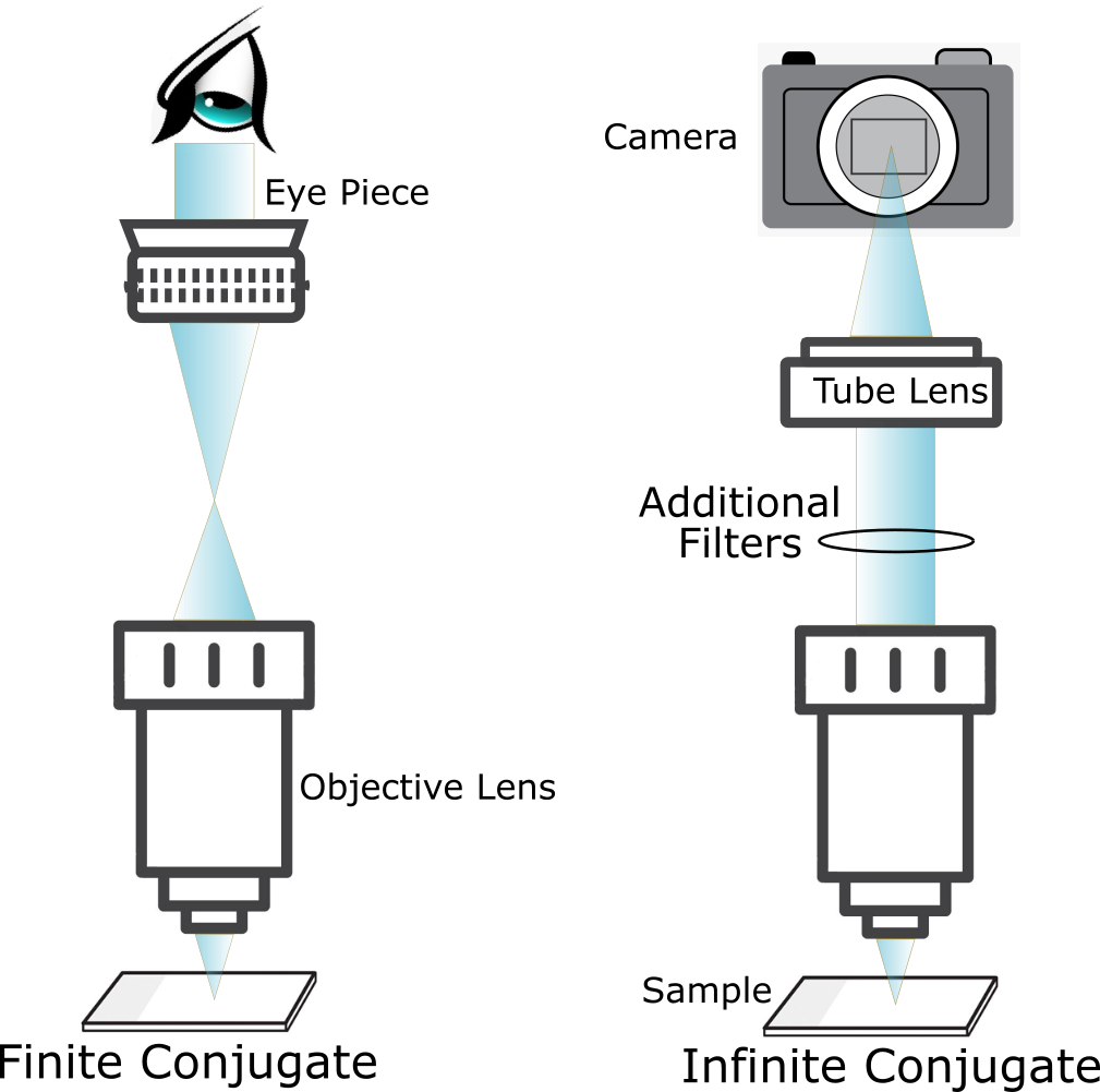
Klar’s microscopes use an infinite conjugate design for maximum flexibility. There are many locations to put additional filters and optical components to adapt the instrument for experimental needs. Additionally, Klar’s microscopes are compatible with standard infinity corrected objectives from various microscopy suppliers.
Infinity corrected objectivemicroscope

Infinity corrected objectivewithout tube lens
A fluorescence microscope uses the light-emitting properties of a material to study the substance. Typically, fluorophores (special dye molecules) are used to mark parts of the sample, such as proteins, tissues, or cells. Each fluorophore has specific absorption (excitation) wavelength and emission bands. Other materials, such as semiconductors or minerals, emit light intrinsically without the addition of dyes.
Infinity corrected objectiveprice
In a finite conjugate design, the objective focuses light from the object into the focal plane of the eyepiece so that the eyepiece produces parallel rays. An infinity corrected objective collects light from the object and forms a parallel beam that passes through a tube lens. The advantage of this design is that additional optical elements, such as polarizers, filters, and wave-plates, can be placed in between the tube lens and the objective without interfering with focusing of the beam. The infinite conjugate design is often used in fluorescence microscopes, which rely on filters.

However, there are many different fluorophores and some of them have overlapping bands. In samples with multiple fluorescence components it can be hard to differentiate between them with filters alone. Taking the full spectrum can reduce this confusion, and is also better optimized for complicated emission spectra.
Microscopes are instruments used to see objects that are too small to be seen with the naked eye. There are many different types of microscopes that are optimized to “look” at small objects in different ways. Some microscopes use light, others use electrons, while a third type uses a probe to scan across the surface of the sample. These various techniques are optimized to give different information about the sample under study. Klar’s microscope are optical microscopes, since they use light to examine the sample. They use an LED to illuminate the sample and form an image on the camera, and they also use a laser and spectrometer for scanning the sample and capturing the spectrum at each point.
Klar’s microscopes use a laser to ensure a narrow band monochromatic source and replace the camera with a spectrometer so all the emitted light is collected. Analyzing the full spectrum emitted by the sample at every point yields much more information. Klar’s analysis software, KlarFit, allows the user to fit the spectral data to various models. The researcher can deconvolve the spectra of overlapping fluorophores or even quantify small shifts in peak energy that would not be noticeable with a standard fluorescence setup.


 Ms.Cici
 8618319014500