
Shop All Nikon Lenses - lenses lens
Apr 30, 2023 — Using the quadratic formula to solve for time yields two solutions: t = 10 s and t = −20 s. A negative value for time is unreasonable, since ...
Utilizing this microscope objective lens is pretty simple. Firstly, you need to adjust the scanning lens to properly focus and center the specimen. Afterward, you need to turn the objective turret clockwise to face the low magnification lens. Lastly, re-center your specimen after you’ve fine-tuned the focus with the coarse focus knob.
The ILT800-UVF measures both dose (mJ/cm2) and irradiance (mW/cm2) and includes profiling as a standard feature. The system has a user-friendly interface that ...
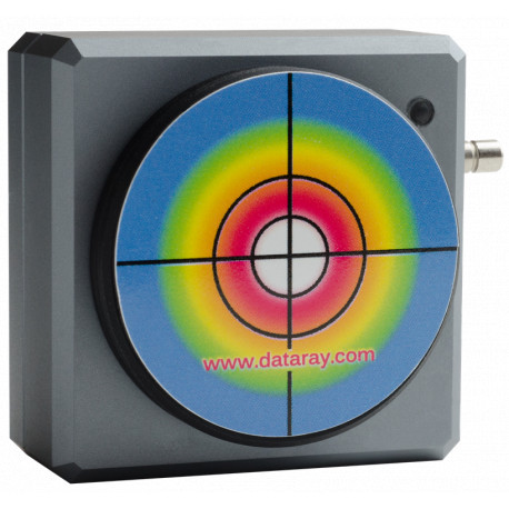
DataRay's WinCamD-LCM laser beam profiler (beam analysis camera) measures pulsed and continuous wave lasers with frame rates of up to 60 Hz at very high resolution (2048 x 2048 pixels). For the operation of the camera, no external power supply is required.
The Laser 2000 website uses cookies to continuously improve and enhance the features and content of our website. Click "Accept All" to get the full user experience, or select the cookies you want to use individually. If you click on the "More Information" button, you will get more details about Laser 2000's privacy policy.
American Science and Surplus offers tools, toys, science kits, educational toys, school supplies, arts and crafts items, hobby tools, scales, lab glass, ...
Dataray camera
Low magnification objective lens typically ranges from 2x to 20x. Using a 10x or 20x eyepiece will magnify objects by 100x or 200x. This lens lets you view tiny specimens such as skin, hair, and fly legs. Furthermore, it has a yellow band that encircles the housing of the lens.
Marketing cookies store information about other websites visited. The cookies are used to display advertising tailored to the user and are therefore relevant for publishers and external (third-party) advertisers.
This laser beam profiler features a CMOS sensor that guarantees frame rates of up to 60 Hz with high dynamics and is suitable for large beam diameters up to 11 mm.
The simplest types of microscopes are magnifying glasses with a single convex lens (meaning both sides are curved outward). This kind of lens usually makes items look 5–10 times bigger by changing how the light gets into the human eye. Compound microscopes are used in schools, homes, and professional labs. They have at least two lenses that work together to magnify an image.
BeamGage
These cookies collect information about the user's browsing behavior in the store in anonymous form in order to obtain reports about the user's interests in the products or services offered by the store.

This lens, in conjunction with the eyepiece lens, will provide the smallest magnification possible. For example, a microscope with a 10x eyepiece lens and a 4x objective lens will have a magnification factor of 40x. The magnification you get from this lens is similar to what you would from a stereo microscope, allowing you to study specimens like leaves and feathers. Also, the lens has a red band that encircles the housing of the lens. Scanning object lenses have low power and are typically used to scan a specimen before using higher magnifications.
Gaussianbeam
Half Ball Lens / Hemispheres. Half ball lenses are made from a wide variety of optical glasses with refractive index ranging from 1.50 to 2.10. More ...
Laguerre Gaussbeam
Microscope lenses come in different types that vary based on the magnification’s power. Here are the types of microscope objective lenses.
You can identify a high magnification lens by the blue band around the housing of the lens. Typically, compound microscopes come with a 40x lens. However, there are cases when this is not true. For example, you might buy a microscope with a high magnification lens of 60x or more.
Necessary cookies are required to enable basic functions such as page navigation or access to restricted areas of the website. Without these cookies, the website cannot be used as intended.
Microscope objective lenses work by changing how light goes through them. Essentially, when light shines on an object underneath a microscope, this light travels through the lens and bends toward your eyes, which makes the object bigger than it is. Remember that magnification power varies based on the type of lens and microscope, with magnification reaching 1000x and above. You can also find specialized objective lenses for advanced experiments.
A reflected darkfield objective works for darkfield microscopy. This technique produces a dark background with a strong contrast to aid in the visibility of translucent specimens. This object is designed to observe samples not dropped inside a covered slide. Reflected darkfield objectives typically have signs like BD, Neo, or BF/DF to help you identify them.
The applications include laser beam analysis of pulsed and CW laser service use in the field of laser systems optical setups, adjustment, monitoring of beam direction stability, quality control and M² measurement with matching M² stage.
The Laser Beam Profiler is supplied with 3 neutral density filters with optical densities OD1, OD2 and OD4. These are magnetically fixed in front of the C-mount opening and can be freely stacked with each other.
ISO 10934 gives the following definitions of the depth of focus: Depth of focus is the axial depth of the space on both sides of the image within which the ...
ThorlabsBeam Profiler
This laser beam profiler features a CMOS sensor that guarantees frame rates of up to 60 Hz with high dynamics and is suitable for large beam diameters up to 11 mm.
This laser beam profiler features a CMOS sensor that guarantees frame rates of up to 60 Hz with high dynamics and is suitable for large beam diameters up to 11 mm.
Phase contrast microscopy makes translucent specimens easier to see by making the difference between the background and the foreground stronger. In a phase contrast objective, a black ring around the lens is used to control and translate changes in the phase of light rays into changes in their amplitude. In addition, the way the light rays are bent and focused gives the image seen through the eyepiece a lot of contrast.
We use the internal website analysis tool Piwik to analyse anonymous visitor behaviour on our website. Your data will not be transmitted to third parties. This gives us the opportunity to evaluate which products and pages are clicked on when you visit our website. By using the Laser 2000 GmbH pages and services, you agree that we use cookies.
Yes! Many of the magnifying lamp, sold by the shops on Etsy, qualify for included shipping, such as: Exquisite Brass Adjustable Magnifying Glass, Home And ...
VIETNAM:Alpha Industrial Park, Tu ThonVillage, Yen My District, HungYen Province 17721+84 221-730-8668sales-vn@avantierinc.com
Chemistry Dictionary. Definition of collimated. forming a highly non-divergent beam. Search the Dictionary for More Terms. Return to top of page.
NanoScan
You can purchase certain specialized microscope objectives when you want to perform advanced microscopy experiments. Here are some of the most common lenses to buy.
Jun 24, 2022 — New review article in Trends in Cell Biology. Wnt signalling in cell division: from mechanisms to tissue engineering. To read the article click ...
An optical microscope comes with lenses that change how rays of light travel through them. When light bounces off an object under a microscope and goes through the lens, it deflects toward the eye. This makes the item seem bigger than it is.
Jun 21, 2011 — Where can I find a scalable vector graphic (SVG file) of USAF 1951 Resolving power target? Dimensions of USAF 1951 are here: ...
Sep 16, 2022 — A magnifying glass causes an image on the retina which is larger than without the magnifier. In principle, the image on the retina can be ...
Microscope lenses are pieces of glass that work in a microscope to aid magnification. Based on the lens type and power, you can magnify a specimen by up to 200x or more. How these tools work is straightforward, and this article will cover everything you need to know about them.
Beamscanner
There is one lens above the object, called the objective lens. Also, there’s another one close to your eye (eyepiece). In some cases, each type of lens consists of various lenses. Compound microscopes can typically magnify by 10x, 20x, 40x, or 100x. However, you can find professional ones that can reach up to 200x magnification or more. There are also modern microscopes like the electron microscope for those who want higher magnification.
This type of lens is usually used for smaller specimens, such as cells and bacteria, which cannot be seen with just the human eye. This includes molds, tardigrades, germs, and others.
The use of differential interference contrast (DIC) lenses in brightfield microscopy helps to visualize transparent samples better. By providing contrast without the need for staining, DIC objectives reduce the amount of staining performed. In most cases, a DIC lens will not be present on a compound microscope for school or home use.
Preference cookies store user-specific information and allow the website to be customized to the user, for example, to the language or region you have chosen.
Most basic microscopes do not come with an oil immersion lens, and this is because most leisure microscopy experiments do not require them. These lenses can reach up to 200x or more magnification with a 10x eyepiece lens and a 200x objective lens. You can find this lens by a white or cream-colored band around the lens.
Thorlabsbeam
Due to the difference between the glass slide and the refractive indices of air, a specific oil is required to help fill the space. Without this oil, the objective lens won’t function correctly. Hence, you won’t get the appropriate magnification and resolution, leaving you with too much distortion.
Long-working distance objectives are made so you can see specimens even when they are farther away than usual. This is usually needed when a sample is stuck in a thick slide or is under a thick glass plate.


 Ms.Cici
 8618319014500