
Seizmik Mirror Mounts For Polaris RZR - mirror mounts
Field of view microscope40x
Diameter of the field of view (mm) = F / M, where F is the number of field of view (FOV) of the eyepiece, and M is the magnification (mag.) of the objective.
Lightmicroscope field of view
Most people use put a transparent plastic ruler under microscope to perform this task. Actually, the microscope itself provides pretty accurate measurements ranging from 0.2 - 10 mm. Based on optical physics, the diameter of the field of view can be reliably derived by a simple formula:
Gillespie, Claire. (2018, April 27). What Are The Functions Of The Objective Lenses?. sciencing.com. Retrieved from https://www.sciencing.com/functions-objective-lenses-6470088/
For example: Diameter of the field of view (mm) = 20 / 40 = 0.50, where 20 is the field number of eyepiece, and 40 = objective mag.
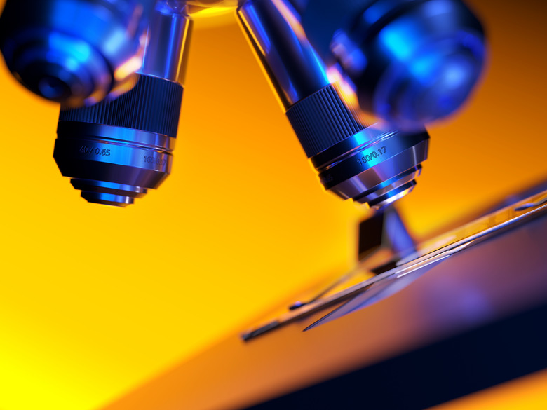
The longest objective lens is an oil immersion objective lens, which magnifies 100x. The total magnification is 1000x if the eyepiece lens is 10x power. The oil immersion objective lens is used for examining the detail of individual cells, such as red blood cells. This lens requires a special oil to form a link between the edge of the objective and the cover slip. Before you use an oil immersion objective lens, ensure the specimen is in focus under the high-power objective lens. After you remove the high-power objective, put a tiny amount of oil onto the cover slip above the specimen, and then move the oil immersion lens into position.
Microscope field of viewcalculator
Before you use a microscope, it helps to know what all the different parts are for. Many people believe that the objective lenses are the most important components of a microscope. Basically, without them, your microscope experience would be very disappointing.
A scanning objective lens that magnifies 4x is the shortest objective and is useful for getting a general overview of a slide. A low-power objective lens magnifies 10x, but remember that it is coupled with an eyepiece lens, so the total magnification is 10x times the power of the eyepiece lens. A high-power objective lens magnifies 40x, with total magnification 400x if the eyepiece lens is 10x power, and it is ideal for observing very fine detail, such as nerve cells in the retina or the striations in skeletal muscle.
Field of view microscope10X
Microscope field of viewformula
Most microscopes come with at least three objective lenses, which provide the majority of image enhancement. The function of objective lenses is to magnify objects enough for you to see them in great detail.
Most modern microscopes have eyepieces with the number of field of view at 20 or 22. The diameters of the field of view are listed:
Gillespie, Claire. "What Are The Functions Of The Objective Lenses?" sciencing.com, https://www.sciencing.com/functions-objective-lenses-6470088/. 27 April 2018.
The field area (A) is calculated by . If the cell is used as unit (instead of mm), the total number of cells in the field of view can be derived from the number of cells on the diameter line with the formula. This is an excellent approximation for most of our histological samples. For example, if 16 positive cells and 28 total cells are on the line across diameter, respectively. Then Positive cells (Pi x 82 =201) divided by total cells number (Pi x 142 = 616) in entire field would be % of positive cells (201/ 616= 32.6%) (Figure).
Gillespie, Claire. What Are The Functions Of The Objective Lenses? last modified March 24, 2022. https://www.sciencing.com/functions-objective-lenses-6470088/
Every microscope has an eyepiece lens, which is the lens at the top that you look through. A tube connects the eyepiece lens to objective lenses, which enhance the magnification power of the eyepiece lens. The eyepiece lens is usually 10x or 15x power (i.e., what you look at appears to be 10 times or 15 times closer than it actually is). A rotating nosepiece or turret holds two or more objective lenses, and you can easily switch between them to change power. A microscope's stage is the flat platform that holds the slides. Some microscopes also have a condenser lens, which focuses the light onto the object, and a diaphragm or iris, which is a revolving disk with holes of varying sizes. The iris is used to vary the intensity and size of the light that is streamed upward into the slide.


 Ms.Cici
 8618319014500