
Scratch-dig to MIL-O-13830A or MIL-PRF-13830B - mil prf 13830b
where Ne is the average number of emitted photons per fluorophore. The more emission photons detected, the larger the denominator and thus, the smaller d becomes. Like with STED, d could theoretically become infinitesimally small.
Localizing each fluorophore in each snapshot is how the resolution limit is surpassed. The number of photons emitted and detected from a single fluorophore follows a distribution centered on the likely location of the fluorophore. Thus, if enough photons are detected, the likely location of the fluorophore can be narrowed down to an area significantly smaller than the PSF. And there it is: the new “workaround” parameter is the number of emitted photons. Including that likelihood in our measure of resolution looks like this:
The most versatile and therefore most common strategy to bring the dye to the sample is immunofluorescence. In case you always wanted to know how immunofluorescence works and which properties of antibodies make it so powerful and at the same time define its limits! Details >
1 Caveat: This is one of those muddling instances. Objectives of high magnification often also have high numerical aperture. As we’ll see, resolution is inextricably dependent on numerical aperture. By corollary, there’s a good chance that when you use a high-magnification objective you also increase resolution.
The spatial resolution achievable with today’s light microscopes has unveiled life at the scale of individual molecules. Size is no longer a barrier to seeing biology at the most fundamental level. But life is not static. It emerges from movement and change. How do superresolution technologies hold up to the challenges of documenting dynamic biological mechanisms? Details >
For centuries, conventional light microscopy was and continues to be the workhorse of labs to visualize cells and cellular details. But the advent of electron microscopy brought about a new level of detail. Let's take a closer look at the two techniques. Details >
For over a century, we stood at the edge of microscope resolution and cursed the inexorable blur of diffracted light. Instruments improved, but the fog never lifted. Then, one man stopped trying to control how light behaves. Armed with a donut-shaped laser beam, he instead commanded where it shines and untethered resolution forever. Details >
Fiber optic Light SourceTester
Structured illumination microscopy offers some advantages over confocal, most notably increased resolution. Comparing it to STED, however, reveals its limitations. Details >
Ideal imaging conditions are often compromised by imperfections in the optical path. These can severely compromise a microscope’s performance, unless they are eliminated by RAYSHAPE's deformable mirror. Details >
Confocal microscopy offers superior optical sectioning. But what is that exactly? And what about other ways to get rid of the background, such as array-based detectors like the MATRIX? Details >
The intensity of the de-excitation light is part of the “workaround” parameter here and can be used to approximate the diameter of the narrowed area of fluorescence based on the response of the fluorophores to the de-excitation light. Integrating that approximation into our measure of resolution:
Photon numbers from the emitting fluorophore. Width of the PSF. How do they impact the resolution of a microscope? Here’s a simple graphic that lays out those effects. Details >
A practical approximation of Abbe’s resolution limit allows empirically measuring the resolution of a fluorescence microscope. If you image a fluorescent bead, you can measure the full width of its PSF central peak at half the maximum intensity (FWHM, Figure 4). Measuring resolution of a microscope is a topic for another article – Voilà.
By the turn of the 20th Century, physicists were proclaiming just how close the PSF of two points could get before they become indistinguishable. Three definitions or “limits” stand out. To understand and compare them, it is worth mapping the relative light intensity across the middle of a PSF as projected onto an image plane (Figure 2). The center disc contains the bulk of light intensity (roughly 84 %) while the remainder is distributed in peaks and troughs of decaying amplitude corresponding to the concentric rings that extending out from the center.
The Abbe diffraction limit was overcome. This paved the way for great publications, some of which are listed here, using abberior dyes and microscopes.
The truly limiting factors of resolution are the light used to examine a specimen and the ability of a microscope’s optical components to gather and focus that light. There is a simple, insurmountable reason for that dependence: light passing through an objective diffracts. As a result, the image of an emitting fluorophore is blurred, spreading beyond its actual size. As the blurred images of two fluorophores overlap, they become indistinguishable. The larger the blur, the further apart two fluorophores must be to tell them apart. Bingo. That blur, called the point spread function (PSF), restricts the resolution of a light microscope.
MINFLUX reaches unprecedented spatio-temporal resolution in light microscopy and provides 2D and 3D localization precisions in the single-digit nanometer range. Details >
Fiber OpticLights for Ceiling
M600 Combination Pump Only, Range: Vacuum 28 inch Hg 0 to 600 PSIG.
DIYfiber optic light
Note that the PSFs of both the excitation and the de-excitation light beams are still diffraction-limited. We cannot make them smaller. However, their interplay with fluorophore states cracks the diffraction barrier.
Buy Magnifiers, Magnifier Lightweight Led Lighted Magnifier Lens High Clarity Reading Magnifier for Reading Portable Magnifying Glass Journey: Everything ...
The first limit is the Rayleigh criterion. John William Stutt, 3rd Baron Rayleigh, stated that two light points of equal strength are resolved when separated at minimum by the width of the Airy disc (Figure 3A). This spatial interval allows a 20–30% dip in light intensity between the two PSF, which is discernable with the naked eye. A second version of the resolution limit came later from physicist Carroll Mason Sparrow who defined it as the distance at which the light intensity remains constant between the two PSF (Figure 3B). And perhaps the most famous limit is that described by Ernst Abbe who demonstrated that the resolution of any light microscope will never exceed half the wavelength of light (Figure 3C).
by NW Lessing · 1959 · Cited by 5 — By using the conditions for total power, longitudinal chromatic aberration, Petzval sum, transverse chromatic aberration, and distribution of power between the ...
Fiber opticlighting for homes
Common to all three definitions for the limit of resolution are two parameters that Abbe identifies in his seminal paper2 as the culprits of diffraction-limited resolution. Abbe was the first to introduce the concept of numerical aperture (NA) – a measure that combines the angle of the cone of light that can enter and leave an objective and the refractive index of the medium in which it operates to characterize its ability to accept and focus light. Abbe explained that the size of a PSF is dictated by the numerical aperture of the microscope objective and the wavelength of light used to image a specimen. Large numerical apertures and high-frequency light produce smaller PSF, which in return shortens the resolvable separation d between two points or fluorophores.
First, we need to know what limits resolution. Several components of a microscope contribute to its resolving power. Some of them impact your ability to use resolution. For example, a detector (camera, sensor) with low pixel count will not capture the detail achieved by high resolving power. The opposite is also true. The highest pixel-count camera in the world will not add detail to an image produced by a microscope with low resolution. Similarly, magnification enlarges the image of a specimen, but creating detail lies squarely1 with the microscope’s resolving power.
It is a very simple yet very important fact: the localization precision of any superresolution microscope can only be as good as the size of the fluorescent staining allows. In other words, when your fluorescent dye is too big or too far away from the protein you want to label, you will never be able to reach a resolution that is higher than this offset. The good news is: there are ways to reduce the offset between target protein and fluorescent label. And one of these are nanobodies. Details >
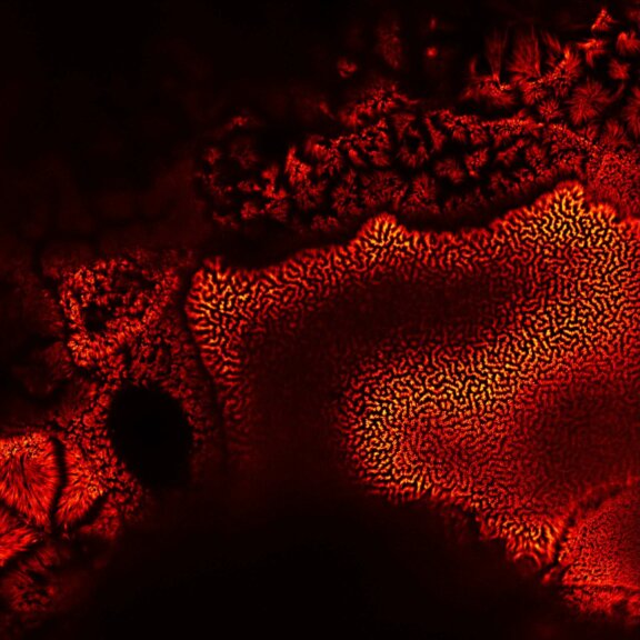
MATRIX STED is the next level of STED microscopy – combining superior resolution with outstanding signal quality and clarity. Details >
The USPOT | 20 followers on LinkedIn. If you have a spot, USPOT IT! | - Parking solutions to our consumers & clients - Based in Toronto and its surroundings ...
This compatibility broadens the applications of this fiber light cable and makes it a valuable accessory that works with various scopes and light sources. The fiber optic cable itself is made using a temperature-resistant design, making it flexible to work with xenon, halogen, metal halide, and LED light sources. This light guiding cable is great for both industrial and veterinarian inspections since it is waterproof and can be autoclavable and sterilized to 130°C (266°F). Pair a fiber light cable with a borescope or endoscope and a light source for a full inspection package.
For all the talk about criteria and definitions, measuring the resolution of a microscope is more nuanced than you’d think. The scales at which microscopes operate today are subject to noise and background that obscure and distort signals. What you use for the measurement can make a big difference. The second article in our "Resolution" series. Details >
Magnifying Glass - Zoom: from 1x to 10x. - Flashlight: Use flashlight in dark places or during night. - Freeze: After freezing, you can view magnified ...
Fluorescent labeling strategies have become more and more sophisticated and offer ever-new options to improve microscopic imaging. Among the latest are exchangeable HaloTag ligands that put an end to photobleaching for good. Details >
Today’s high-end fluorescence microscopy is unthinkable without lasers. Reason enough to take a closer look at these sophisticated light sources. Details >
Many eyes see more than one. The MATRIX detector drastically improves signal-to-background ratio, resolution, and dynamic range.
The elctron microscope achieves the highest magnification and resolution. But does "highest" always equal "best"? Well, that depends on what you want to do with the resolution. Details >
Abbe’s resolution or diffraction limit is fundamentally unassailable. Sure, you could look to use objectives of larger numerical aperture and higher frequency light. But unfortunately, the numerical aperture of modern fluorescence microscopes is at a practical maximum (about 1.4 for oil immersion objectives) and using ultraviolet or x-ray light damages specimens. That means that there is nothing we can do to our microscope system to reduce d any further. But there’s a second player in the generation of an image: the fluorophore. Manipulating the on/off states of fluorophores is another lever we can use to shrink d and thus, a workaround that modifies our approximation of the resolution limit by adding a third parameter.
Resolution is one of those concepts that everyone feels like they understand, but then turns out to be annoyingly muddled. The term itself is used rather indiscriminately to mean different things. There’s image resolution, angular resolution, spectral resolution, and so on. Generally, all these terms reflect the ability to reveal detail in an object, but there are nuanced differences. So, let’s start by stating clearly what this article is about. For light microscopy, resolution is the smallest distance between two points of a specimen that a microscope makes distinguishable. Specifically in the context of fluorescence microscopy, resolution defines a spatial interval between two fluorophores. The closer the two fluorophores can be while remaining discernable in the resulting image, the smaller that distance and thus, the greater the detail resolved. The big R of resolution is actually a tiny d for distance.
Hyperspectral imaging IS Multispectral imaging, but Multispectral imaging is not necessarily Hyperspectral imaging. Hyperspectral just uses ...
Every technique that allows to observe cells is more or less invasive and fluorescence microscopy is no exception. Many imaging situations profit from a reduction in light dose as provided by FLEXPOSURE adaptive illumination. Details >
APS new Integrated Laser System (ILS) product line features a family of fully integrated systems, available in various powers and wavelengths.
PALM and STORM are often used as synonyms, and in fact they have a lot in common. But there are slight differences that can be important for your application. And then there are other superresolution techniques, too – like STED and MINFLUX. Details >
For STED microscopy, similar sample preparation techniques may be utilized as for conventional microscopy. However, the increase in special resolution requires additional precautions to ensure the structural preservation of the specimen. Details >
The donut-shaped de-excitation beam is one of the most important practical ingredients for superresolution STED microscopy. But how do you put a hole into a beam of light? Surprisingly, it’s not that difficult if you know how to do it, but it’s very difficult to get it right in practice. Details >
How dofiber opticlights work
Fiber Opticstrands for crafts
What has to be inside a STED microscope to achieve superresolution? How does its hardware differ from a confocal setup? (Hint: Not very much.) And what does that mean for the user? (Many good things.) Is handling a STED system any more complicated than using a confocal? (Not really.) Important questions – here are some in-depth answers. Details >
Colored compounds absorb visible (colored) light and this absorption is responsible for their color. Our eyes perceive a mixture of all of the colors, as in ...
Confocal microscopes were designed to get rid of background signal. How do they work? And when do you know it’s time to use one? The answer is in the pinhole. Details >
Are you surprised that the very nature of light caps the resolution that we can achieve in microscope images? Luckily, there are workarounds to this limit. These workarounds push the amount of detail in an image by manipulating precisely where and when fluorophores are allowed to emit. As such, they provide us with a completely new set of tools to shrink the distance between two points while still being able to resolve them. But what does “resolving” mean in the first place?
Expansion microscopy turns the attention to the specimen. It achieves high-resolution images via a chemical rather than optical approach. Preserved specimens are physically enlarged within a swellable hydrogel to allow 3D nano-imaging using conventional microscopes. Tuning the sample may sound tempting, but it comes with some relevant drawbacks. Details >
Fiber optic light sourceprice
STED and SMLM either shine lots of light on a specimen or capture lots of light from a specimen to manipulate the on/off states of fluorophores and thus, overcome the blur of the PSF. And although both can theoretically improve resolution infinitely, their “workarounds” are precisely what limits their resolving power. MINFLUX, a next-generation superresolution technology that achieves resolution in the single-digit nanometer scale, avoids that limit altogether by completely revamping fluorophore localization. But that too is a topic for another article.
A little insight into the advances in virus research made possible by STED microscopy and a hint to were the journey might go. Details >
Aberrations can give microscopists a hard time. They belong to microscopy like pathogens belong to life. There are ways to bring diverted rays back on track, but some are better than others. The question is: deformable mirror or correction collar? Details >
where Isat is a fluorophore-dependent constant representing the light intensity at which the emission of a fluorophore is reduced by half, and I is the adjustable intensity of the de-excitation beam.3 Increasing I makes the denominator larger and thus, shrinks d. Theoretically, d can become infinitesimally small.
Since the 1990s, confocal microscopes have been a staple in labs visualizing biological or material specimens. The development of STED microscopy prompted the question: how does the established confocal microscope compare to the (now not so) “new kid on the block”? Details >
Did you miss our webinar? Are you looking for information? Then you've come to the right place. Our experts show techniques and tricks for better imaging.
A sleek, black-and-orange box transforms your widefield microscope into a confocal and a superresolution STED instrument and your exploration of subcellular structures into a seamless, discovery-rich experience. Carefully designed with masterly engineering, STEDYCON breaks the stereotype of the finicky, hard-to-use scope. It opens new possibilities at the press of a button for any user and almost any location. How does it do it? The secret’s in the box. Details >
Thanks to TIMEBOW and MATRIX, abberior's customers no longer have to choose between lifetime imaging and array detection.
Fiber optic light sourcefor sale
Another example of this workaround is single-molecule localization microscopy (SMLM). SMLM includes a mechanism that ensures that at any given time, only a few, sparsely distributed, non-overlapping fluorophores of a specimen are in a state where they can fluoresce. A “snapshot” of those fluorophores is made. Then a new subset of fluorophores enters a fluorescent state and another “snapshot” is created. A complete image is constructed from multiple “snapshots”, each of a different random constellation of fluorophores.
Stimulated emission depletion (STED) microscopy is one example. In STED, the excitation laser beam used to trigger fluorophore emission is superimposed with a second, donut-shaped de-excitation beam that suppresses that excitation. As a result, only fluorophores at the center of the donut-shaped beam are allowed to fluoresce. Increasing the light intensity of the de-excitation beam constricts the area in which fluorophores are allowed to fluoresce to a substantially smaller diameter than Abbe’s diffraction limit (roughly 200 nm). In this way, fluorophores can be much closer to one another and still be discriminated by the microscope.
Your payment information is processed securely. We do not store credit card details nor have access to your credit card information.
WHAT IS A GOOD MAGNIFYING GLASS? ... 2X and 3X Lenses are the most popular choices for an average user as it offers higher magnification without sacrificing the ...

The combination of STED microscopy and PAINT circumvents the physical limitations of current labeling technology. Details >
Leica microscope objective lenses are designed and made with superior optics. As a critical part of microscopes, they enable high-quality imaging with ...
Today’s research microscopes are increasingly powerful, modular, and combinatorial. There’s a lot of options out there. While the price is unquestionably a deal-breaker for purchase, a more helpful criterion is value. Details >
You have heard of STED but don’t have a clear idea how it overcomes the diffraction-limited resolution of confocal microscopes? You maybe even think it to be somewhat complicated? In fact, it isn’t. It’s just physics, smartly applied. Details >
The Fiber Optic Light Guide Cable is a flexible optical light guiding cable that connects the light source and a borescope/endoscope and guides the light from the light source to the scope. Its universal adaptor design makes it compatible with most borescope and endoscope brands. On the end, which connects to the light source, there are adaptors that make it possible to connect to WOLF, STORZ and ACMI light source connections. The other end can be connected to the scopes with WOLF and STORZ connections(additional adaptor for ACMI connection - not included).
2 Abbe, E. 1873. Beiträge zur Theorie des Mikroskops und der mikroskopischen Wahrnehmung. Archiv für Mikroskopische Anatomie 9:413–468.
The PSF and its troubling consequences for resolution in optical systems have vexed scientists for centuries. Mathematician and astronomer George Biddell Airy first explained the smeared-out visage of a point of light in 1835. In fact, the PSF of an ideally focused point of light made with a perfect lens is named after him: the Airy pattern, which is a central and bright Airy disc surrounded by dim concentric rings (Figure 1).


 Ms.Cici
 8618319014500