
Rust: All CCTV Camera Codes (& How To Use Them) - camera ids
Achromatic lenses are used to diminish chromatic and spherical aberrations which are the loss of color and focus that can happen when light wavelengths refract in direct light. These aberrations can be controlled by using an objective lens that contains both a convex and concave lens inside. Mounting these two different types of lenses to each other can bring wavelengths of red and blue light closer together, which puts them in the same focus and cancels out chromatic aberration. Another type of lens used to correct for both color and spherical aberration is the plan (or planar) lens. These produce a flatter field and can also give you a much larger working distance. However, they can be more expensive than achromatic lenses, so choosing between the two depends largely on how much power you need in your objective lens, and whether or not you need to adjust for field curvature, which only plan lenses can do. Achromatic lenses and plan lenses both come in dozens of magnifications and types, accommodating a wide variety of microscopy needs.
The Eye Clinic of Edmonds is stocked with a wide variety of trial contact lenses from Daily to Multifocal varieties and we sample nearly a dozen different ...
Microscopes are made up of lenses for magnification, each with its own magnification powers. Depending on the type of lens, it will magnify the specimen according to its focal strength.
Infinity objective lenses did not become common until the 1980s but have since carved out a permanent spot in the microscope objective market. Previously, all microscopes had a standard tube length–the distance from the eyepiece to the objective lens was always 160 mm. Once microscope manufacturers began developing microscopes with varying tube lengths, lens manufacturers had to catch up with the changing technology. New tube lengths meant that microscopy equipment developers needed to adjust for these changes in their accessories, including objective lenses. Infinity optical systems use multiple sets of lenses within the lens house to correct a wide range of tube lengths–typically from 160-200 mm. This enables the lenses to be more versatile between microscopes of varying tube lengths.
Ans. A microscope is an optical instrument with one or more lens systems that are used to get a clear, magnified image of minute objects or structures that can’t be viewed by the naked eye.
Ans. Rack stop is included in the microscope for preventing the specimen slide from coming too far up and hitting the objective lens.
I did NOT like this website sourse. Wanna know why I didn’t like it??? I don’t like it BECAUSE my school wants me to use this website sourse. My new science teacher wants us to answer those 10 questions. I think its pretty dumb. No Offensen to anyne out there, because I am a nice person not a mean one.
Their ability to function is because they have been constructed with special components that enable them to achieve high magnification levels. They can view very small specimens and distinguish their structural differences, for example, the view of animal and plant cells viewing microscopic bacterial cells.
There are hundreds of unique objective lenses to choose from, but once you have a greater understanding of the most common types, you can make a more informed decision regarding which lens is right for you. Whether you are a hobbyist or whether you require the use of a microscope in your day-to-day research, it’s important to gain an understanding of the strengths and weaknesses across the spectrum of objective lenses. Once you know exactly what you’re looking for, you’ll be well on your way to obtaining the best results and having an optimal viewing experience.
Jun 30, 2023 — Emerging optical components are often referred to as flat optics, since they are manufactured on flat silicon or glass substrates. One advantage ...
Thanks a lot for this wonderful note: It is really helpful, Really appreciate the way all the detail about microscope have been explained
Typesof objectivelenses
Ans. The coarse adjustment knob moves the stage up and down to bring the specimen into focus. The fine adjustment knob brings the specimen into sharp focus under low power and is used for all focusing when using high-power lenses.
The diameter of the laser beam should be significantly smaller than the clear aperture of the mirror (Figure 1). A general rule restricts the diameter of the ...
A beam of light is passed through the condenser to the specimen. The light transmitted from the specimen enters the objective lens. While passing through the objectives, the transmitted rays are spread so that they appear to come from the bigger objects.
Specialized microscopes, such as metallurgical microscopes, require their own specific metallurgical objective lenses. These devices are most often used to examine structural detail of ceramics, metals and other non-living materials. Another common microscope objective accessory is a Barlow lens. These can be added to the bottom of an objective lens to either increase or decrease its working distance, field of view or magnification. Since they can be interchanged between lenses, they are a cost-effective way to change the power and magnification of lenses you already own. Lastly, if all these lenses are starting to seem overwhelming, remember one quick trick for determining magnification at a glance: look at the band of color near the bottom of your objective lens. While the magnification number is usually written right on the lens, you can also quickly determine its strength by the color ring. Red indicates 5x magnification, while yellow means 10x, light blue means 40x and white can mean 100-250x.
Microscopes are instruments that are used in science laboratories to visualize very minute objects, such as cells and microorganisms, giving a contrasting image that is magnified.
Alternative distributions can be specified by providing three functions: (1) a function fitting the parameters of a distributions and providing a vector of ...
Thanks much for this. We just did microscopy as a topic and the write-up has really helped me to understand better. Thanks again
What is the objective lens of a microscopeexplain
1. which objective lens focuses closest to object 2. what controls the light entering the binocular lenses 3. how can you enhance the resolving power of a microscope 4. what is useful and false magnification PLEASE CAN YOU HELP ME IN ASWERING THOSE QUESTIONS
Eyepiece Tube holds the eyepieces in place above the objective lens. Binocular microscope heads typically incorporate a diopter adjustment ring that allows for ...
Microscopeparts
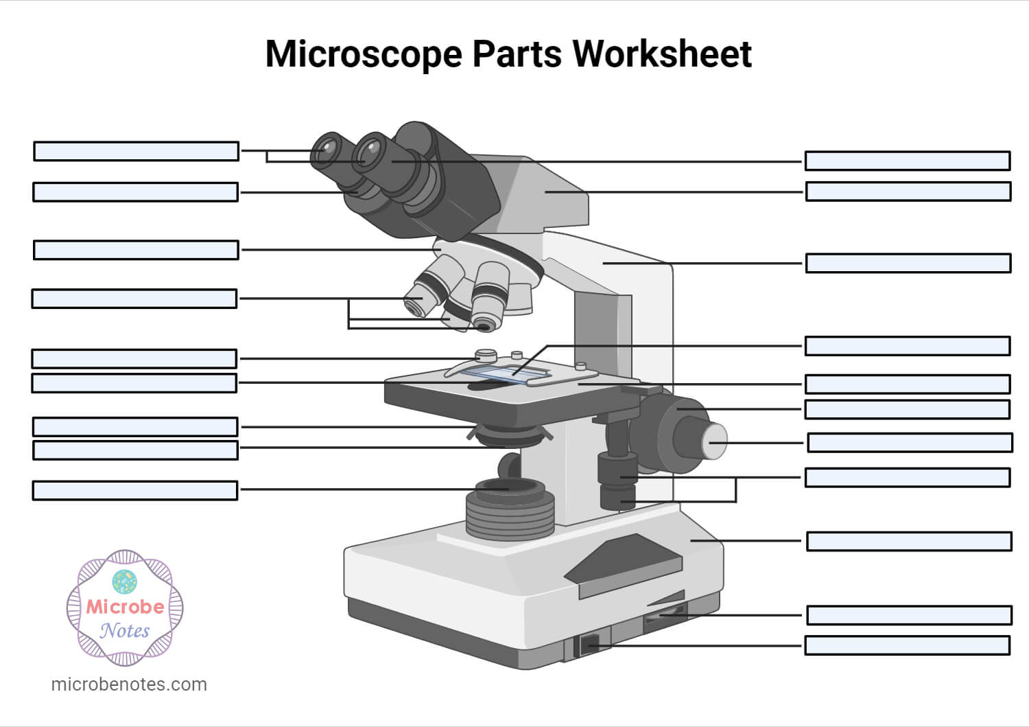
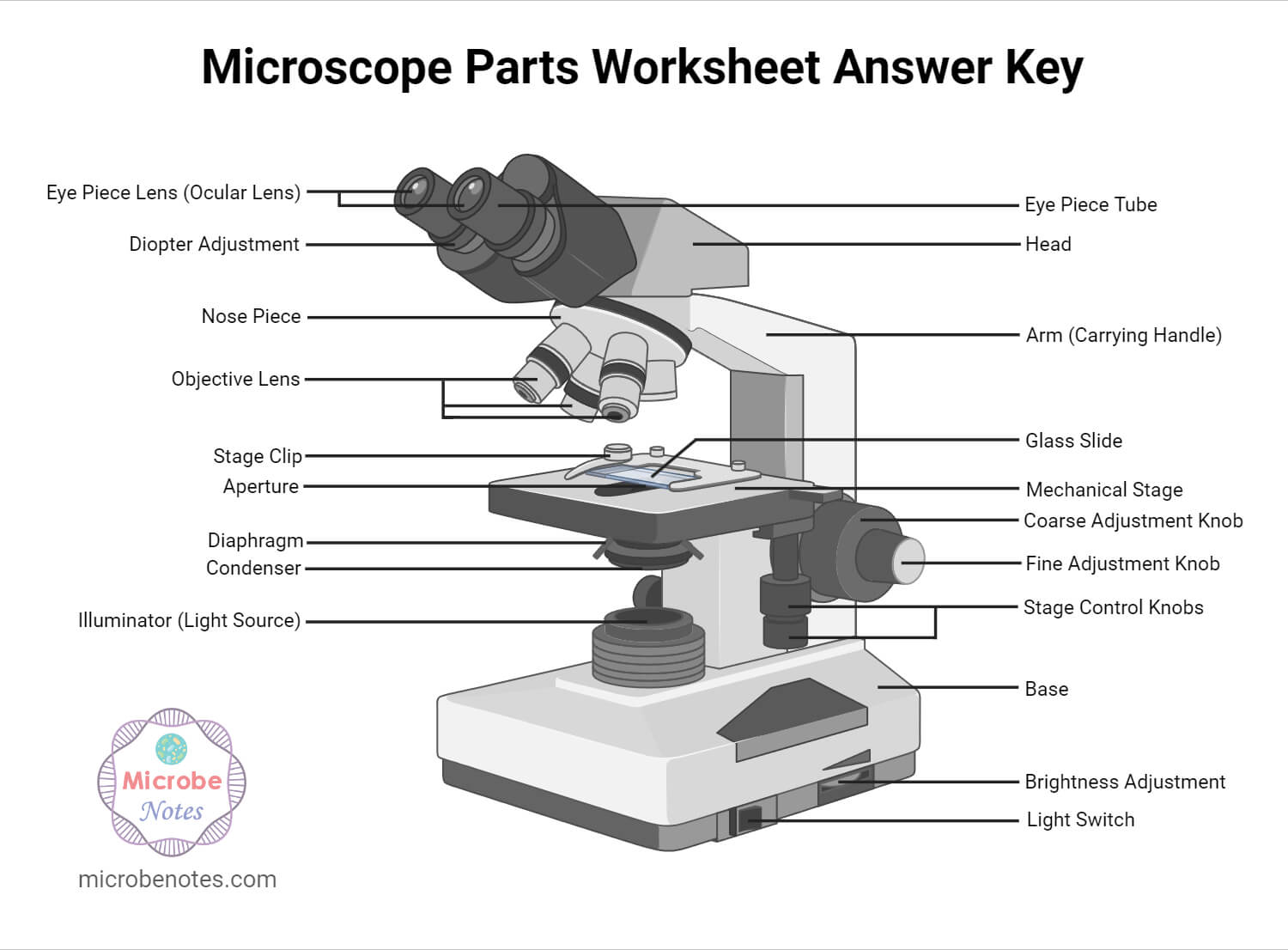
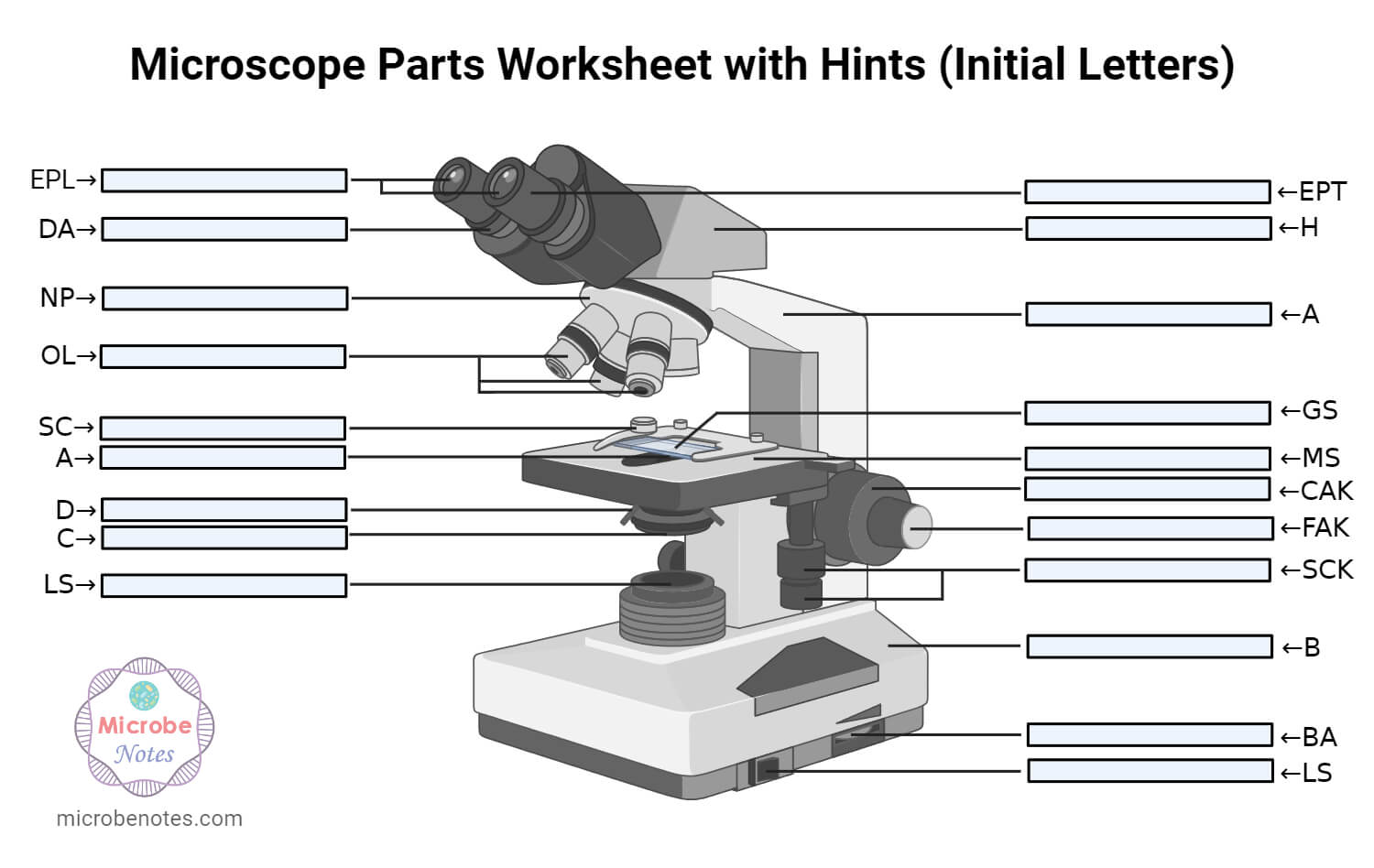
1. Ocular Lens (Eye Piece)2. Diopter Adjustment3. Head4. Nose Piece5. Objective Lens6. Arm (Carrying Handle)7. Mechanical Stage8. Stage Clip9. Aperture10. Diaphragm11. Condenser12. Coarse Adjustment13. Fine Adjustment14. Illuminator (Light Source)15. Stage Controls16. Base17. Brightness Adjustment18. Light Switch
AmScope exclusive ALL-IN-ONE 3D DIGITAL INSPECTION MICROSCOPE. View different angles and perspectives of objects with ease.
Ans. The eyepiece, also known as the ocular is the part used to look through the microscope. Its found at the top of the microscope. Its standard magnification is 10x with an optional eyepiece having magnifications from 5X – 30X. Objective Lens are the major lenses used for specimen visualization. They have a magnification power of 40x-100x. There are about 1- 4 objective lenses placed on one microscope, in that some are rare facing and others face forward.
Objective lensfunction
The objective lens is the most important optical component of the microscope. It’s the part that sits in closest proximity to the specimen being examined, gathering light to produce optimal images for observation and analysis. This lens creates the first magnification by spreading out the light’s rays to make the object appear considerably larger by the time it meets your field of view at the other end of the eyepiece. Such a critical piece of equipment doesn’t come in a one-size-fits-all package. Below, we will discuss some of the different types of microscope objective lenses and the unique roles they play in microscopy.
it very good website i use in 4 grade right after i plai amog us and they vote me out using orang strat witch mad me sad 🙁
this is a really good artical i used it to study my science i just wanted to point out to you that tere are a few spelling errors but other than that it is a 100% rating from me
Overcoming this distortion using technology distanced from the camera to process live image feeds is now possible with the AlphaEye realtime ...
Thanks alot of your help and knowI can draw it well in my exams and write the functions.Thankyou very much for your help
by R Su · 2016 · Cited by 31 — A novel approach for active coherent beam combining of a large number of fiber lasers based on cascaded phase control is demonstrated.
Having been constructed in the 16th Century, microscopes have revolutionized science with their ability to magnify small objects such as microbial cells, producing images with definitive structures that are identifiable and characterizable.
DLC (Diamond-Like Carbon) Coating involves a multistep process: · Precleaning · Placement of parts in a planetary fixture using single-, double- or triple-axis ...
Seriously, if i am not grateful, i am lying. This note is really helpeful to me to differet ways to different methology.
Objective lens microscopefunction
Thanks for helping me to know the parts and functions of a light microscope. THANKS AGAIN AND I HOPE THAT I WILL DRAW IT IN MY EXAM
What is the objective lens of a microscopegive
Obtaining high-contrast images of transparent specimens is difficult, especially when your specimen is alive and moving on a slide. Phase-contrast lenses allow you to observe microorganisms without having to fix and stain them. When your specimens are kept alive, a variety of biological functions can be examined and analyzed in real-time. Phase plates at the top of the objective lens diffract light, allowing these specialized lenses to tap into tiny changes in wavelength amplitude, which appears to the viewer as starker contrast on the slide. This makes the specimen much easier to view and observe.
Thank you for the support u have done may the Holy Spirit from the Almighty shine upon you to have more knowledge 2 continue making more notes from various topics in microbiology????✍️
EDMUND OPTICS LTD. - Free company information from Companies House including registered office address, filing history, accounts, annual return, officers, ...
Whatarethe3objectivelenses ona microscope
Thank you very much it really helped me with my science home work since i in 8th grade and this my home work to draw a microscope label all the parts and the function thank may the holy father of holy spirits bless you and give more wisdom thanks love you all keep up the good work and thank you again bye.
Objective lensmagnification
Discover Hollywood's source for motion picture and digital video equipment including grip and lighting equipment to expendables.
Ans. Condensers are lenses that are used to collect and focus light from the illuminator into the specimen. They are found under the stage next to the diaphragm of the microscope. They play a major role in ensuring clear sharp images are produced with a high magnification of 400X and above. Abbe condenser is a condenser specially designed for high-quality microscopes, which makes the condenser to be movable and allows very high magnification of above 400X. High-quality microscopes normally have a high numerical aperture than objective lenses.
The light is then focused on the eyepiece lens. This lens further magnifies the pre-magnified image coming from the objectives.
There are different types of microscopes like light microscope, dark-field microscope, phase contrast microscope, electron microscope, fluorescent microscope, etc.
Ans. The magnification of a lens is defined as the ratio of the height of an image to the height of an object. Microscope magnification measures the total enlargement of the image of an object. Magnification power is the product of eyepiece lens power and objective lens power.
1. Illuminator (Light Source)2. Diaphragm (Iris)3. Condenser4. Condenser Focus Knob5. Rack Stop6. Stage7. Stage Control Knobs8. Nose Piece9. Objective Lens10. Tube (Head)11. Eyepiece (Ocular Lens)12. Diopter Adjustment13. Adjustment Knobs (Fine Adjustment Knob and Coarse Adjustment Knob)14. Arm15. Base16. Light Switch17. Brightness Adjustment
Thank you so much for the note that you have given to me i was so grateful to know that you are so bright people that extend your help to a student
Thanks very much dear and please continue doing so, am Gerald M from Uganda East Africa doing diploma in nursing at Mulago school of nursing and midwifery
Microscopes are generally made up of structural parts for holding and supporting the microscope and its components and the optical parts that are used for magnification and viewing of the specimen images. Modern microscopes have additional electronics and display devices. This description defines the parts of a microscope and the functions they perform to enable the visualization of specimens.
The optical parts of the microscope are used to view, magnify, and produce an image from a specimen placed on a slide. These parts include:


 Ms.Cici
 8618319014500