
Ring Light | Up to 50% Off On Sale | Photographic LED Light - circular camera light
Compound microscopes are suited for detailed examination of microscopic structures, while stereo microscopes are more appropriate for observing larger objects in three dimensions and for tasks that involve manipulation and dissection.
Polaroid filters consist of tiny crystallites of iodoquinine sulfate oriented in the same direction and embedded in a polymeric filter. This embedding prevents migration and changes in the orientation of the crystals. The device that selects plane-polarized light from natural or unpolarized light is called a polarizer.
Illuminate your subjects with brilliance. Our microscopes feature advanced lighting technologies, providing the perfect balance for optimal observation, even in low-light conditions.
P, Surat. 2024. What are Polarized Light Microscopes and How Do They Work?. AZoLifeSciences, viewed 25 November 2024, https://www.azolifesciences.com/article/What-are-Polarized-Light-Microscopes-and-How-Do-They-Work.aspx.
Dr. Surat graduated with a Ph.D. in Cell Biology and Mechanobiology from the Tata Institute of Fundamental Research (Mumbai, India) in 2016. Prior to her Ph.D., Surat studied for a Bachelor of Science (B.Sc.) degree in Zoology, during which she was the recipient of an Indian Academy of Sciences Summer Fellowship to study the proteins involved in AIDs. She produces feature articles on a wide range of topics, such as medical ethics, data manipulation, pseudoscience and superstition, education, and human evolution. She is passionate about science communication and writes articles covering all areas of the life sciences.
Opticalmicroscope
P, Surat. "What are Polarized Light Microscopes and How Do They Work?". AZoLifeSciences. https://www.azolifesciences.com/article/What-are-Polarized-Light-Microscopes-and-How-Do-They-Work.aspx. (accessed November 25, 2024).
A specimen is a sample or example used for scientific study. It can be anything from biological tissues to materials, examined under a microscope or other instruments for analysis.
In this interview, Kyle James from ERWEKA highlights the company's commitment to supporting pharmaceutical sciences through advanced equipment and continuous innovation.
Fluorescence imaging visualizes intracellular structures, particularly protein and molecular structures, using fluorescent proteins or dyes. To acquire high- ...
Used in fields like biology, geology, entomology, electronics assembly, and manufacturing for tasks requiring manipulation and examination of objects in three dimensions.
Your questions, but not your email details will be shared with OpenAI and retained for 30 days in accordance with their privacy principles.
The fixed focal length lens can be focused for different working distances to obtain differently sized FOV, even though the viewing angle remains constant. Thus ...
A phase contrast microscope is an optical microscope designed to enhance the contrast of transparent and colorless specimens without the need for staining. It works by exploiting differences in the refractive index of different parts of the specimen, transforming these differences into variations in light intensity.
Any light which vibrates in more than one direction is called ‘unpolarised light’; whereas, a light wave that vibrates in a single direction is called ‘polarised light’. The human eye is not sensitive to the direction of vibration of light.
Objective lensmicroscope

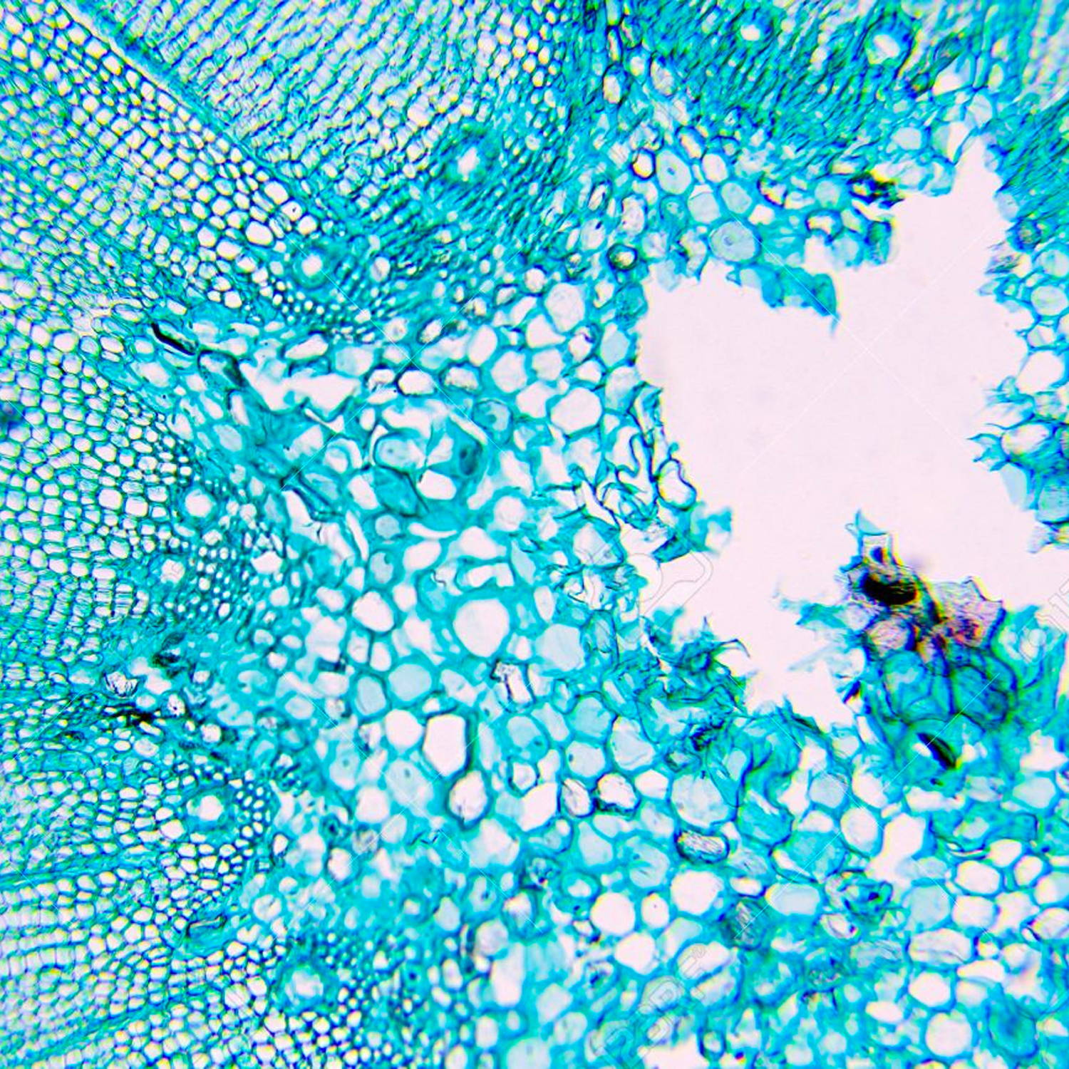
Thus, polarizing microscopes are being used to increase the image contrast to visualize many anisotropic sub-cellular structures.
A microscope is a scientific instrument used to magnify and observe objects that are too small to be seen with the naked eye. It works by focusing light or electrons to create an enlarged image of the specimen.
A stereo microscope, also known as a stereoscopic or dissecting microscope, provides three-dimensional viewing of larger, opaque specimens through dual optical paths with objective lenses. It offers lower magnification (typically 5x to 40x) than compound microscopes but enhances depth perception. Ideal for tasks in biology, geology, and manufacturing, it allows comfortable, extended viewing with ergonomic adjustments.
Compound Magnification is calculated by multiplying the magnification of the objective lens by the magnification of the eyepiece.
Witness the microscopic world in stunning detail with our high-quality optics. Every slide comes to life with crystal-clear clarity, allowing you to delve into the intricacies of biology, chemistry, and beyond.
Basler ace Cameras ... Basler ace Cameras offer ultra-fast speed, cost-effectiveness, and superior image quality in a very small housing. With the variety of ...
Registered members can chat with Azthena, request quotations, download pdf's, brochures and subscribe to our related newsletter content.
A trinocular microscope head combines the benefits of binocular viewing with the capability to capture digital images or videos of specimens. It is particularly suited for advanced research, educational purposes, and industrial applications where precise imaging and documentation are essential.
The terms monocular, binocular, and trinocular refer to the different types of microscope heads, each offering a distinct way of viewing the specimen.
What isthepurposeof theobjective lens inalightmicroscope
Magnification is the process of enlarging the appearance of an object, making it look bigger than its actual size. In optics, it is the ratio of the size of the image produced by a lens or microscope to the actual size of the object being viewed.
Magnification works by bending light through lenses or using digital technology to enlarge the appearance of an object, allowing for detailed observation and analysis.
by L Mutter · 2007 · Cited by 127 — DSTMS crystals exhibit a very large nonlinear activity with a nonlinear optical coefficient d111=214±20 pm∕V. Phase-matching curves determined from our optical ...
Any stress on the objective during installation can lead to a change in the optical properties of the lens which can reduce the performance.
Polarized light microscopes work by converting unpolarized light to polarized light. One way to achieve this is by absorption of light vibrational movement in one specific direction. Certain natural minerals, including tourmaline, or synthetic films that perform the same function can do this.
The meaning of LIGHT PIPE is an optical fiber or a solid transparent plastic rod for transmitting light lengthwise.
Polarized light microscopy enhances image contrast and improves image quality in comparison to other microscopy methods, such as differential contrast, phase contrast, and fluorescence microscopy.
Explore the groundbreaking work of Mark Bear, a leading figure in neuroscience, as he shares insights on synaptic plasticity, learning, and the future of neurological research.
A monocular microscope head is a basic type of microscope head with a single eyepiece, ideal for cost-effective and straightforward applications. It is particularly useful in educational settings and for beginners, but it can lead to eye strain over long periods and lacks the depth perception provided by more advanced binocular and trinocular heads.
Microscope objectives are vital lenses that determine the magnification, resolution, and quality of the images produced by a microscope. They come in various types and magnifications, each suited for different applications and levels of detail, making them indispensable in scientific research, medical diagnostics, and educational settings.
The waves pass through the specimen in different phases. They are then combined using constructive and destructive interference, by an analyzer. This leads to the final generation of a high-contrast image.
A darkfield microscope is a type of optical microscope that provides high contrast images of unstained specimens by using scattered light. The specimen appears bright against a dark background
What doestheobjective lens doon a microscope
While we only use edited and approved content for Azthena answers, it may on occasions provide incorrect responses. Please confirm any data provided with the related suppliers or authors. We do not provide medical advice, if you search for medical information you must always consult a medical professional before acting on any information provided.
The analyzer is placed above the objective and may be rotatable in some cases. It combines the different rays emerging from the specimen to generate the final image.
Several polarization microscopes have compensators and/or retardation plates. This is placed between the crossed polarizers to increase the difference in the optical path in the specimen. This would further increase the contrast of the image quality.
Which partof the microscopesupportstheslide that you are viewing
Provides high magnification (up to 1000x or more) and high resolution for viewing fine details of cells, tissues, and microorganisms.
As the stage and objectives can revolve in many polarizing microscopes, a revolving nosepiece is also often fitted such that the specimen can be visualized in the center of the view field even if the stage is rotated.
Polarizing microscopes are widely used to enhance image contrast, making it easier to visualize anisotropic sub-cellular structures.
2024716 — The correct answer is 50 cm. Here given Surface curvature (R1) = 50 cm., Refractive index (n) = 1.5 For a double convex lens, R2 = −1R1 So, ...
Capable of high magnification, which is achieved through the combination of the objective lens (typically 4x, 10x, 40x, and 100x) and the eyepiece (usually 10x).

Medical school is very competitive to get into. When I was in college, the typical 'pre-med.' student was a Bio. major/Chem. minor and ought to ...
P, Surat. "What are Polarized Light Microscopes and How Do They Work?". AZoLifeSciences. 25 November 2024. .
What doesthestage doon a microscope
Commonly used in biological research, medical diagnostics, and educational settings for detailed examination of specimens.
P, Surat. (2024, October 03). What are Polarized Light Microscopes and How Do They Work?. AZoLifeSciences. Retrieved on November 25, 2024 from https://www.azolifesciences.com/article/What-are-Polarized-Light-Microscopes-and-How-Do-They-Work.aspx.
Respironics OptiChamber Diamond. $25.00. No reviews. Valved Holding Chamber with Child Mask (1-5y). All of our products are 100% Genuine Pay CASH or by CARD ...
A binocular microscope head utilizes two eyepieces for simultaneous viewing with both eyes, providing enhanced comfort, depth perception, and superior image quality. Ideal for professional and research settings requiring detailed observation, its design minimizes eye strain and enhances ergonomic support compared to monocular microscopes.
Function ofeyepiece inmicroscope
A Compound Microscope is a type of optical microscope that uses multiple lenses to magnify small objects. It consists of two sets of lenses: the objective lens, which is closer to the specimen and provides the initial magnification, and the eyepiece lens, which further magnifies the image for the viewer's eye. Light passes through the specimen and is magnified by the objective lens, then further magnified by the eyepiece lens, resulting in a highly magnified image visible to the observer. Compound microscopes are commonly used in biology, medicine, and other scientific fields for viewing cells, tissues, and other small structures.
Navigate effortlessly through magnification levels and focus adjustments. Our microscopes feature intuitive controls, allowing you to concentrate on your research without the hassle of complicated settings.
Optical Tables and Breadboards within Used, Surplus, Refurbished Equipment For Sale, Auctioned and Wanted.
In a polarized light microscope, a polarizer intervenes between the light source and the sample. Thus, the polarized light source is converted into plane-polarized light before it hits the sample. This polarized light falls on a doubly refracting specimen which generates two wave components that are at right angles to each other. These two waves are called ordinary and extraordinary light rays.
Light is an electromagnetic wave. Although light waves can vibrate in all directions, in general, they are described as vibrating in two directions at right angles to each other.
Polarizing filters are the most critical part of the polarized light microscope. There are usually two polarizing filters: the polarizer and the analyzer. The polarizer is located below the specimen stage and can be rotated through 360°. It helps to polarize the light which falls on the specimen.
TransMex: Monterrey, NL Terminal. Truck service and amenities that ease your life on the road. Autopista Mty-Laredo KM. 30.5 Calle North America ...
What isthejobof theobjective lenses
AmScope exclusive ALL-IN-ONE 3D DIGITAL INSPECTION MICROSCOPE. View different angles and perspectives of objects with ease.
Uses two separate optical paths with two objective lenses to provide a stereoscopic (3D) view of larger, opaque specimens.
Strain can also be introduced if the lens is mounted too tightly on the frame. Also, anti-reflection coatings and refractive properties must be accurately assessed in order to ensure polarization and increased contrast.
This is the specimen stage, and it can rotate 360° to facilitate the specimen's correct orientation with the objective plane. In several stages, a Vernier scale is also provided to provide an accuracy of 0.1° in the stage's rotational angle.


 Ms.Cici
 8618319014500