
Right-Angle Prisms - Java Tutorial - right angle prism
These reflection probe bundles are made entirely of stainless steel. The patch cables have reinforced stainless steel tubing for durability, and the connectors have a metallic ferrule.
SEDI-ATI designs strong and reliable fiber-based solutions that are involved in current data security techniques through optical encryption.
SEDI-ATI took part in the INSPECT project by supplying a bundle of 16-channel optical fibers for instrumentation to measure the level of radioactivity in nuclear equipments.
Single mode fiber bundle
SEDI-ATI took part in the ITER project by producing a multi-channel base based on hermetic optical fibers for use in radiative and cryogenic environments.
Linear fiber array
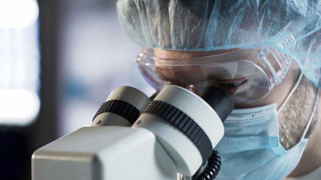
Most people have used some form of compound microscope before, usually in a science or biology class in school. Doctors and scientists use compound microscopes to look at bacteria, plant cells, animal cells, chromosomes and even thin layers or slices of organs and tissues. Many veterinarians use compound microscopes in a similar matter typically looking at animal cells, organs, tissues, bacteria and more.
DSLR Cameras are also known as Digital SLR Cameras. These cameras require two adapters, a c-mount adapter, and a camera-specific SLR adapter; working best on Trinocular microscopes because of the weight. DSLR cameras have the finest image quality and are a great option when needing to capture images of your subjects.
SEDI-ATI has developed a bundle of scintillating plastic optical fibers to do real-time detection of nuclear contaminants in drinking water.
Fused fiber bundle
Simple yet extremely functional, you can easily use your smartphone as a camera for your microscope with the phone mount. Phone mounts usually mount to the top of the scope. You would simply place your smartphone in the mount, so your camera is looking into the eyepiece. You should then see the specimen you looked at through the eyepiece on your smartphone, allowing you to photograph or video it.
Use an eyepiece camera by attaching the small camera to the eyepiece of your microscope. Using a USB port, the eyepiece camera will connect to a computer monitor where you can view and save the images of your microscopic material.
Stereo microscopes work with a sample in real-time and can really be used by anyone who needs to take a closer look at a small object. Veterinarians and their technicians generally use stereo microscopes for dissection or micro-surgery.
A stereo, stereoscopic, or dissecting microscope is an optical microscope designed for low-magnification observation. Stereo microscopes typically use reflected light that bounces off the surface of an object, rather than light being transmitted through an object.
Tapered fiber bundle
Once you know what specimens you will be looking at from day to day, you need to consider the different features every microscope offers. No microscopes nor their features are going to be the same. One main feature to look at when purchasing a new microscope is the head. Most commonly, you will find Monocular, Binocular, and Trinocular microscope heads. Another feature to consider is what focus the microscope comes with. Some scopes come with just a coarse focus dial while others have both a coarse and fine focus dial. Lastly, another common feature to consider is the light source.
What this really means, is that a stereo microscope shows a 3D or three-dimensional image of whatever microscopic material you are looking at. Commonly, the specimen is visible to the naked eye when using a stereo microscope.
SEDI-ATI took part in the INSPECT project by supplying a bundle of 16-channel optical fibers for instrumentation to measure the level of radioactivity in nuclear equipments.
Imaging fiber bundle
Choosing a new microscope for your veterinary practice? You’re likely weighing options between stereo and compound microscopes, while also considering camera compatibility and various other features. Selecting the right microscope is crucial but can be overwhelming. In Intriquip’s guide, we’ll highlight the differences and key factors to consider, helping you make an informed decision for your clinic. Let’s start by understanding what a stereo microscope is and how it differs from its compound counterpart..
SEDI-ATI has participated in the design of the LIFU and one MOS by the design, the setting up of manufacturing processes and the assembly of fibered sets.
Each leg uses Ø200 μm core silica/silica multimode fibers. The fibers are selected according to the desired wavelength range: VIS-NIR as a standard, or UV-VIS upon request.
Microscopes can have LED, Halogen, Tungsten/Incandescent, Fluorescent, or Natural/Sunlight to illuminate a subject. Again, many specimens require different light forms, so knowing what light source is going to work best for those subjects is necessary when purchasing a new microscope.
SEDI-ATI took part in the project by supplying specific patchcords, fiber-optic bundles, and feedthroughs with our FCXtreme® connector, a compact FC connector for defense and aerospace applications.
Another factor to consider is knowing who is going to be using the microscope and what qualifications or training they have. Microscopes can be relatively expensive and require adequate maintenance and cleaning to keep it running smoothly and for a long time. Someone without proper training may not realize this, which can cause damage to an expensive piece of equipment. Providing sufficient training on a new microscope to any and all staff who are going to be using it can help ensure a long lifetime for your microscope.
Fiber bundle sizes
SEDI-ATI took part in the project by supplying specific patchcords, fiber-optic bundles, and feedthroughs with our FCXtreme® connector, a compact FC connector for defense and aerospace applications.
SEDI-ATI supplies a set of high-directivity couplers and ruggedized fiber patchcords for in-line thickness control of glass bottles.
After determining what specimens you are going to be looking at from day to day, you’ll be better equipped in choosing a vet microscope with the necessary features. With the numerous options available, considering who will operate the microscope and their skillset, along with whether a microscope camera is essential, are crucial steps in the purchasing process.. Reach out to Intriquip today to talk more about what microscope is going to best suit your practice.
Both the light source leg and the detector leg are terminated with an ST or SMA connector for compatibility with most spectrometers and light sources.
When purchasing a new microscope, there are many things you need to consider to make sure you are getting the right scope for your practice. Knowing what application you need the microscope for is going to be the first crucial thing to consider.
SEDI-ATI has developed a bundle of scintillating plastic optical fibers to do real-time detection of nuclear contaminants in drinking water.
SEDI-ATI supplies a set of high-directivity couplers and ruggedized fiber patchcords for in-line thickness control of glass bottles.
Also known as a Charge-Coupled Device Camera, you can easily attach it to a Binocular microscope with a c-mount adapter or a Trinocular microscope with its built-in c-mount adapter. Similar to eyepiece cameras, CCD Cameras will connect to your computer monitor via a USB port. Unlike eyepiece cameras or phone mount cameras, CCD Cameras produce better image quality because of the higher megapixels.
Assembled optical fibre bundlesfor sale
SEDI-ATI has participated in the design of the LIFU and one MOS by the design, the setting up of manufacturing processes and the assembly of fibered sets.
Thorlabs fiber bundle
Compound microscopes obtain higher levels of magnification and help to diminish chromatic aberration. Compound microscopes use two lenses; one being found in the objectives and one in the eyepieces. Objectives on a compound microscope have a short focal length and are located quite close to the subject, aiding in collecting light to focus on the image.
The second lens, located in the eyepiece, has a much longer focal point. This allows the image to appear much more enlarged. Compound microscopes are often referred to as biological microscopes, phase contrast microscopes, polarizing microscopes, metallurgical microscopes and fluorescence microscopes. Breaking that down, compound microscopes are used to see a 2D or two-dimensional image.
SEDI-ATI designs strong and reliable fiber-based solutions that are involved in current data security techniques through optical encryption.
Some subjects require unique magnification, distinct lighting, and some even require a heated stage. Viewing specimens that require a microscope with particular features can be challenging or even impossible. Knowing roughly what material you will be viewing day to day can aid in your decision about what microscope is going to be the one for your clinic.
Microscope cameras record whatever specimen is being viewed under the microscope in real time. Images or recordings of your microscopic material can be displayed on a digital screen, stored for record-keeping, or shared over the internet in research papers or blog articles. Typically, being used on compound microscopes, there are four common types of microscope cameras.
We offer bifurcated fiber-optic reflection probe bundles for on/off sensors, with 100% inorganic materials. They are optimized for working at high temperatures up to +400 °C.
SEDI-ATI took part in the ITER project by producing a multi-channel base based on hermetic optical fibers for use in radiative and cryogenic environments.
Unlike stereo microscopes, compound microscopes use light to see in or through a subject which is called light transmission. Most compound microscopes have three or four objectives ranging in magnification from 4x, 10x, 40x, and 100x. Having the second lens in the eyepieces, the total magnification is 40x, 100x, 400x, and 1000x.


 Ms.Cici
 8618319014500