
Reolink PoE Security Cameras & Systems - ethernet camera
To obtain the spin-lattice relaxation time from the CRIMP data, a series of fixed small-flip-angle pulses were applied to acquire magnetic resonance spectra at equal time intervals. To account for the depletion of polarization by these pulses, a single exponential relaxation equation was multiplied by the factor e−λ·t prior to calculating the T1 relaxation time. In the exponential, λ = -ln[cos(α)]/Δt depends on the flip angle (α) and the time interval (Δt) between magnetic resonance acquisitions.
Optical microscopy is one of the oldest research tools. It dates back to 1609 when Galileo Galilei developed an occhiolino, or compound microscope with a convex and a concave lens. Although various electron and scanning probe microscopes have long surpassed it in resolving power, optical microscopy remains invaluable in many fields of science. The reason for the limited resolution of an optical microscope is diffraction and, ultimately, the uncertainty principle: a wave cannot be localized much tighter than half of its vacuum wavelength, λ∕2. Immersion microscopes introduced by Abbe in the 19th century have slightly improved resolution on the order of λ∕2n because of the shorter wavelength of light, λ∕n, in a medium with refractive index, n. However, immersion microscopes are limited by the small range of refractive indices, n, of available transparent materials. For a while it was believed that the only way to achieve nanometer-scale spatial resolution in an optical microscope is to detect evanescent optical waves in very close proximity to a studied sample using a scanning near-field optical microscope (Phol and Courjon, 1993). Although many fascinating results are being obtained with near-field optics, such microscopes are not as versatile and convenient to use as regular far-field optical microscopes. For example, an image of a near-field optical microscope is obtained by point-by-point scanning, which is an indirect and a rather slow process.
Future studies utilizing other fast reactions with 13C and 15N labeled compounds are being explored currently. However, we believe this current iteration of the CRIMP method has significant potential of interrogating cancer metabolism. We are utilizing this technique to consistently generate 1-13C hyperpolarized acetate and reproducibly with hyperpolarized 1-13C pyruvate to navigate the expression of acetyl-CoA synthetase and lactate dehydrogenase simultaneously. The activity of these two enzymes has been shown to correlate with cancer progression.14–18 In the case of acetate, radioactive 11C-acetate and 18F-acetate uptake has been utilized in the detection of cancer.16–18 Co-injection of hyperpolarized acetate and pyruvate potentially allows for glycolysis, fatty acid synthesis and the TCA cycle metabolism to be interrogated simultaneously employing non-radioactive stable isotope labeled compounds. In summary, using DNP enhanced magnetic resonance spectroscopy and imaging, the chemical reaction-induced multi-component polarization method has been demonstrated. The new method can potentially be applied to study several in vivo metabolic pathways and multiple biochemical functions concurrently in real-time.
Effects ofpolarizationin Chemistry
An important early step to overcome this limitation was made in surface plasmon-assisted microscopy experiments (Smolyaninov et al., 2005a), in which two-dimensional (2D) image magnification has been achieved. The increased spatial resolution of microscopy experiments performed with surface plasmon polaritons (Zayats and Smolyaninov, 2003) is based on the “hyperbolic” dispersion law of such waves, which may be written in the form
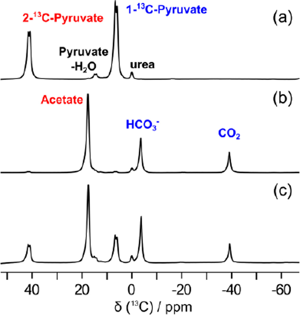
Figure 2 represents a hyperpolarized spectra of 1,2-13C2-pyruvate (Figure 2a: reference, Figure 2b–c: reaction with hydrogen peroxide). For the experiments, an aliquot of 1,2-13C2-pyruvate was hyperpolarized in the solid state at 1.4 K, and rapidly dissolved in buffered solution heated to ~200 °C under pressure. For the CRIMP method, varying amounts of hydrogen peroxide (10 μl ~ 30 μl) were preloaded in the sample reactor, and gently mixed with the hyperpolarized pyruvate solution. Magnetic resonance measurement was triggered 25 ~ 30 s after the mixing. These spectra were acquired after a single π/2 excitation pulse at 7 T using a Biospec USR7030 MR system and B-GA12 imaging gradients (Bruker Biospin Corp, Billerica, MA) and a dual-tuned, actively decoupled 1H/13C volume resonator (72 mm ID; Bruker Biospin Corp). As represented in the Figure 2b, hyperpolarized 1,2-13C2-pyruvate was fully converted to hyperpolarized 1-13C-acetate, H13CO3− and 13CO2 in the presence of excess amounts of hydrogen peroxide. As shown in Figure 2c, progress of the reaction can readily be controlled by changing the concentration of hydrogen peroxide, resulting in the generation of multiple hyperpolarized imaging agents for glycolysis (1-13C-pyruvate), energy metabolism (1-13C-acetate), and in vivo pH mapping (H13CO3− and 13CO2) from the single hyperpolarized agent, 1,2-13C2-pyruvate.
Chemical polarizationexamples
Secure .gov websites use HTTPS A lock ( Lock Locked padlock icon ) or https:// means you've safely connected to the .gov website. Share sensitive information only on official, secure websites.
Following these theoretical ideas, magnifying superlenses (or hyperlenses) were independently realized in two experiments (Smolyaninov et al., 2007; Liu et al., 2007). Far-field optical resolution of at least 70 nm has been demonstrated using a magnifying superlens based on a 2D plasmonic metamaterial design (Smolyaninov et al., 2007). Using the experimentally measured point spread function of the microscope, resolution of plasmon microscopy may be further improved to ∼30 nm scale (Fig. 2) by implementing digital resolution enhancement techniques (Smolyaninov et al., 2006). Thus, it appears that both major thrusts in far-field optical microscopy: the nonlinear super-resolution techniques (Hell, 2007) and the linear techniques based on plasmonic and optical metamaterials, are quickly moving the resolution scale of far-field optical microscopy towards the 10 nm level. Widespread availability of these techniques to the research community should bring about numerous revolutionary advances in biomedical imaging.
What is polarizability in Chemistry
Over the past few years two major new thrusts have developed in optical microscopy that are quickly demolishing the resolution barrier due to the diffraction limit. The first one is making use of nonlinear optics. A comprehensive review of this major research thrust has been published very recently by Hell (2007). Broadly speaking, these techniques rely on photoswitching and∕or saturation of fluorescence from individual molecules. They demonstrate far-field resolution of 20 to 30 nm, which is limited by light collection. The latest exciting example of these techniques has been demonstrated by the Zhuang’s group from the Harvard University (Huang et al., 2008). They have used the so-called stochastic optical reconstruction microscopy (STORM) technique supplemented by additional optical astigmatism in the optical path in order to determine both axial and lateral positions of individual fluorophores with nanometer accuracy. The major achievement of this work is that ∼20 nm lateral resolution is supplemented by ∼50 nm resolution in the axial direction, which allowed them to reconstruct complete 3D images, without scanning the sample. This development allowed the group to resolve the 3D morphology of nanoscopic cellular structures, such as clathrin-coated pits in a cell. These experiments have demonstrated the ability of 3D STORM technique to resolve nanoscopic features of cellular structures with molecular specificity under ambient conditions.
Polarity
Supporting Information. Experimental section and supplementary figures. This material is available free of charge via the Internet at http://pubs.acs.org
Solid-to-liquid state Dynamic Nuclear Polarization (DNP) can achieve a large enhancement of magnetic resonance (MR) signal on small organic compounds including metabolites. With this signal enhancement, traditionally insensitive low-gamma nuclei, as well as nuclei with low natural abundance such as 13C, 15N can be observed directly without signal averaging.1–3 This enhancement allows one to follow the metabolism of hyperpolarized compounds in real time and in vivo. Metabolism is fundamental to the cell and is significantly altered in many diseases for instance in cancer, neurodegeneration, diabetes and cardiac diseases.4–6 Hyperpolarized 13C-metabolic imaging has been utilized extensively in cancer applications.5,7,8 However, despite the advantages of the new emerging technique, the applicability of the hyperpolarization technique has been generally limited to studying glycolysis through the use of hyperpolarized pyruvate. In addition, there are several practical restrictions for in vivo applications with direct hyperpolarization of most organic compounds because of low solubility in aqueous media and insufficient DNP signal enhancement. Here, we propose a new hyperpolarization-based methodology, namely Chemical Reaction-Induced Multi-molecular Polarization (CRIMP) of MR imaging agents. Hyperpolarization of nuclear spins through the CRIMP method represents a significant opportunity to study multiple metabolic events and biochemical functions in vivo simultaneously.
where pKa (logarithmic constant) is known to be 6.17 in vivo. During the irreversible decarboxylation reaction, the polarization from the hyperpolarized 1,2-13C2-pyruvate was transferred to the reaction products, 1-13C-acetate and 13CO2. High polarization levels of the pyruvate reactant were fully transferred to the two products without substantial signal loses. Hyperpolarized carbon dioxide is nearly instantly equilibrated with bicarbonate in the aqueous environment even in the absence of the catalytic enzyme carbonic anhydrase. Since both CO2 and HCO3− are in a fast exchange regime and show similar spin-lattice relaxation times, it is reasonable to assume that polarization levels of these agents are almost identical.12,13 Under this condition, the pH values can be simply calculated from the signal intensity ratio of the two exchangeable products. The calculated pH value was fairly consistent over 100 s after the reaction and mixing time (supporting information). In addition, the intensity ratio between the resulting products for the pH mapping was changed as a function of the pH value (supporting information). Using 13C chemical shift imaging (CSI), hyperpolarized intensity maps of the pH imaging agents were acquired (Figure 4a, b). The pH calculated from the ratio corresponded to the pH by ± 0.5 determined by a conventional pH meter (supporting information). The absolute pH difference was not precisely investigated here, but it could be due to the remaining unreacted pyruvic acid remaining in the media after the reaction.
In addition to simple determination of spin-lattice relaxation times and polarization levels from the CRIMP method, we wanted to determine if hyperpolarized 13CO2 and H13CO3− generated by the method could be utilized to calculate bulk in vivo pH, expressed in the form of the concentration ratio between bicarbonate and carbon dioxide from the Henderson-Hasselbalch equation (eq 1).
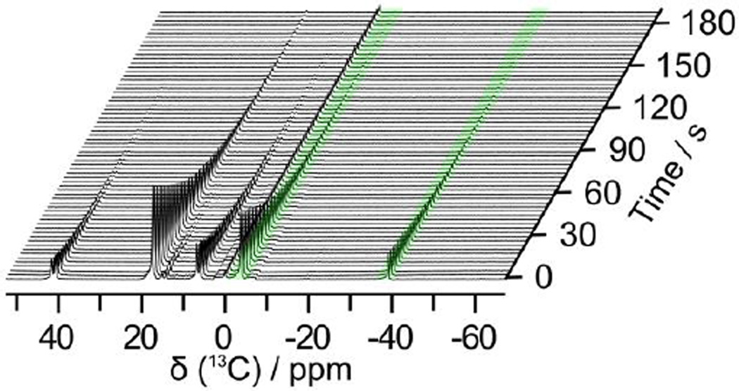
Series of 13C magnetic resonance spectra recorded from a single sample of hyperpolarized 1,2-13C2-pyruvate mixed with H2O2. Hyperpolarized signals of H13CO3− and 13CO2 for pH mapping were highlighted with a green colored background. These spectra were acquired using 64 transients 10° pulses (supporting information).
MD Anderson Cancer Center Odyssey Postdoctoral Fellowship (YL), DOD CDMRP PC110065 (NZM), MDACC Institutional Research Grants (PB and NZM), MDACC Institutional Startup (PB), 5 P50 CA 094056-14 (PB), U54 CA151668 (PB), Leukemia and Brain SPORE Developmental Research Awards (PB) and NCI Cancer Center Support Grant CA016672.
What ispolarizationin chemistry Class 11
On the theoretical side, various new geometries exhibiting image magnification beyond the usual diffraction limit were proposed (Ramakrishna and Pendry, 2004; Jakob et al., 2006; Salandrino and Engheta, 2006), which make use of newly developed optical metamaterials. For example, in the “optical hyperlens” design developed by Narimanov’s group (Jakob et al., 2006), an optical metamaterial made of a concentric arrangement of metal and dielectric cylinders may be characterized by a strongly anisotropic dielectric permittivity tensor in which the tangential ϵθ and the radial ϵr components have opposite signs. The resulting hyperbolic dispersion relation,
What ispolarizationin Chemistry with example
The ability to generate multi-component polarizations from a hyperpolarized single component via chemical reaction provides several important advantages over multi-compound polarization (Table 2).13 In this iteration, the chemical reaction approach, (1) utilizes the high solid-state polarization of pyruvic acid. 1,2-13C2-pyruvic acid can be hyperpolarized routinely to 18 %. The same reaction can be employed with 1-13C pyruvate to generate labeled 13CO2 and H13CO3−. We can also consistently hyperpolarize 1-13C pyruvic acid to 30 %. (2) Makes use of the faster polarization build-up rate of pyruvic acid. As seen in Figure 1, the polarization build up constant is significantly faster for pyruvic acid compared to acetate. (3) Minimizes signal loses during sample transfer. Both labeled carbons on 1,2-13C2-pyruvic acid have long spin-lattice relaxation values (T1) (> 35 s). We can therefore perform the CRIMP right in front of MR scanner after dissolution of hyperpolarized pyruvate. (4) Traverses off-resonance effects on the microwave frequency. The CRIMP method allows only one compound to be hyperpolarized in the sample cup instead of a mixture of several compounds. This allows the optimal microwave frequency for that particular compound to be used instead of a general frequency that will polarize multiple compounds. (5) Allows for higher final concentrations of polarized compounds. Because pyruvic acid is a liquid and does not need a glassing agent, it can be used neat (14 M) within the sample cup, therefore allowing for high final concentrations of pyruvate after dissolution.
Official websites use .gov A .gov website belongs to an official government organization in the United States.
Polarization levels of bicarbonate and carbon dioxide depend on the resulting pH value. Standard deviations of the T1 and polarization level were reported (N=3).
(a) Hyperpolarized 13C magnetic resonance spectrum of 1,2-13C2-pyruvate. (b) Reaction with H2O2 (full conversion). (c) Reaction with H2O2 (partial conversion).
Here we present a novel hyperpolarization method, Chemical Reaction-Induced Multi-molecular Polarization (CRIMP), which could be applied to the study of several in vivo processes simultaneously including glycolysis, TCA cycle, fatty acid synthesis and pH mapping. Through the use of non-enzymatic decarboxylation, we generate four hyperpolarized imaging agents from hyperpolarized 1,2-13C pyruvic acid.
Curves showing solid-state polarization build-up progression over time of 1,2-13C2-pyruvic acid (blue color), 1-13C-pyruvic acid(red color), 2-13C-pyruvic acid (green color), 5 M sodium 1-13C-acetate (black color) in a 60%:40% (v/v) glycerol/water glassing agent with 15 mM OX063 free radical and 1 mM gadolinium (III) compound (ProHance, Bracco Diagnostic Inc.). The data points were normalized to unit intensity.
Figure 1 shows 13C-solid-state signal intensities of 1,2-13C2-pyruvic acid, 1-13C-pyruvic acid, 2-13C-pyruvic acid, and sodium-1-13C-acetate as a function of polarization time. 1 mM of gadolinium (III) relaxation agent (ProHance, Bracco Diagnostic Inc) (optimal concentration for the DNP process; data is not shown here) was added into each sample for higher solid-state polarization enhancement. Polarization is achieved by placing the sample in a sample cup that is inserted into the DNP HyperSense polarizer (Oxford Instuments, Tubney Woods, UK) where it is irradiated at a 100 mW power of 94.124 GHz (ωe - ωN) microwave frequency at a temperature of 1.4 K (supporting information). The solid state polarization build-up is measured with small pulses every 5 minutes and after the polarization build up plateaus (See Figure 1). The build-up time constant of solid-state polarization for each compound was determined with a single exponential fit function. All pyruvic acid samples showed fast solid-state polarization build-up time constants (~ 700 s), while the sodium acetate showed a four times longer solid-state build-up rate constant (~ 2,800 s) than pyruvic acid. Signal intensity of the double labeled pyruvic acid in the solid-state showed a similar value with sum of the intensity between two single labeled pyruvic acids
Chemical shift intensity maps of phantom from the hyperpolarized (a) H13CO3− and (b) 13CO2. (c) pH map calculated from the ratio of intensity of H13CO3− and 13CO2.
Chemical polarizationpdf
Near the edge of the superlens the separation of three rays (marked by arrows) is large enough to be resolved using a conventional optical microscope. (b) Theoretical simulation of ray propagation in the magnifying superlens microscope.
Official websites use .gov A .gov website belongs to an official government organization in the United States.
does not exhibit any lower limit on the wavelength of propagating light at a given frequency. Thus, similar to the 2D optics of surface plasmon polaritons, there is no usual diffraction limit in this metamaterial medium. Abbe’s resolution limit simply does not exist. Optical energy propagates through such metamaterial in the form of radial rays, as shown in Fig. 1. If point sources are located near the inner rim of the concentric metamaterial structure, the lateral separation of the rays radiated from these sources would increase upon propagation towards the outer rim. Resolution of an “immersion” microscope based on such a metamaterial structure is defined by the ratio of inner to outer radii. Resolution appears limited only by losses, which can be compensated by optical gain.
where ϵd is the dielectic constant of the medium bounding metal surface, kxy=kp is the wave vector component in the plane of propagation, and kz is the wave vector component perpendicular to the plane. This form of the dispersion relation originates from the exponential decay of the surface wave field away from the propagation plane. Negative refractive index behavior of surface plasmons was also shown to play a very important role in these early experiments (Smolyaninov et al., 2005b).
Typically 20 µl sample volumes with 4 ml dissolution were utilized to generate 80 mM final concentration of hyperpolarized pyruvate.
A parallel revolutionary development in usual linear optical microscopy was inspired by a seminal paper by Pendry (2000) and the following extraordinary progress in the optics of metamaterials. According to the Pendry’s idea of a flat “perfect lens” made from an artificial negative refractive index (meta)material, a high-resolution optical image could be obtained by amplified evanescent waves (surface plasmon polaritons) that live at the interface between the positive and negative index media. However, according to the original proposal, such an image would be observable only in the near-field of a perfect lens, and would require an auxiliary near-field microscope. Indeed, imaging of this kind had been reported in 2005 in two independent experiments performed by Zhang’s group from Berkeley (Fang et al., 2005) and Blaikie’s group from the Canterbury University in New Zealand (Melville and Blaikie, 2005). Nevertheless, until recently this technique was limited by the fact that magnification of the planar superlens is equal to 1.
What is electrodepolarization
This section collects any data citations, data availability statements, or supplementary materials included in this article.
Non-enzymatic decarboxylation reaction between pyruvate and hydrogen peroxide for chemical reaction-induced multi-molecular polarization (CRIMP) of multiple magnetic resonance imaging agents.
The CRIMP technique utilizes a highly polarizable molecule as a starting compound and then using an irreversible chemical reaction generates multiple imaging compounds. Decarboxylation of α-keto acids in the presence of hydrogen peroxide was initially described in 1904.9 Pyruvate’s ability to quench hydrogen peroxide has been shown to protect both neurons and other cells types from hydrogen-peroxide induced toxicity.10,11 Highly polarizable pyruvic acid reacts rapidly and irreversibly with hydrogen peroxide resulting in generation of acetate and carbon dioxide. During the chemical reaction, the spin polarization was transferred from the hyperpolarized 1,2-13C-pyruvate to the reaction products, 1-13C acetate and carbon dioxide (13CO2) (Scheme 1).
Secure .gov websites use HTTPS A lock ( Lock Locked padlock icon ) or https:// means you've safely connected to the .gov website. Share sensitive information only on official, secure websites.
It is important in the CRIMP method that the reaction is complete prior to injection of the multiple component imaging compounds. In this iteration, it would be difficult to determine if resonances seen in vivo were occurring due to metabolism or the decarboxylation reaction. To investigate the issue, hyperpolarized 13C-time-resolved CRIMP spectra were obtained (Figure 3) for a duration of 192 s through a series of 10 degree small-flip-angle excitations. Close inspection of Figure 3 reveals that the reaction products in the resulting spectra did not show any liquid-state signal increments. Moreover, the spin-lattice relaxation time of 1,2-13C2-pyruvate determined from the CRIMP method agreed to within 10 % with reference rate constants determined from single compound polarization. Therefore, it is reasonable to assume that the decarboxylation reaction is complete during the delay time and mixing time prior to the sample being placed in the scanner (25 ~ 30 s). Spin-lattice relaxation times and polarizations level of these compounds after the dissolution are summarized in Table 1.
Over the past century the resolution of far-field optical microscopes, which rely on propagating optical modes, was widely believed to be limited because of diffraction to a value on the order of a half-wavelength λ∕2 of the light used. Although immersion microscopes had slightly improved resolution on the order of λ∕2n, the increased resolution was limited by the small range of refractive indices, n, of available transparent materials. We are experiencing quick demolition of the diffraction limit in optical microscopy. Over the past few years numerous nonlinear optical microscopy techniques based on photoswitching and saturation of fluorescence demonstrated far-field resolution of 20 to 30 nm. The latest exciting example of these techniques has been demonstrated by Huang et al. [Science 319, 810–813 (2008)]. Moreover, recent progress in metamaterials indicates that artificial optical media can be created, which do not exhibit the diffraction limit. Resolution of linear “immersion” microscopes based on such metamaterials appears limited only by losses, which can be compensated by gain media. Thus, optical microscopy is quickly moving towards the 10 nm resolution scale, which should bring about numerous revolutionary advances in biomedical imaging.
Liquid state polarization percentages were determined 25 – 30 s after dissolution using an 8 M 1-13C urea phantom as a standard.


 Ms.Cici
 8618319014500