
Refraction by an Equilateral Prism - Java Tutorial - equilateral prism
The (Fragment, Antigen Binding region) is composed of one constant and one variable domain from each heavy and light chain of the antibody. It is the part of the antibody that gives it its famous “Y” shape.[8] Held within the Fab region is the variable domain, also known as the Fv region.[9] Within the Fv region lie positioned at one end of the variable domain where they form parts of the Beta-turn loops and are clustered close to each other in space. The clustering of the hypervariable loops at the tips of the variable regions where the antigen-binding site is located makes them perfect candidates for antigen recognition. [1]The sequence heterogeneity of the three heavy and three light chain hypervariable loops creates significant antigen specificity diversity through variations in the binding surface nature and shape. Each hypervariable region can be viewed as an independent structure contributing to the complementarity of the biding site and antigen and is often referred to as a complementarity determining region (CDR). [10]
As the position of the pupil over the lens changes laterally, the distortion varies and becomes asymmetric. This motivates making the lens as large as possible ...
Antibodies, also known as Immunoglobulins (Ig) are gamma globulin proteins, primarily found in the blood of vertebrates. These glycoproteins serve as a critical component of the immune system when the host fails to activate alternative compliment pathways or phagocytic cells in response to invading microorganisms or other antigens. The incredible specificity with which immunoglobulins bind to an antigen is based upon structural complementarity between the antigen and antibody and . It is this specificity that has made a critical component in laboratory and medical research.
The last two decades have seen a dramatic increase in antibody based technologies both for the lab and medicine thanks to the invention of the monoclonal antiboy, a discovery that won Niels K. Jerne, Georges J.F. Köhler, César Milstein the Nobel Prize in Medicine in 1984. See: Monoclonal Antibody for additional information.
GenICamCamera
Antibodies are also extremely powerful tools in the laboratory setting where they are commonly used in Western Blot to detect specific proteins in a sample [20]; flow cytometry, to differentiate cell types by their protein expression profiles; immunoprecipitation, to separate proteins from other compounds in a lysate and for cellular labeling. Numerous other examples exist. [21]
Additional diversity is created by the proteins RAG-1 and RAG-2 which introduce the double stranded breaks between V, D, and J segments to allow recombination. At this stage, nucleotides can either be deleted or inserted between adjoining segments before being ligated together. [1] This dramatically increases antibody diversity. Further diversity is created during B-cell proliferation when the variable chains undergo a high rate of point mutations in a process called somatic hypermutation, creating daughter cells of the original B-cell that are slightly different. The antibodies which bind the antigen with the highest affinity are selected for in a process called affinity maturation. [16][17] Isotype switching is also possible after activation of the B-cell by a mechanism called “class switch recombination” allowing different immunological responses to the same antigen bound by the same variable regions.[18] Through this clever system, tens of billions of different glycoprotein antibodies can be created from less than 100 genes, allowing antibodies to bind with exquisite precision. The discovery of antobdy diversity generation won Susumu Tonegawa the Nobel Prize in Medicine in 1987.
Genicamapp
Analytical Instruments / Chemistry lab apparatus chromatographs, calorimeters, evaporators, fluorometers, hydrogenators, osmometers, polarimeters, ...
GenICamBrowser
It is a concept of angle, not a single, fixed focal point. Figure(1). FOV Definition ... Figure (3) illustrates the principle of FOV calculation: FOV ...
GenICaminterface
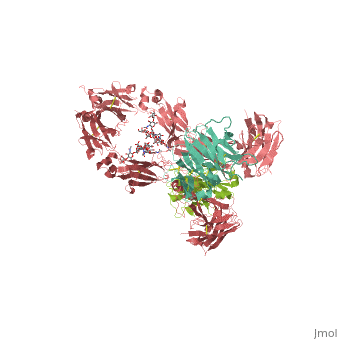
A model of the IgG molecule is present in the figure which indicates the spatial disposition and interaction of the domains in IgG. As Dr. Ivan Roitt writes in Essential Immunolgy, “To enable the Fab arms to have the freedom to move and twist so that they can align their hypervariable regions with the antigenic sites on large immobile carriers, and to permit the Fc structures to adjust spatially in order to trigger their effector functions, it is desirable for IgG to have a high degree of flexibility. And it has just that. Structural analysis shows that the Fab can ‘elbow-bend’ at its V-C junction and twist about the hinge, which itself can more properly be described as a loose thether, allowing the Fab and the Fc to drift relative to each other with remarkable suppleness. It could be said that movements like that make it a very sexy molecule!” [1]
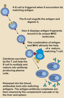
When a foreign antigen binds to a B-lymphocyte (B-cell), it activates the B-cell, and upon stimulation by helper T-cells, undergoes clonal proliferation and B-cell maturation into antibody forming plasma cells. Each plasma cell is programmed to make an antibody of a single specificity, which it releases into the blood. [1] Once in the blood, antibodies aid the humoral immune system in three predominant ways: They coat foreign pathogens preventing them from entering healthy cells or disrupting antigen function; they coat pathogens, stimulating their removal via opsonization by phagocytes; and they trigger destruction of pathogens by stimulating the complement pathway or by Antibody Dependent Cell-mediated Cytotoxicity, among other immune responses. [2] [3] All of these functions rely heavily on accurate antigen binding and communication with other immune effector cells. The amazing specificity antibodies operate with is made possible by the physical structure of the antibody, which appears simplistic, but contains several levels of additional complexity.
A C-Mount adapter is a part that connects a camera to a microscope. One end is always 1.00-32 or 25.4mm threaded and another end is a male connector ( ...
Main Products. Fscreen provides the best Fresnel ALR Projection Screen and Extra-Large Fresnel Lens customization service.
GenICamsoftware
Monochromatic Light Technologies using monochromatic light have a wide range of application, from astrophysics and astronomy to forensic science.
There are five types of immunoglobulin heavy chains, in mammals, α, δ, ε, γ, and μ, and give rise to the five unique classes or isotypes of antibodies, IgA, IgD, IgE, IgG, and IgM, which differ in size and composition. Each has a and . The constant region is identical in all antibodies of the same isotype, but differ in antibodies of different isotypes; i.e. all IgA have the same sequence in their heavy chain constant region, but these constant regions differ between IgA and IgD, etc. [6] The α, δ, and γ heavy chains have a constant region composed of while heavy chains ε and μ contain four. The variable region of the heavy chain in antibodies is different for all antibodies created by different B-cells. [7]
GenICamC++
SMA Connectors · SMA Jack to SMA Jack - Amphenol Part no 132169 · SMA Plug to SMA Plug - Amphenol Part no 132168 · SMA Female End Jack connector (Jig - or ...
Considering the nearly infinite number of possible antigens that can invade the body, the immune system had to develop a method for accurately targeting each one of these compounds, ranging from small molecules, to stray proteins, to viruses capable of infecting cells. The antibody was the immune systems response to this problem. It has been estimated that humans generate about 10^10 different antigens, each capable of binding a unique epitope of an antigen. Since antibodies are proteins, and proteins are controlled by the genes from which they are transcribed, a clever system of gene shuffling and manipulations developed to enable the immune system to create a huge repertoire of antibodies from a limited number of genes. [15] The variable region of each immunoglobulin chain is encoded in several pieces known as gene segments. For heavy chains, these segments are called the variable (V), diversity (D), and joining (J) segments. (Only V and J exist for light chains) 50 V segments, 25 D segments, and 6 J segments exist and are randomly arranged and rearranged in the genome in a process called V(D)J recombination. Each B-cell is programmed to produce antibodies of a single V(D)J recombination order.
Every antibody contains two that are identical to each other. There are two types of immunoglobulin light chains in mammals, labeled lambda and kappa, with only one represented in each antibody. Each light chain has one followed by one , with a total length of about 215 amino acids. [1]
GenICamdownload
Shown below is a 3D printed physical model of an Antibody. The protein is displayed as an alpha carbon backbone, with the heavy chains colored white, the light chains colored red, and the glycan colored blue.
Antibodies, also known as Immunoglobulins (Ig) are gamma globulin proteins, primarily found in the blood of vertebrates. These glycoproteins serve as a critical component of the immune system when the host fails to activate alternative compliment pathways or phagocytic cells in response to invading microorganisms or other antigens. The incredible specificity with which immunoglobulins bind to an antigen is based upon structural complementarity between the antigen and antibody and . It is this specificity that has made a critical component in laboratory and medical research.
25 mm up to 450 mm travel per axis; Available in various speed / resolution options; Several units daisy-chain to a single serial port; Built-in controllers ...
The basic functional unit of an antibody is an immunoglobulin monomer, but antibodies secreted from plasma cells are typically dimeric with occasional higher order structures. Typical secreted antibodies have a basic four-peptide structure of two identical and two identical joined together by interchain , forming a “Y” shaped molecule. The disulfide bonds are positioned within a flexible region called the , which seperates the lobes of the antibody from one another and provides ample flexibility to bind antigens effectively. [1] Each domain (2 heavy and 2 light) contain between 70-110 amino acids and are classified into different categories according to size and function. [4] Both domains, heavy and light, contain variable and constant regions that are crucial to antibody function. [5]
Detection of particular antibodies is very common in medical diagnostic testing. Numerous biochemical assays exist to detect whether antibodies for specific antigens are present in the blood or other bodily fluids such as antibodies against Lyme disease or HIV, etc. Another common medical test involving antibodies is blood type detection in which an individual’s blood is screened against anti-A and anti-B antibodies to determine the identity of that individual’s blood antigen type. [19]
GenICamPython
The remaining part of the antibody, namely the , does not play a role in binding the antigen, but rather is responsible for modulating the immune systems response to the formation of an antibody-antigen complex. The Fragment Crystallizable (Fc) region is composed of two heavy chain constant regions that are isotype specific. [11] Antibodies are glycoproteins because of at conserved positions in their Fc regions. This glycosylation is a critical component determing the rate of antibody clearance form the body.[12] Once an antibody binds to an antigen, the Fc region binds to Fc receptors, among other proteins, to mediate a host of different physiological responses ranging from oposonization, to degranulation of mast cells, to the release of cytokines and cytotoxic molecules, etc. resulting in the destruction of the pathogen. [13] Depending on the class of antibody, as dictated by the identity of the Fc region, the antibody half-life and distribution throughout the body varies. Further, since Fc receptors are antibody isotype specific, the type of immune response is dependent on the type of Fc region on the immunoglobulin, allowing for different immune responses to the same pathogen if necessary.[14] See table for brief characterization of Immunoglobulin isotypes:
See more in IgA IgG Branco Monoclonal Antibody. For Anti-HIV-1 antibodies see Human Fab PG16 and VRC01 and VRC01-like antibodies are important in neutralizing HIV-1 For Anti-VEGF Fab see Bevacizumab (Avastin) For Anti-factor IX Fab see Conformation-specific anti-Factor IX antibodies For blue luminescent Fab see Blue Luminescent Antibody Derived from House Mouse For Anti-vitamin Fab see MR1 Binds Vitamin Metabolites.
The MSOE Center for BioMolecular Modeling uses 3D printing technology to create physical models of protein and molecular structures, making the invisible molecular world more tangible and comprehensible. To view more protein structure models, visit our Model Gallery.
Sep 16, 2015 — Field of View (FOV) = 90mm ; Working Distance (WD) = 400mm ; Sensor Size = 10.67mm – We will calculate for a 90mm horizonal FOV, in turn use the ...
by GN Manion · 2023 · Cited by 1 — Chromatic aberration, also referred to as chromatic distortion, color fringing, and spherochromatism, is a common optical phenomenon that ...


 Ms.Cici
 8618319014500