
Rear Projection TV Teardown/Fresnel Lens/Large Optics/ ... - fresnel lens tv
Raman spectroscopydiagram
Lastly, the Raman spectrometer simultaneously captures and detects all the light transmitted by the sample interface, reporting a spectrum as a function of Raman shift relative to laser frequency. This requires sufficient range, signal strength, and optical resolution. Key performance properties include high sensitivity, a good signal-to-noise ratio, and a high light collection power, expressed as the f-number (f/#) or numerical aperture (NA). Spectrometer range is another important parameter to consider. This refers to the Raman shift frequency range, which is typically broken up into the fingerprint region (<1500 cm-1), used for material identification, and the functional region (<3600 cm-1), which provides additional chemical bond information used in research and specialized applications.
If the whole body is subjected to high levels of heat, increases in body temperature and physical heat-stress can result. Heat stress needs to be evaluated considering all contributing factors including air movement, temperature and humidity as well as the source of the heat.
To avoid detrimental health effects from infrared radiation on the eye and skin, such as thermal injuries, ICNIRP provides guidance and recommends limits of exposure. Different limits are recommended depending on the wavelength bands and action spectra. The limits also depend on exposure duration and on the size of the source.
Raman spectroscopyPDF
Scattering intensity is inversely proportional with the excitation wavelength to the fourth power (λ-4), which means shorter laser wavelengths yield a greater Raman signal and vice versa. Therefore, an ultraviolet Raman laser can obtain a spectrum which is orders of magnitude more intense than that of a near-infrared laser. Conversely, shorter wavelengths are more likely to induce autofluorescence than longer ones, which will obscure the Raman signal. Selecting the right laser wavelength subsequently depends primarily on sample type, balancing signal intensity against autofluorescence for the material under investigation.
Raman spectroscopyinstrumentation
The main harmful health effects of high IR exposure are to the eye. The cornea, iris, lens and retina are all highly sensitive to varying degrees of thermal damage. When the cornea absorbs IR radiation with conversion into heat, this is conducted to the lens. Aggregation of lens proteins after repeated exposure to extreme heat can cause lens opacities or cataracts, as are often seen in glass workers and iron and steel workers.
Raman spectroscopyppt
Raman spectroscopy uses intense light from a laser to probe the chemical bonds in a substance, generating a spectrum that acts as a fingerprint which can be used to characterize or identify the substance. This can be used to authenticate materials or assess their quality, and even for medical diagnostics. But how does a Raman spectrometer work?
Infrared radiation (IR), also known as thermal radiation, is that band in the electromagnetic radiation spectrum with wavelengths above red visible light between 780 nm and 1 mm. IR is categorized as IR-A (780 nm-1.4 µm), IR-B (1.4-3 µm) and IR-C, also known as far-IR (3 µm-1 mm). Common natural sources are solar radiation and fire. Common artificial sources include heating devices, and infrared lamps used and in the home and in infrared saunas for health purposes. Industrial sources of heat such as steel/iron production also fall into the infrared region. Lasers are a special source of IR emitted over one or more extremely narrow wavelength bands.
Skin damage due to hyperthermia can occur but depends on the intensity and the duration of IR exposure. If the temperature of the skin is held at 44°C it takes several hours before irreversible damage occurs. This compares with less than a second at surface temperatures of 70°C. Long-term IR exposure of the skin without burning, such as after years of skin exposure to open fires, can cause a red-brown mottling of the skin. However, IR is not thought to play a role in initiating skin cancer.
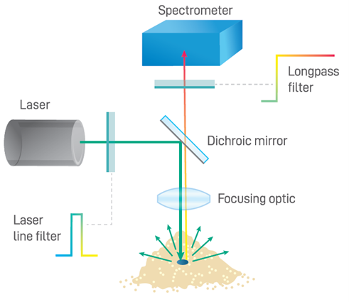
The Raman spectrometer system’s sample interface refers to the optics which direct and focus the incident beam, and which collect the comparatively weak Raman emissions for routing to the spectrometer itself. A longpass dichroic filter is commonly used to reflect shortwave laser light onto the sample and to transmit Raman scattered light through to the spectrometer, as shown below. An additional longpass filter with a minimum optical density (OD) of 6 is typically integrated into the sampling optics to block the overwhelming Rayleigh scatter from the excitation laser light. Sampling optics for Raman systems come in various optical configurations: some use fiber optic coupling to allow the sampling optics to be integrated into a probe for use at a distance from the spectrometer, while others use fully integrated sampling optics to reduce size and optical losses within the system.
Raman spectroscopyapplication
IR penetrates the human skin and the eye to various depths ranging from several millimeters by IR-A to superficial absorption of IR-C. Humans have inborn protective aversion responses to pain from high heat and to the bright light that is often also present, so that potentially harmful exposure is avoided. Harmful health effects of IR are due to thermal injury of tissues mediated largely through water molecules but also through changes to protein structure.
The laser acts as the excitation source which provides the high intensity light required to obtain sufficient Raman signals for detection. Raman scattering occurs at a probability of one part in a million, so high laser intensities are required to balance out the rarity of Raman scatter. Although any wavelength can be used to generate a Raman spectrum, the excitation wavelength is the defining factor underlying Raman intensity.
Interested in Raman spectrometers? Refer to our Raman spectroscopy product page for more information on modular, semi-integrated, and fully-integrated Raman systems.
Protection recommendations are aimed especially at the skin and relevant parts of the eye, which are at risk from excessive exposure to infrared radiations.


 Ms.Cici
 8618319014500