
Raman Scattering - Raman Spectroscopy, Applications of ... - raman scattering effect
1. Eyepiece or Ocular lens: Eyepiece lenses are attached to the top end of the body tube and have a small amount of magnification of their own. The magnified image is seen through this lens.2. Objective lens: Objective lenses are the closest to the slide and are embedded in the nosepiece. They can be of varying magnification. Light enters into the body tube through the objective lens.3. Mirror: Many microscopes do not come with a light source. Hence, the user has to rely on an outer source of light (e.g., sunlight). We adjust the mirror to align the source of light.Bonus: Although compound microscopes are easy to transport, they can never fit in a pocket. Foldscopes are origami-based microscopes that are small enough to fit in a pocket. These are made out of paper, and the lenses are 3D printed. According to creator Manu Prakash, “The capabilities of Foldscope are equivalent to conventional microscopes that cost thousands of dollars.”
Magnifyinglensglasses
Essentially, a microscope is a series of magnifying glasses housed in a tube. Because there are more lenses, and the lenses have varied magnifying power, microscopes can show an incredible amount of detail.
In these situations, corrective lenses - either in the form of glasses or contact lenses - are added on top of the eye to help properly focus and represent images of things around you.
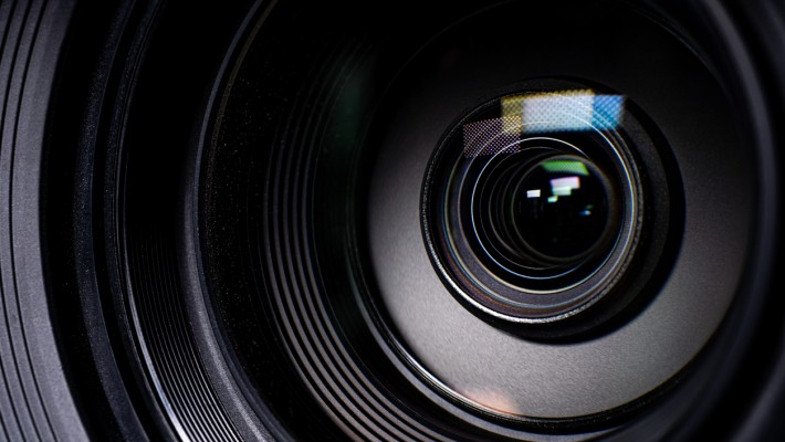
All the lenses inside the camera have to work together to focus on the image to create the clearest, most true representation of whatever you’re taking a picture of, or else you end up with a blurry photo. When the lenses work together properly, you get a sharp, clear photo to help you remember a moment.
In their long tubes, telescopes house lenses with powerful magnification properties that allow scientists and space enthusiasts here on Earth to see distant planets, galaxies, and objects floating millions of light years away from us.
Magnifyinglenscamera
6. Condenser lens: Some microscopes have a light source under the diaphragm. Condenser lens focuses the light on the specimen to render a detailed image. However, it is mainly found in microscopes with 400x magnification.
4. Rotating nosepiece: The nosepiece is connected to the lower end of the body tube. The objective lenses are embedded in this nosepiece. We can change the magnification by rotating the nosepiece.
Copyright @smorescience. All rights reserved. Do not copy, cite, publish, or distribute this content without permission.
Magnifyinglensconcave or convex
Without the microscope, we could have never detected that germs cause diseases and food spoilage. We would have been in the dark about the presence of cells in our bodies. A whole other world in biology would have been left undiscovered without microscopes.
Thanks to microscopes, scientists have discovered microscopic organisms such as bacteria, and they’ve been able to see small parts of tiny organisms, such as the heart cavities of insects and microorganisms.
MagnifyingLensprice
Magnifyinglensapp
Arm: Arm connects the head to the base of the microscope. One can hold the arm and carry the microscope to the workstation.
The aperture is an opening that lets light into the camera. The lenses are then moved back and forth to help the camera focus on the image you’re trying to capture, then the final image is captured when you press the shutter button.
Individual parts of a microscope are further divided into mechanical and optical parts. Mechanical parts are mainly used to maneuver the mounted slide and focus the lenses on the specimen. Optical parts consist of the lenses and the mirror that align the light at an angle, providing a bright field of view.
3. Clips: We use clips to hold the slide in place on the stage. Clips come with two knobs used to move the slide sideways and up and down.

Head: The head is a hollow cylindrical tube connecting the eyepiece lens to the objective lens. The light bends inside this tube before reaching the eyepiece lens. It is connected to the nosepiece. It is also known as the body tube.

Magnifyinglensmicroscope
Cameras use a system of lenses to reflect and magnify the image you’re trying to take a picture of, using light to render the photograph on film or in digital format.
1. Stage: Slides are mounted on the stage. It is a flat, rectangular part attached to the arm’s lower end with inclination joints. It has a hole called an aperture in the middle through which light can pass into the body tube and then to the eyepiece.
A magnifying glass is nothing more than a large convex lens held in a frame. It’s possibly the simplest application of a lens in common use.
These easily portable, biodegradable microscopes can be used in areas struck with poverty and also for on-site checking of water samples.
Depending on how poor your vision is, you may need lenses of a higher power to help you drive, read, and cook without too much difficulty. And, if your eyes need different lens powers to focus on objects that are far away and those that are up close, corrective lenses can be made into bifocals and trifocals allowing you to switch between magnifying powers without having multiple pairs of corrective lenses.
While microscopes magnify tiny things that are fairly near, telescopes use the same system of lenses to show us large structures that are very far away.
Magnifyinglensfor mobile
If you’re looking to see the tiny details on something really small, for example, you will need to get a magnifying glass that has a higher-powered lens and ensure you’re holding it close enough to the subject to see those details.
7. Coarse adjustments: Coarse adjustments are knobs that move the body tube up and down to focus on the specimen. These adjustments are reasonable enough for 10x magnification.
Base: The foundation on which the microscope stands. It provides stability to a microscope. Electronic devices like lights and switches are fitted into the base. One can hold the base and the arm for additional stability while carrying a microscope.
Microscopes connect our realm to that of the microbes, which we can’t see with our naked eyes. It is much like a complex magnifying glass, and to operate it, one needs to know its parts. In this blog, I will discuss about the parts of a microscope.
Magnifyinglensonline
At CBS Inc., we have years of experience in optics and photonics simulation software sales, consulting, support, and training. We are the exclusive Canadian distributor for Photon Design and Photon Engineering’s FRED software products, and provide the superior support you deserve. Contact us today!
Without the invention of the telescope, our knowledge of space would be limited to the stars and structures, such as the moon, that can be seen with the naked eye.
2219 Millbourne Rd West NW Edmonton, AB T6K 0Y3 Phone: (780) 641-0850 Toll Free: 1-888-641-0850
The lens is used to magnify small objects such as insects or individual leaves on plants so that the viewer can better see them and the details on them. The amount of magnification depends on the power of the lens used, as well as the distance between the lens and the object.
2. Inclination joint: Sometimes, we need to tilt the microscope to view the specimen, usually if the workstation is a bit high. The inclination joint helps us to adjust the tilt of the microscope.
5. Diaphragm: The diaphragm controls the intensity of light that passes through the microscope. It is attached just below the stage. There are two types of diaphragm: disc and iris diaphragm.
8. Fine adjustments: These are smaller than coarse adjustments and are used to fine-focus on the specimen. They are usually used in magnification equal to or more than 40x.


 Ms.Cici
 8618319014500