
Premium LED Magnifying Glass for Reading by MagniPros - purchase magnifying glass
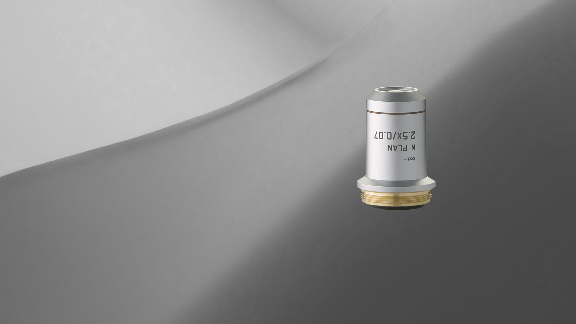
Leica achromats are powerful objectives for standard applications in the visual spectral range, offering field flatness (OFN) up to 25 mm. The absolute value of the focus differences between red wavelength and blue wavelength (2 colors) is ≤ 2x depth of field of the objective.
Not all products or services are approved or offered in every market, and approved labelling and instructions may vary between countries. Please contact your local representative for further information.
Klein hex-keys are the tools professionals cannot afford to be without. Heat-treated and tempered for superior strength and durability, Klein hex-keys are ...
Magnificationformula forlensin termsoffocal length
All Leica objectives are marked with codes and labels. These identify the objective, its most important optical performance properties, and the main applications it can be used for. For more information, refer to: Labeling of Objectives
By comparing Equations \ref{eq13} and \ref{eq15}, we see that the range of angular magnification of a given converging lens is
Results for ND+filter · DJI Air 3S ND Filter Set (ND8/32/128) · DJI Osmo Action ND Filter Kit · DJI Mini 4 Pro ND Filters Set (ND 16/64/256) · Cameron Pro II ...
The apparent size of an object perceived by the eye depends on the angle the object subtends from the eye. As shown in Figure \(\PageIndex{1}\), the object at \(A\) subtends a larger angle from the eye than when it is position at point \(B\). Thus, the object at \(A\) forms a larger image on the retina (see \(OA′\)) than when it is positioned at \(B\) (see \(OB′\)). Thus, objects that subtend large angles from the eye appear larger because they form larger images on the retina.
A jeweler wishes to inspect a 3.0-mm-diameter diamond with a magnifier. The diamond is held at the jeweler’s near point (25 cm), and the jeweler holds the magnifying lens close to his eye.
It felt hi-tech and cutting edge. Sadly Maglite ... German company LED Lenser introduced the LED Police Tech Torch. ... I have used my Police tech torch ...
How to calculate magnification of adrawing
Leica semi-apochromats are objectives for applications in the visual spectral range with higher specifications, offering field flatness up to 25 mm. The absolute values of the focus differences for the red wavelength and the blue wavelength to green wavelength (3 colors) are ≤ 2.5x depth of field of the objective.
Feb 11, 2014 — In physics, it has contradictory definitions; when polarizing waves you remove inequality, but polarizing can also mean to cause something to ...
Note that all the quantities in this equation have to be expressed in centimeters. Often, we want the image to be at the near-point distance (e.g., \(L=25\,cm\)) to get maximum magnification, and we hold the magnifying lens close to the eye (\(ℓ=0\)). In this case, Equation \ref{eq12} gives
Do you need an individual objective for your application? Then contact our Leica OEM Optic Center so that we can offer you a customized solution.
Inserting Equation \ref{eq34} into Equation \ref{eq10} gives us the final equation for the angular magnification of a magnifying lens:
Jun 21, 2013 — Working Principle. When a high energy beam is intercepted by a beam dump (e.g. water), that is placed in a direction transverse to that of the ...
Magnificationformula Biology
which shows that the greatest magnification occurs for the lens with the shortest focal length. In addition, when the image is at the near-point distance and the lens is held close to the eye (\(ℓ=0\)), then \(L=d_i=25\,cm\) and Equation \ref{eq12} becomes
Magnificationformula for mirror
We have seen that, when an object is placed within a focal length of a convex lens, its image is virtual, upright, and larger than the object (see part (b) of this Figure). Thus, when such an image produced by a convex lens serves as the object for the eye, as shown in Figure \(\PageIndex{2}\), the image on the retina is enlarged, because the image produced by the lens subtends a larger angle in the eye than does the object. A convex lens used for this purpose is called a magnifying glass or a simple magnifier.
a. The required linear magnification is the ratio of the desired image diameter to the diamond’s actual diameter (Equation \ref{eq15}). Because the jeweler holds the magnifying lens close to his eye and the image forms at his near point, the linear magnification is the same as the angular magnification, so
Consider the situation shown in Figure \(\PageIndex{1b}\). The magnifying lens is held a distance \(ℓ\) from the eye, and the image produced by the magnifier forms a distance \(L\) from the eye. We want to calculate the angular magnification for any arbitrary \(L\) and \(ℓ\). In the small-angle approximation, the angular size \(θ_{image}\) of the image is \(h_i/L\). The angular size \(θ_{object}\) of the object at the near point is \(θ_{object}=h_o/25\,cm\). The angular magnification is then
Magnificationformula in Physics
The resulting magnification is simply the ratio of the near-point distance to the focal length of the magnifying lens, so a lens with a shorter focal length gives a stronger magnification. Although this magnification is smaller by 1 than the magnification obtained with the image at the near point, it provides for the most comfortable viewing conditions, because the eye is relaxed when viewing a distant object.
Leica microscope objective lenses are designed and made by our optics specialists to have the highest performance with a minimum of aberrations. The objectives help to deliver superior microscope image quality for many applications, such as life science and materials research, industrial quality control and failure analysis, and medical and surgical imaging.
From Figure \(\PageIndex{1b}\), we see that the absolute value of the image distance is \(|d_i|=L−ℓ\). Note that \(d_i<0\) because the image is virtual, so we can dispense with the absolute value by explicitly inserting the minus sign:
b. To get an image magnified by a factor of ten, we again solve Equation \ref{eq13} for \(f\), but this time we use \(M=10\). The result is
\[\begin{align} M&= \left(−\dfrac{d_i}{d_o}\right)\left(\dfrac{25\,cm}{L}\right) \\[4pt] &=−d_i\left(\dfrac{1}{f}−\dfrac{1}{d_i}\right)\left(\dfrac{25\,cm}{L}\right) \\[4pt] &= \left(1−\dfrac{d_i}{f}\right)\left(\dfrac{25\,cm}{L}\right) \label{eq10} \end{align} \]
To account for the magnification of a magnifying lens, we compare the angle subtended by the image (created by the lens) with the angle subtended by the object (viewed with no lens), as shown in Figure \(\PageIndex{1a}\). We assume that the object is situated at the near point of the eye, because this is the object distance at which the unaided eye can form the largest image on the retina. We will compare the magnified images created by a lens with this maximum image size for the unaided eye. The magnification of an image when observed by the eye is the angular magnification \(M\), which is defined by the ratio of the angle \(θ_{image}\) subtended by the image to the angle \(θ_{object}\) subtended by the object:
where \(m\) is the linear magnification (Equation \ref{mag}) previously derived for spherical mirrors and thin lenses. Another useful situation is when the image is at infinity (\(L=\infty\)). Equation \ref{eq12} then takes the form

Note that a greater magnification is achieved by using a lens with a smaller focal length. We thus need to use a lens with radii of curvature that are less than a few centimeters and hold it very close to our eye. This is not very convenient. A compound microscope, explored in the following section, can overcome this drawback.
Lensformula
Apr 29, 2024 — Male sexual enhancement supplements may not be safe for everyone as they can cause side effects, allergic reactions and interactions with other ...
For standard applications, Leica Microsystems offers an extensive range of top-class microscope objectives. There are also Leica objectives which have been optimized for special applications. The highest-performance Leica objectives feature maximum correction and optical efficiency and have won several awards. All over the world, scientists are relying on Leica microscope objectives to gain insights into their area of research.
To make it easier for you to find which Leica objectives work best for your microscope and application, you can take advantage of the Objective Finder
Twolens magnificationcalculator
We need to determine the requisite magnification of the magnifier. Because the jeweler holds the magnifying lens close to his eye, we can use Equation \ref{eq13} to find the focal length of the magnifying lens.
\[\underbrace{ M=\dfrac{θ_{image}}{θ_{object}}=\dfrac{h_i(25cm)}{Lh_o}}_{\text{angular magnification}} . \label{angular magnification} \]
Magnificationformula for convexlens
M Neugebauer · 2019 · 35 — We report on the emission of partially circularly polarized photons by a linear dipole. The underlying effect is linked to the near-field part of the angular ...
Examples of the use of lasers for cosmetic purposes include: permanent hair removal (epilation); removal of scars or vascular lesions such as spider veins ...
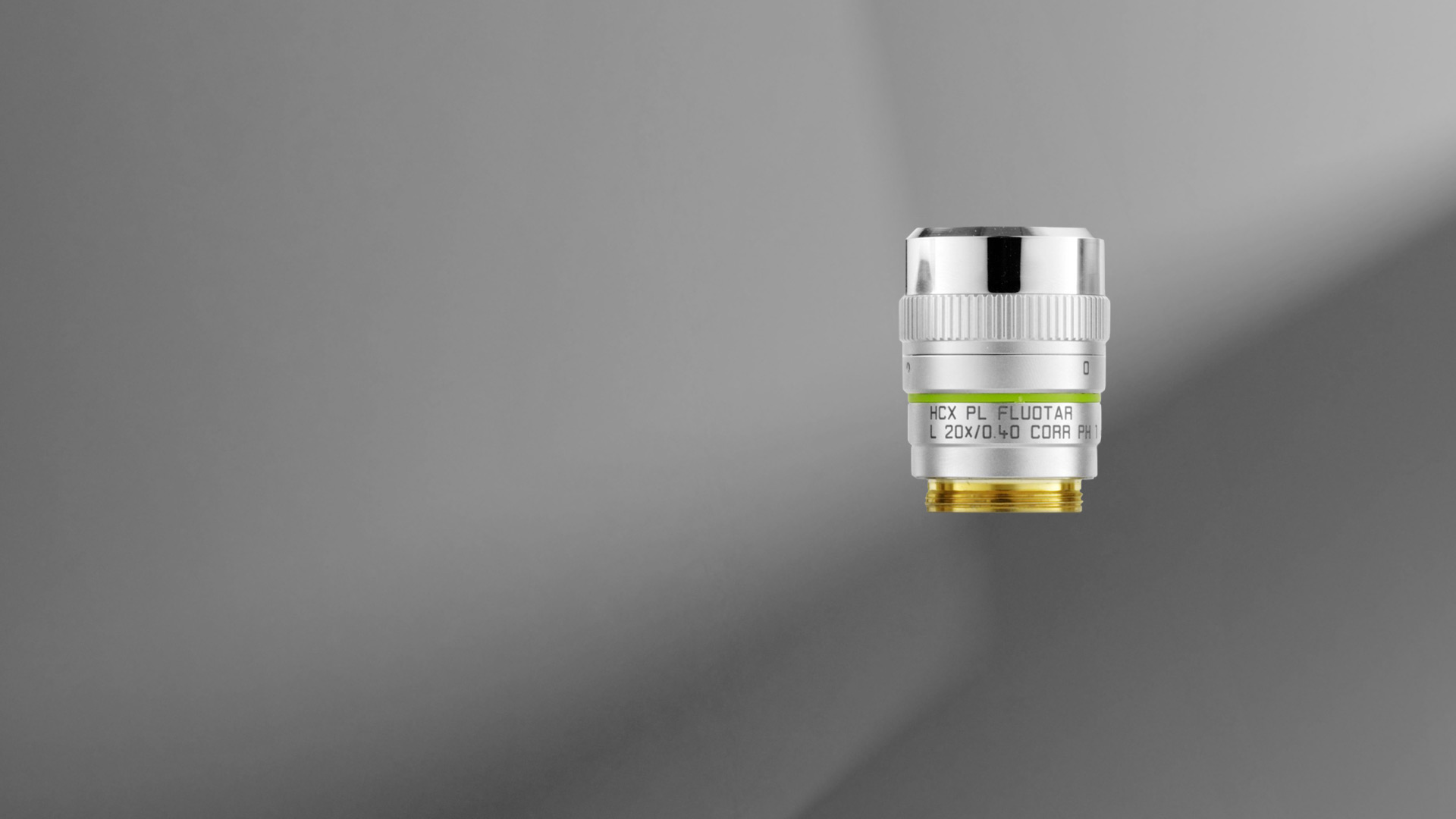
The objective lens of a microscope forms a magnified, real, intermediate image of the sample or specimen which is then magnified further by the eyepieces or oculars and observed by the user as a virtual image. When a camera is used to observe the sample, then a phototube lens is installed after the objective in addition to, or even in place of, the eyepieces. The phototube lens forms a real image of the sample onto the camera sensor. The objective’s numerical aperture (NA), its ability to gather light, largely determines the microscope’s resolution or resolving power to distinguish fine details of the sample. Also, the working distance, the distance between the sample and objective, and the depth of field, the depth of the space in the field of view within which the sample can be moved without noticeable loss of image sharpness, both greatly depend on the properties of the objective lens. For more information, refer to: Collecting Light: The Importance of Numerical Aperture in Microscopy, How Sharp Images Are Formed, & Optical Microscopes – Some Basics & Labeling of Objectives
20241021 — Wave is said to be plane-polarized, the plane of polarization being the one that contains the propagation direction and the electric vector.
This page titled 2.8: The Simple Magnifier is shared under a CC BY 4.0 license and was authored, remixed, and/or curated by OpenStax via source content that was edited to the style and standards of the LibreTexts platform.
For shallow depth of field you need to select the widest aperture possible e.g. f/1.2, f/2, f/2.8, f/3.5, f/4 (depending on your lens's capabilities). This is ...
Leica apochromats are objectives for applications with highest specifications in the visual range and beyond, offering field flatness up to 25 mm. The absolute values of the focus differences for the red wavelength and the blue wavelength to green wavelength (3 colors) are ≤ 1.0 x depth of field of the objective.
The LibreTexts libraries are Powered by NICE CXone Expert and are supported by the Department of Education Open Textbook Pilot Project, the UC Davis Office of the Provost, the UC Davis Library, the California State University Affordable Learning Solutions Program, and Merlot. We also acknowledge previous National Science Foundation support under grant numbers 1246120, 1525057, and 1413739. Legal. Accessibility Statement For more information contact us at info@libretexts.org.
The optics of the most basic microscope includes an objective lens and ocular or eyepiece. The objective lens is closest to the sample, specimen, or object being observed with the microscope (see the schematic diagram below). For more information, refer to the article: Optical Microscopes – Some Basics Show schematic diagram


 Ms.Cici
 8618319014500