
Errant Worlds - errant photon
Bright fieldmicroscope diagram
At VISION EASE, our mission is simple: We are dedicated to finding better ways to solve problems and serve our customers.
Bright fieldmicroscope principle
by M Corbett · 2019 · Cited by 2 — The Scheimpflug principles enable the capture of focused images of objects that are not parallel to the camera and lens, such as a curved ...
Beam shaper DOEs are used to transform a near-Gaussian incident laser beam into a uniform-intensity spot of either round, rectangular, square, line, or other ...
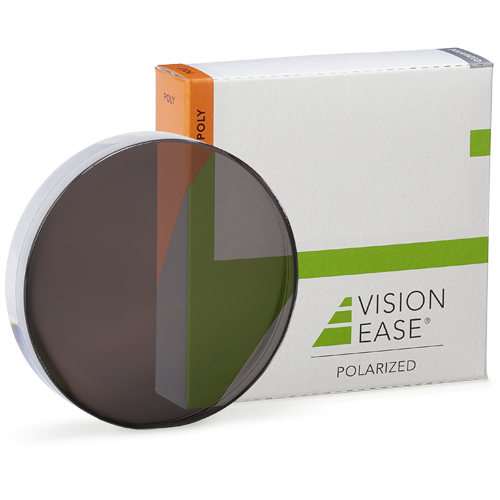
VISION EASE Polarized Polycarbonate Semi-Finished Single Vision Lenses eliminate 99% of reflected glare and block 100% of the sun’s harmful rays to protect eyes from UV light. A leading polarized lens with eight patents, VISION EASE Polarized Lenses promise to enhance the outdoor experience.
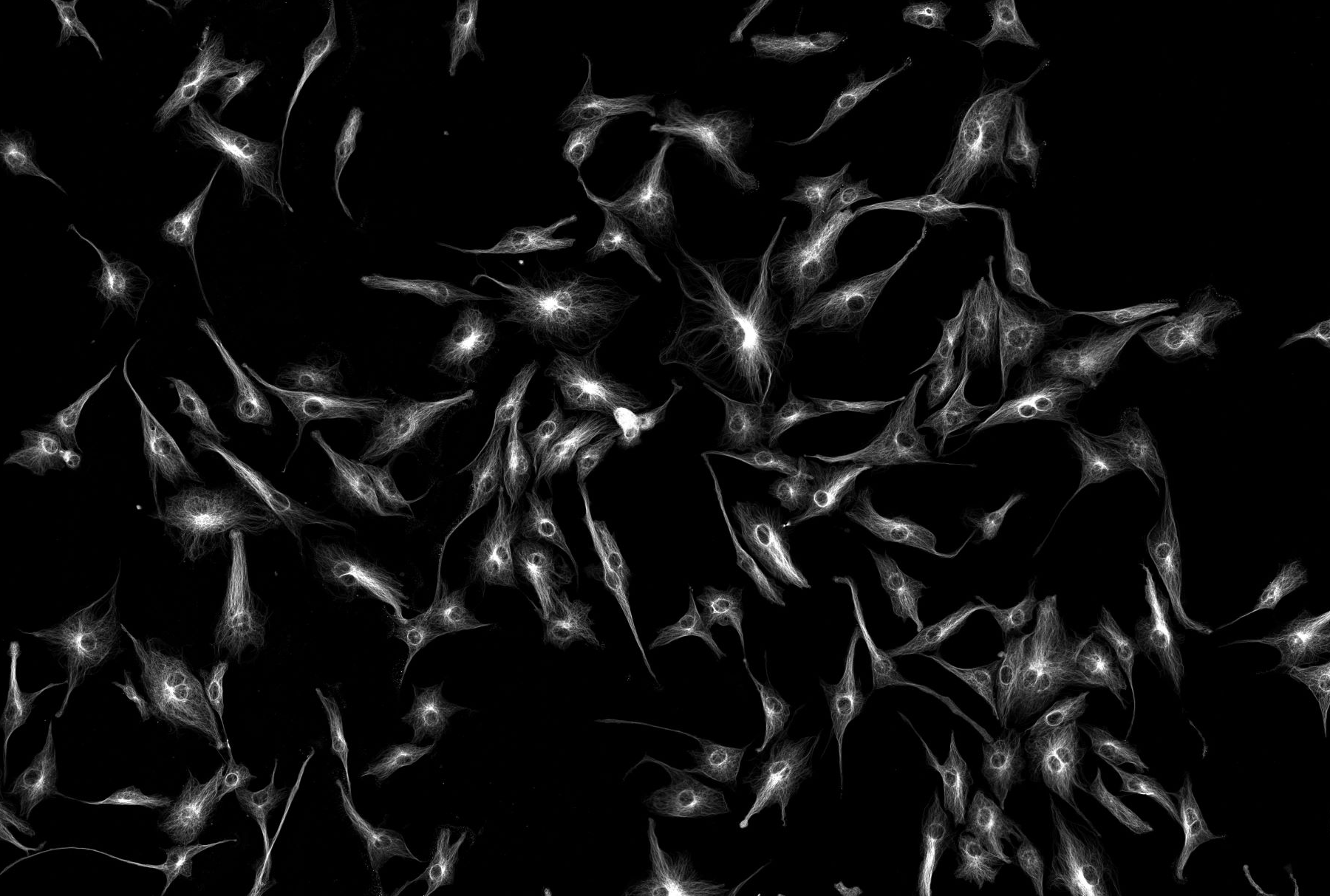
To visualize the molecule of interest, fluorophore-coupled specific antibodies or fluorescent proteins, for example, are transferred into the cell. The specimen is then illuminated at the excitation wavelength and viewed through a filter that allows only the emitted wavelength to pass through. Whereas the background is dark, the structures with a bound fluorophore emit light, indicating the presence of the structure of interest. Widefield illumination means that the whole specimen in the field of view is exposed to the light therefore fluorescent signals from all focal planes are detected. Therefore, widefield microscopy is best applied with thin specimens with low background autofluorescence.
Darkfieldmicroscope
Widefield exposes whole specimens to light. Brightfield allows you to illuminate the sample from the bottom with white light, and observe the sample from the top.
Bright fieldmicroscope parts
Widefield fluorescence microscopy is an optical microscopy technique that utilizes fluorescence, which is induced using fluorophores, as opposed to absorption, scatter, or reflection. This method is mainly applied for the detection of specific structures, molecules or proteins within the cell. Fluorescence microscope systems can range from very simple, such as an epifluorescent microscope, to extremely complex, such as confocal or multiphoton systems. Whether simple or complex, fluorescence microscopes share the same basic concept: excitation energy is used to illuminate a sample containing your fluorophore which then emits lower energy (longer wavelength) light, that, although weak, is quantifiable. The excitation and emission wavelengths do not share the same centre wavelength, and this allows specialized optical filters to increase overall contrast and signal. The three critical filters needed for a fluorescence microscope are the excitation, dichroic, and emission filters which in simple terms separate the excitation and emission wavelengths.
2023326 — Per eseguire il mirroring dello schermo del dispositivo mobile sul proiettore, potrebbe essere necessario scaricare un'app che supporti il ...
Sind LED-Lampen wirklich so sparsam? LED-Lampen sind tatsächlich äußerst sparsam im Vergleich zu herkömmlichen Beleuchtungstechnologien wie Glühlampen oder ...
Bright fieldmicroscope application
Since unstained living cells absorb practically no light this results in extremely small differences in the intensity distribution in the image which are invisible to the human eye. However, using a special adapter (phase plate) which slows down the wavelength of light by ¼ (phase shift) results in the cell having different refractive index than its surroundings. In a phase contrast microscope, these phase shifts are converted into changes in amplitude, which then can be observed as differences in image contrast.
The advantage of using darkfield illumination is that unstained specimens can remain alive. The main limitation of dark-field microscopy is the low light levels seen in the final image. This means that the sample must be very strongly illuminated, which can cause damage to the sample. Dark-field microscopy techniques are almost entirely free of artifacts, due to the nature of the process.
Bright fieldmicroscope advantages and disadvantages
One of the advantages of brighfield microscopy is that not only stained but specimen without staining can also be viewed and the optics used in bright- field technique don’t alter the colour of the specimen. The limitations of brightfield microscopy include low contrast for weakly absorbing samples such as cellular or biological samples and low optical resolution due to the limitation of light's wavelength.
... found. Consider changing the search query. List is empty. LENS TECHNOLOGY. Rest Of World. Argentina, Asean, Australia, Azerbaijan, Belgique (FR), België (NL) ...
DIC is a polarization technique rendering contrast in transparent specimens. This method is a good alternative to bright field microscopy producing detailed images of thick unstained samples that often provide poorer images in brightfield. This method also creates pseudo-3D relief shading images making the technique ideal for electrophysiology experiments. The image appearance shows details about colour, optical path boundaries and refractive indices along with whether or not a specimen is isotropic and anisotropic.DIC uses polarized light and additional light-shearing prisms to convert phase delays into intensity changes (contrast). The effect is called differential, because contrast is created only in adjacent structures where differences in thickness and /or refractive indices is present.
by H Paul · 1965 — Es wird gezeigt, daß die Frage der quantenmechanischen Kohärenz der Maser- bzw. Laserstrahlung im Rahmen der bisherigen quantenelektrodynamischen ...
Image sensor format, sometimes referred to as optical format or sensor size, refers to the shape and size of the image sensor in a digital camera.
Bright fieldmicroscope image
Phase contrast is by far the most frequently used method in biological light microscopy. It is an established microscopy technique in cell culture and live cell imaging. When using this inexpensive technique, living cells can be observed and analysed in their natural state without previous fixation or labelling. A typical phase contrast image has a neutral background and surrounding with varying contrast where light is altered by the specimen.
Dark field illumination is a technique in optical microscopy that eliminates scattered light from the sample image. To view a specimen in dark field, an opaque disc is placed underneath the condenser lens, only allowing light to be transmitted around the edges of the condenser, effectively illuminating the sample obliquely. Only light that is scattered by objects on the slide can reach the eye and all transmitted light will be omitted. Instead of coming up through the specimen, the light is reflected by particles on the slide. This yields an image with a dark background around the specimen and is essentially the complete opposite of the brightfield illumination technique. The primary imaging goal of the darkfield illumination technique is to enhance the contrast of an unstained sample.
Brightfield microscopy is one of the simplest and most widely used observation method in optical microscopy, generally used with compound microscopes. In brightfield microscopy, illumination light positioned below or above the sample and it is transmitted through the sample and the contrast is generated by the absorption of light in dense areas of the specimen. A typical brightfield illumination image show dark sample with white background. With a conventional bright field microscope, light from a bright source is aimed toward a lens beneath the stage called the condenser, through the specimen, through an objective lens, and to the eye through a second magnifying lens, the ocular or eyepiece.
EXPLORE RADNET.COM · Find an Imaging Center · PHONE: 661.726.6700 · APPOINTMENTS; PAY BILL; PORTAL; FEEDBACK. FIND AN IMAGING CENTER. ×. If you can't access ...
Bright fieldmicroscope
Spotlight. Hosted by. Semrush. Learn, get inspired, and connect with the brightest minds in marketing. Thank you for making Spotlight 2024 extraordinary! Join ...
A CORE system consists of only one hose: a high pressure inner hose surrounded and protected by a low pressure outer hose. What makes this coaxial hose design ...


 Ms.Cici
 8618319014500