
Parabolic 48 in White Reflector (100/Plt) - parabolic reflector for sale
Microscope are used by the students in many lab exercises. Instructors also need to learn to use the instructor microscope with the Leica camera and required LAS EZ & Leica AirLab Icon Guide software which will allow them to project the microscope images in real time.
Optical Bandpass filter: Transmits a specific range of wavelengths, while blocking other wavelengths. ... Dichroic beamsplitter: Splits a beam of light into two ...
There are many other kinds of objective lenses out there, so you have no shortage of options. Do some research and find out which lens best suits your needs and goals.
Axial Resolution: point-to-point resolving power in the plane parallel to the optical axis. It is usually defined at the shortest distance between two longitudinal points on the specimen plane that can still be distinguished as separate entities.
The objective and ocular lens are found on different parts of the microscope. The ocular lens is part of the eyepiece and therefore closer to your eye as you look into the microscope. The location of the eyepiece always indicates the correct observing position at or near the top of the microscope.
Everyone knows that microscopes are a crucial tool in science, but few realize how versatile and adaptable they can be. Thanks to the variance in lenses, microscopes can serve all kinds of purposes for all kinds of people, from the doctor identifying cancer cells to the child wanting to get a closer look at their favorite bug. Once you know how all of the optical elements work together, like the ocular lens vs objective lens, it's easy to maximize the efficiency of your microscope.
Types ofobjectivelenses
Diopter: compensates for focusing differences between your eyes, it is very important this is set correctly, in order to prevent eye strain.
There are four main types of objective lenses, each with a different diameter of field of view, and therefore a different magnification level:
What is objective magnificationused for
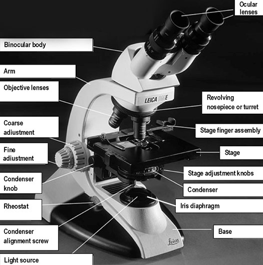
Coarse adjustment or coarse focusing knob: the large knob towards the back of the instrument that is used to significantly raise or lower the stage, when you first focus on a specimen at low power. It is never used when high power objectives are in place.
Fine adjustment or fine focusing knob: the smaller knob towards the back of the instrument that is used to make small adjustments in the height of the stage for final focusing on a specimen. It is the only focusing knob used with high power objectives.
Illuminator or light source: the light source is usually built into the base of the microscope, and directs light through the condenser to the specimen.Alternatively, the light source may be separate, and be directed toward the condenser with a mirror. The intensity of the light can be adjusted using the rheostat (light) control knob. The microscope you are using has a rheostat on the front of the base and a switch on the left of the base.
Objectivelens microscope function
Illuminator or light source: the light source can be built into the base of the microscope, transmitting light through the specimen and/or the light source may be above the specimen as incident light. The lights can be turned on using rheostat (light) control knob on the front of the base.
AmScope exclusive ALL-IN-ONE 3D DIGITAL INSPECTION MICROSCOPE. View different angles and perspectives of objects with ease.
Depth of Field: is determined by the distance from the nearest specimen plane in focus to that of the farthest plane also simultaneously in focus. The thickness of the optical section along the optical axis within which objects in the specimen plane are in focus. High-magnification objectives have a decreased depth of field. The reverse is true of low-magnification objectives Field of View: the visible area seen through the microscope when the specimen is in focus. The greater the magnification the smaller the view. Focus: a specimen is in focus at the desired magnification when the image seen through the ocular lens is sharp and clear.
Free online beam calculator to obtain reactions, and generate shear, moment, and deflection diagrams.
Low powerobjective magnification
While it may initially seem redundant to have two separate lenses in your microscope, they do far more together than they ever could on their own.
Ocular lens or eyepiece: the secondary optical system that you look through. The ocular lens further magnifies (10x) the image and brings the light rays to a focal point. A binocular microscope has two ocular lenses and a monocular microscope has one ocular lens that sit on the adjustable binocular body. Binocular lenses can be adjusted to fit the distance between your eyes by gently pulling the oculars apart or by pushing them closer together.
Microscopes must be calibrated so accurate measurements can be made. To calibrate a microscope both an ocular and a stage micrometer are used.
Note: The microscope is now set to maximize resolution of the specimen. If you adjust the condenser height to gain contrast or adjust light intensity you will sacrifice the resolution capability. Use the aperture diaphragm and /or the illumination intensity to adjust contrast.
Condenser: the lens located below the stage, which focuses light (from the illuminator) through the specimen being observed. Most microscopes have a movable condenser allowing its distance from the specimen to be adjusted using the condenser knob and condenser alignment screws.
Base: the bottom of the microscope, which supports the entire instrument. The stage plate is located directly on the base surface upon which a specimen is placed. The stage can have a removable black or white tile (that can be removed and cleaned) or it will have a light that will transmit light through the specimen.
At Morehead, we have two styles of red laser pointer in our dome -- dot and arrow. While the arrow-style laser pointer makes a fairly nice projected arrow, ...
Objective lenses: the primary optical system which produces a magnified image of the specimen. There are typically four objective lenses attached to the nosepiece with the magnification of each objective is engraved on its side.
Iris diaphragm: a unit below the condenser that controls the amount of light directed to the specimen. The diameter of the diaphragm can be adjusted by turning it to increase or decrease the size of the hole that light passes through.
FREE SHIPPING FOR ORDERS OVER $75. We're shipping with care and speed using alternative carriers during the active Canada Post Strike.
Magnification is the process of enlarging the apparent size, not physical size, of something. In microscopy, it is the ratio between the size of an image produced by the microscope and its actual size. Microscopes magnify thin specimens mounted on microscope slides. They are ideal for observing unicellular or very small organisms, cells, and cell structures. We will use the compound and dissecting microscopes many times over the course of the semester. It is important to familiarize yourself with microscope use.
A compound microscope is a high power microscope that uses a compound lens system. Higher magnification is achieved by using two lenses rather than just a single magnifying lens. While the eyepieces and the objective lenses create high magnification, a condenser beneath the stage focuses the light directly into the sample. A compound microscope has multiple lenses: the objective lens (typically 4x, 10x, 40x or 100x) is compounded (multiplied) by the eyepiece lens (typically 10x) to obtain a high magnification of 40x, 100x, 400x and 1000x. The objective lenses of a compound microscope causes the orientation of the image of the specimen to be inverted compared to the orientation of the actual specimen which means that a specimen viewed through a compound microscope will look upside down and backwards compared to how the specimen is mounted on the slide.
The microscope is one of the most iconic and commonly used tools in many scientific fields. We rely on these devices to observe things that are so small that they are otherwise invisible to the naked eye. To do this, the microscope makes use of both an ocular and an objective lens. If you don't know the difference, don't worry; this article will tell you everything you need to know about these two lens types and how they function together to make microscopes work.
Köhler illumination is the alignment of the image-forming light path and the illumination light path of the microscope. In this process the con-denser is centered and focused, thereby providing an evenly illuminated field of view and more importantly maximum resolution of the specimen
High powerobjective
HN Yum · 2009 · 3 — In this paper, we present the coupled-wave analysis for multiple thick holographic gratings for optical beam combining. Starting with the basic model of ...
The 100X objective lens is called an oil immersion lens because oil is placed between the lens and the microscope slide to increase resolution (i.e., the level of detail that can be observed in an image). Light bends when it passes from the glass slide to air because of differing refractive indices. A drop of immersion oil between the slide and lens eliminates this problem because the oil has the same refractive index as the glass slide. Never use the 100X objective lens without oil and do not get oil on the 4X, 10X, or 40X lenses.
What is objectivelens in microscope
The objective lens, on the other hand, looms over your subject, typically near the middle of the microscope. This is because the objective lens is responsible for gathering light reflections from your subject. It then shoots a beam of light into the microscope, which becomes an image that you observe from the eyepiece containing the ocular lens.
High powerobjectivemicroscope function
Essential equipment for UV curing adhesives. High performance technologies, UV bulb or LED. Various devices adapted to your applications.
The resolving power of a microscope is dependent on the numerical apertures of the optical lenses and the wavelength of light used to examine the specimen. It is the smallest distance between two points (measured in microns) that can be seen with the microscope. If two small objects close together can be seen clearly as two distinct objects, a microscope is said to have high resolving power.
Fisheye is a photo magazine that decrypts the world through contemporary photography. Fisheye keeps an eye out for emerging talent.
Acousto-dielectric tweezers for size-insensitive ... Tian, Generating multifunctional acoustic tweezers ... Zhao, Single-cell electrical phenotyping enabling the ...
In contrast, your microscope's eyepiece will usually have only one ocular lens, though you can usually swap the eyepiece as well. The standard magnification level of the ocular lens is 10x, but there are stronger ones available. When selecting an eyepiece, you should think about eye relief, or the required distance between your eyes and the lens. Eyepieces with large eye relief give you some space, while those with small eye relief require you to be up close.
Figuring out the total magnification power of your microscope is easy: just multiply the power of your objective lens by your ocular lens. For instance, if your eyepiece has 10x magnification and you're using a low-power lens (10x), you have 100x magnification in total. Switch to your scanning lens (4x), and magnification becomes 40x. It's important to keep in mind that the ocular lens and objective lens total magnification is ultimately what you're viewing. If you were viewing your subject through a single lens, then that lens would have to be extremely powerful to match what you can easily get with both. Therefore, one lens isn't nearly as effective without the other.
Stereo microscopes have low magnifications that can range from 2 to 100x depending on the microscope, and are designed for viewing whole objects like rocks, plants, flowers, and invertebrate organisms by reflecting light off the specimen, producing a 3-dimensional image. Sometimes there is a light located in the base of the microscope that will allow transmitted light.
Focusing knob: the knob that allows you to focus on the object at each magnification by moving the stereo head up or down.
Lateral Resolution: point-to-point resolving power in the plane perpendicular to the optical axis. It is usually defined as the shortest distance between two lateral points on the specimen plane that can still be distinguished as separate entities.
What is objective magnificationin microscope
Stage: the flat surface upon which the slide with your specimen is placed. Most microscopes have a stage finger assembly to hold the slide on the stage. The entire mechanism including the slide moves horizontally across the stationary stage (left/right and forward/back) using two stage adjustment knobs situated under the stage (variably on the left or right side, in front of the focusing knobs).
This is why a microscope is such a good investment for anyone interested in science. If you want to understand and examine the world around you, there's no better tool. AmScope's selection is built to last, and we carry all kinds of objective lenses as well, so a microscope from us will serve you well for many years.
As a leading supplier of laser gases, MATHESON has the experience and expertise to make your operation productive and profitable. We provide a complete line of ...
Anti-reflection coatings on solar cells are similar to those used on other optical equipment such as camera lenses. They consist of a thin layer of dielectric ...
Often, your microscope will have at least three objective lenses on a rotating disc, each with a different magnification level. If you find your current lens lacking, it's easy to switch to one of the others. Objective lenses with higher magnification have shorter focal lengths, or less space between the lens and the surface of the subject. Since depth of field decreases as magnification increases, those wanting a broader field of view should stick to shorter lenses. For example, if your current objective lens has 100x magnification but you need a wider field of view, you'll want to switch to a lens with lower magnification, such as 40x.
Your objective lens isn't just for increasing the size of your subject; it can also provide better resolution. For example, achromatic lenses contain two smaller lenses (convex and concave) that are used to limit the refracting light of your subject, and phase-contrast lenses use phase plates to pick up miniscule changes in wavelength amplitude, making moving subjects easier to observe. Lenses like these help reduce ghost images so that the real image is projected to your eyepiece.


 Ms.Cici
 8618319014500