Overview of diffraction gratings technologies for spaceflight ... - grating space
fov是什么
The integration of SWIR cameras, such as the Princeton Infrared Technologies MVCam SWIR camera, represents a significant advancement in microscopy. By extending imaging capabilities beyond the visible spectrum, SWIR technology provides enhanced contrast, detail, and penetration, opening new possibilities for research and analysis across various fields.
The field of view of an optical imaging instrument is often limited by an intentionally created field stop. This is an optical aperture, e.g. in the form of a diaphragm, which is placed in an image plane or close to such a plane, such that the edges of the field of view are sharply defined. However, in some cases one obtains vignetting effects, i.e., a gradual decrease of image brightness towards the edges of the field of view. This happens when the field of view is limited by an aperture which is not in an image plane. An example for that situation can be found in the article on field lenses, where we vignetting in an optical telescope is explained. In cases with vignetting, one may define different values for the field of view – for example, the field without any vignetting, the field up to the point of half-vignetting or (is the very maximum) up to the point where the intensity vanishes.
Please do not enter personal data here. (See also our privacy declaration.) If you wish to receive personal feedback or consultancy from the author, please contact him, e.g. via e-mail.

fov参数
For small angles, the full-angle field of view in radians is approximately the sensor diameter divided by the focal length; for obtaining a value in degrees, one has to multiply that with <$180\textdegree / \pi$>. For wide angle cameras, one has to use a more accurate formula:
The field of view gets particularly small when using a tele-objective, while wide field objectives are by definition made for a large field of view. Extreme versions are called fish-eye objectives; they produce substantial image distortions, which are hardly avoidable in that regime.
By submitting the information, you give your consent to the potential publication of your inputs on our website according to our rules. (If you later retract your consent, we will delete those inputs.) As your inputs are first reviewed by the author, they may be published with some delay.
The advantages of SWIR in microscopy include enhanced contrast and detail, improved penetration and reduced scatter and noise. SWIR imaging provides superior contrast and detail for distinguishing between materials and structures that are difficult to differentiate using visible light. SWIR light can penetrate deeper into biological tissues and materials, allowing for imaging of internal structures that are opaque to visible light. SWIR wavelengths are less scattered by particles and imperfections, leading to clearer images in challenging conditions.
fov和焦距的关系
Viewangle
Enter input values with units, where appropriate. After you have modified some inputs, click the “calc” button to recalculate the output.

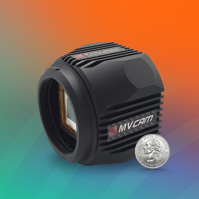
The MVCam SWIR camera represents the latest advancements in SWIR imaging technology, specifically designed to enhance microscopy applications. Key features of the MVCam include high resolution (1280x1024 megapixels), fast frame rate (up to 90 frames per second), InGaAs sensor technology with a 12µm pixel pitch, 0.4-1.7µm wavelength range covering both visible and SWIR for versatile imaging. The MVCam also has an integrated single-stage thermoelectric cooler for consistent sensor temperature, minimizing thermal noise and medium and base Camera Link™ configurations for flexible data output and system integration.
The field of view of a telescope or a photo camera, for example, is usually understood as the range of angular directions in which objects can be observed for a fixed orientation of the instrument. (One may of course observe a wider range by combining images from different instrument orientations.) It can be quantified in different ways:
Microscopy is a fundamental tool in scientific research, allowing scientists to observe and analyze the minutiae of biological specimens, materials, and devices. While visible light microscopy provides valuable insights, the integration of shortwave infrared (SWIR) cameras introduces a new dimension to imaging. SWIR technology, particularly through Princeton Infrared Technologies' MVCam SWIR camera, enhances microscopy capabilities and addresses the limitations of conventional imaging methods.
Field of view
The term field of view is not only used for that field itself, but also for its magnitude. In some cases, for example for telecentric lenses, it is used for the viewed area on the object plane.
FOV to focal length
A large field of view is particularly relevant, for example, for astronomical telescopes which are used in stellar surveys.
FOV to focal length calculator
In some cases, the term angle of acceptance is used instead of field of view – particularly for non-imaging optical instruments.
Here you can submit questions and comments. As far as they get accepted by the author, they will appear above this paragraph together with the author’s answer. The author will decide on acceptance based on certain criteria. Essentially, the issue must be of sufficiently broad interest.
Note: the article keyword search field and some other of the site's functionality would require Javascript, which however is turned off in your browser.
Telescopes provide some magnification for viewing distant objects. The higher the magnification, the smaller is typically the field of view. However, there are optical designs which provide a larger field of view for given magnification. For example, a simple Keplerian telescope has a small field of view, which can be expanded by inserting an additional field lens.
In material science, the MVCam SWIR cameras offer valuable advantages for analyzing materials and detecting defects. In material characterization, the MVCam can examine material surfaces and internal structures with high resolution, revealing hidden defects and characteristics. The MVCam can also assist with quality control by monitoring and inspecting materials in real-time, ensuring consistent quality and performance.
Further, we have many interesting case studies on the same page, with topics mostly in fiber optics. Concrete examples cases, investigated quantatively, often give you much more insight!
The field of view of the human eye is also not precisely defined. Maximum image resolution is only achieved in the central area, and the peripheral regions exhibit a substantially lower image quality. This is largely compensated by rapid eye movements for covering a wider angular range and accurately viewing objects of particular interest.
field ofview中文
Standard photographic objectives are made such that their field of view is similar to that of the human eye – with a full horizontal angle around 50°, when considering the range with reasonable sharp imaging.
Shortwave infrared (SWIR) imaging captures light in the wavelength range of approximately 0.4 to 1.7 µm. Unlike visible light, SWIR can penetrate deeper into materials and tissues, providing unique contrasts and details that are not visible with traditional visible light microscopy.
Note: this box searches only for keywords in the titles of articles, and for acronyms. For full-text searches on the whole website, use our search page.
As mentioned above, the field of view of an instrument is often intentionally limited. This is often not motivated by its application, but rather because the image quality would be too much degraded by optical aberrations when permitting a larger field of view. Advanced optical designs, e.g. based on aspheric lenses or more refined combinations of lenses, can offer a wider field of view with good image quality. However, they are not always employed, e.g. for reasons of higher cost, size or weight of the instruments.
SWIR imaging with the MVCam can significantly advance biological research by providing deeper insights into cellular and tissue structures by observing cellular processes and structures with enhanced contrast and detail. The MVCam is also able to visualize internal tissue structures in tissue analysis and study tissue morphology with improved penetration and reduced scattering.
The field of view of the eye may be restricted by various kinds of devices, for example by correction glasses of small size or by magnifying glasses. In some cases, the peripheral view is completely blocked, e.g. with some laser safety glasses; that can introduce additional hazards, e.g. of bumping into items which could not be seen.
The MVCam SWIR Camera’s Camera Link™ interface ensures compatibility with a wide range of microscopy systems, allowing for seamless integration. The flexible data output options accommodate different frame rates and resolutions, catering to various application needs. To maximize the benefits of SWIR imaging, it is essential to optimize camera settings and integrate the SWIR camera effectively into your microscopy system.
The field of view of a photo camera depends on both the used photographic objective and the size of the photographic film or the image sensor. This is explained in Figure 1, which shows the optical configuration is a greatly simplified way: the objective is represented by a single lens, although it is usually a system containing multiple lenses. One can simply consider rays coming from object points and going through the center of the lens, where no ray deflection occurs.


 Ms.Cici
 8618319014500