
Optical Tube Assemblies (OTA) | Best Deals - telescope optics
LUCIDHelios2
Helios is a compact Time of Flight (Tof) camera with superior depth precision with a working range of up to 6m. Unlock the potential of Time of Flight in a variety of industrial applications including robotics, 3D inspection and logistics.
The compound microscope (with two convex lenses) was invented in Holland around 1590 by two spectacle makers, Hans Jannsen and his son, Zacharias. During the early 1600’s, Galileo (1564–1642) made several telescopes and microscopes that he called “occhialino.” Also in Italy, James Faber, a physician, coined the word “microscope” in 1625; and the first association of microscopists was formed.
Features: Resolution: 640 x 480 pixels (0.3 MP) in monochrome. Frames per second: 30 fps at full resolution. Sensor: CMOS Global Shutter sensor Sony DepthSense IMX556PLR. Video Interface: 4-Lane MIPI D-PHY CSI-2 (FFC) Lens Mount: Integrated lens with 6mm focal length (not user changeable)
Helios cameraprice
This is the first illustration of the microscope in use for clinical examinations in medicine. The microscope designed by Joseph Campani of Bologna is standing on a table (in an enlarged form, left of the picture); and a hand-held microscope is shown in actual use to examine a wound on the leg of the recumbent patient. Note the woman who holds a candle and a mirror for optimal illumination. A second observer with a microscope (standing on the left) seems confused about whether he is using a microscope or a telescope. (Figure from page 372 of Acta Eruditorum, 1686, ref. [4]).
Despite the countless people, including royalty, who paid tribute to early microscopists, the medical world, practicing clinicians, and academic physicians generally ignored or ridiculed them. The microscope was not appreciated as a useful scientific instrument by leaders in morbid anatomy such as Morgagni (1682–1771), John Hunter (1728–1793), and Mettew Baillie (1761–1823). The first atlas of pathology [12], written by Baillie and published in 1799, contains not even one microscopic illustration among more than 100 engravings.
ToFcamera

Embedded model of the compact Time of Flight camera with monochromatic Sony DepthSense IMX556PLR sensor and GigE Vision interface, which allows to capture high-quality images with depth data.
Malpighi (1628–1694) a microscopist, histologist and embryologist, was the first person to see the anastomosis between arterial and venous capillaries [10]. His descriptions of the Malpighian bodies of the kidney, the Malpighian corpuscles of the spleen, and the Malpighian layer of the epidermis are known to every student of medicine [11].
Model name HLS003S-001ETX2 Product family Helios Product type Area scan 3D ToF Sensor name Sony DepthSense IMX556PLR Sensor type CMOS Sensor size (H x V), format 6.40 mm x 4.80 mm, 1/2" Pixel size (H x V) 10.0 µm x 10.0 µm Color/Mono Mono Resolution (H x V), MP 640 x 480 px, 0.3 MP Frame rate 30 fps ADC Dynamic Range Shutter Global Exposure Range Synchronization Software trigger, hardware trigger, PTP (IEEE 1588) Inputs/Outputs Image Buffer Lens Mount Integrated lens with 6mm focal length (not user changeable) Output Formats 3D Point Cloud, Intensity and Confidence Working Ranges Near mode: up to 1.5m, Far mode: up to 6m Accuracy Less than 5mm (0.3m to 1.5m) Precision Standard deviation less than 2mm at 1m Lens Field of View 59⁰ x 45⁰ (nominal) Illumination 4 x VCSEL laser diodes @ 850nm Interface 4-Lane MIPI D-PHY CSI-2 (FFC) Machine Vision Standard GigE Vision v2.0 Power Requirements Power Consumption (Maximum) Pavg <15W Housing Type None Dimensions (L x W x H) 55 mm x 55 mm x 43.7 mm Weight Camera head: 113 g Camera adapter board: 15 g FFC cable: 3 g Power cable: 6 g AC power supply: 380 g Temperature (Operating) -10° to 60°C Temperature (Storage) Conformity CE, FCC, RoHS, REACH, WEEE, Eye Safety Class 1 IEC/EN 60825-1:2014, GenICam, GigE Vision Warranty
Basler ToFCamera
Helios cameramanual
Clinical microscopy had a slow beginning; more than two centuries passed before the value of microscopes began to be appreciated by clinical and laboratory scientists. In 1800, Bichat (1771–1802), a young pathologist, published a book in which, for the first time, morbid anatomic and histopathologic changes of various organs of the body were discussed and illustrated [13]. Soon thereafter the microscope became an indispensable laboratory tool at medical schools all around the world.
In 1653, Petrus Borellus [1] wrote the first publication on the use of microscope in medicine. He described 100 observations and applications, including how to remove ingrowing eyelashes that are invisible to the naked eye. In 1646, Athanasius Kircher [2] (or “Kirchner, as it is often spelled), a Jesuit priest, wrote that “a number of things might be discovered in the blood of fever patients.” In 1658, in his Scrutinium Pestis, Kirchner [3] described microscopic “worms” in plague victims which he suspected caused the disease that killed millions of people in Europe during the 17th century. Most likely, he was viewing pus cells, or perhaps red blood cells, since he could not possibly see the Bacillus pestis with his 32-power microscope. Another early microscopist was Joseph Campini of Bologna. His microscope was the first that was depicted in clinical use in medicine (Fig. 1⇓) [4].
time-of-flightcameraprice
Helios2cameraprice
Sony combined Tof with backside illuminated sensor (BSI) technology to create the new DepthSense ToF sensor. The BSI technology provides better light collection efficiency in NIR wavelengths. The new Sony IMX556PLR iw a 1/2in sensor running at 640 x 480px at 30 fps.
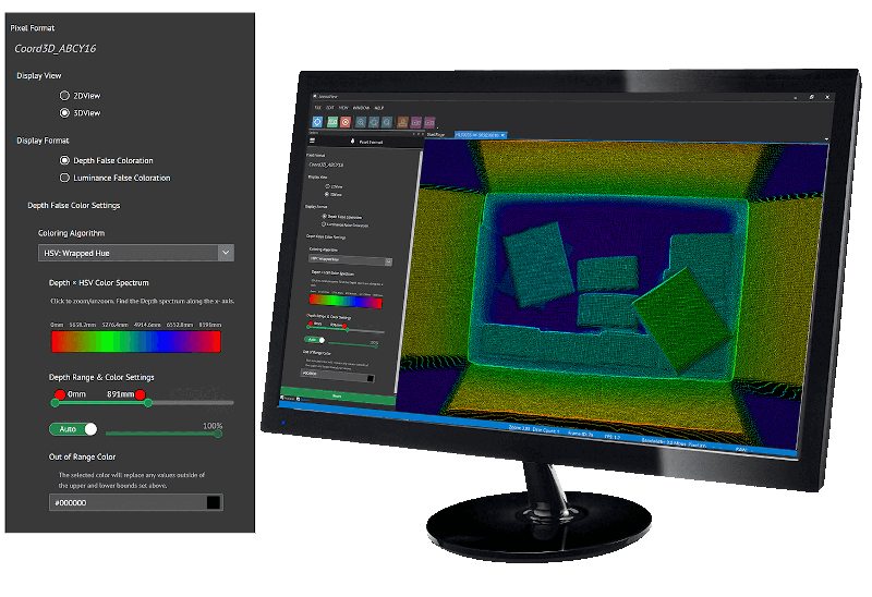
The Arena SDK includes easy to use controls for the Helios ToF camera. ArenaView allows for 2D and 3D views: 2D view allows one to see the intensity and depth of the scene from the camera’s perspective. 3D view displays a point cloud of the scene and allows users to manipulate the orientation in real-time. Additionally, settings can be changed in real-time such as false color overlay, depth range adjustments, out of range color settings and more.
This Helios Embedded model (P/N: HLS003S-001ETX2US1) is designed to be connected to a NVIDIA® Jetson TX2. The Helios Embedded camera module streams raw data via a MIPI connection. Depth information is processed on the Jetson TX2 using included Arena SDK. HLS003S-001ETX2US1: 640 x 480 at 30 FPS, MIPI Connection, Working Range: up to 6m
Helios cameralens
The microscope is undoubtedly one of the greatest inventions that men have ever made. The use of lenses for spectacles (eyeglasses), distant vision (telescopes), and high magnification (microscopes) required early lens makers accurately to grind lenses with different focal lengths. During the 16th and 17th centuries, Holland and Italy were the principal countries for the construction and use of telescopes and microscopes.
While there were many botanists and zoologists who used microscope in the 17th century, there were few physicians. The observations of Leeuwenhoek (1632–1723), a Dutch drapery maker, excelled all other microscopists, because of his skill in making high quality lenses. The red blood cell was described in 1667 by Swammerdam (1637–1680) [5] and in 1673 by Malpighi (1628–1694), but it was Leeuwenhoek in 1695 who first illustrated red blood cells in his Arcana [6]. In the 190 letters that he wrote to the Royal Society in London over a period of 50 years, Leeuwenhoek gave descriptions and illustrations of bacteria from the human mouth, protozoa, spermatozoa, striations of skeletal muscles and epithelial cells from a wart on the trunk of an elephant in the Amsterdam Zoo [7–9].
The Helios industrial camera detects 3D depth of objects. It features Sony’s DepthSense ToF sensor, along with 4 VCSEL laser diodes operating at 850nm. The camera calculates depth data by measuring the time it takes for light emitted from the diodes to reflect off objects in the scene and return to the sensor for each point of the image. This design results in high accuracy of < 5 mm with a precision (standard deviation) of 2 mm at 1.0 m distance.
This Helios camera model (P/N: HLS003S) is designed to be connected to a host PC running either Windows or Linux. Depth information is processed on the Helios ISP and streamed through the Gigabit Ethernet connection. HLS003S: 640 x 480 at 30 FPS, Gigabit Ethernet, Working Range: up to 6m


 Ms.Cici
 8618319014500