
Optical Tables – mounting holes, honeycomb core, ... - optics table
Microscope objectives are vital lenses that determine the magnification, resolution, and quality of the images produced by a microscope. They come in various types and magnifications, each suited for different applications and levels of detail, making them indispensable in scientific research, medical diagnostics, and educational settings.
SchärfentiefeAbbildungsmaßstab
AmScope exclusive ALL-IN-ONE 3D DIGITAL INSPECTION MICROSCOPE. View different angles and perspectives of objects with ease.
Schärfentiefe (II) Die Magie der Schärfentiefe in der Fotografie Die Schärfentiefe ist – wie schon im ersten Blogpost zu diesem Thema gesagt – eines der wichtigsten (und auch faszinierendsten) Gestaltungsmittel in der Fotografie. Hier geht es jetzt etwas ausführlicher...
A microscope is a scientific instrument used to magnify and observe objects that are too small to be seen with the naked eye. It works by focusing light or electrons to create an enlarged image of the specimen.
Schärfentieferechner für immer dabei
A trinocular microscope head combines the benefits of binocular viewing with the capability to capture digital images or videos of specimens. It is particularly suited for advanced research, educational purposes, and industrial applications where precise imaging and documentation are essential.
Thomas Kupas Lavesstraße 20/21 30159 Hannover Telefon 0511 23504422 oder Telefon 0511 3947397 Mobil 0179 5380490 mail@fotoschule-hannover.de
SchärfentiefeTabelle Excel
Illuminate your subjects with brilliance. Our microscopes feature advanced lighting technologies, providing the perfect balance for optimal observation, even in low-light conditions.
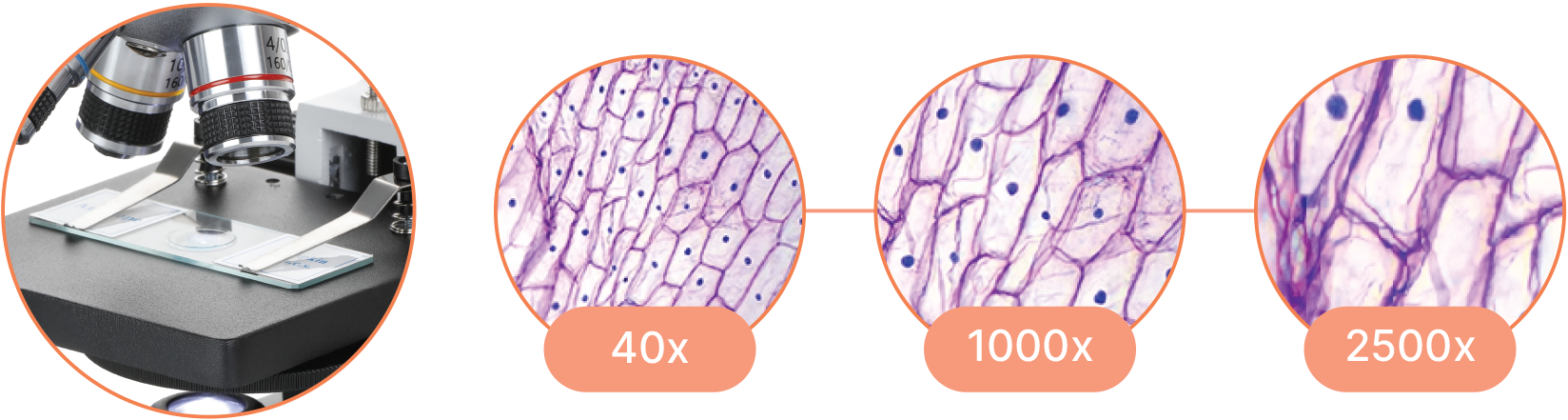
Provides high magnification (up to 1000x or more) and high resolution for viewing fine details of cells, tissues, and microorganisms.
Capable of high magnification, which is achieved through the combination of the objective lens (typically 4x, 10x, 40x, and 100x) and the eyepiece (usually 10x).
Spiegelreflex-Kameras als Bausatz Für den Workshop „Fotografie: analog!“ habe ich neulich zwei inzwischen ein wenig eingestaubte, aber immer noch und wieder sehr spannende Fotoapparate aus dem Regal genommen. Nämlich die „Konstruktor“ als einäugige und eine zweiäugige...
SchärfentiefeTabelle für die Hosentasche
Compound Magnification is calculated by multiplying the magnification of the objective lens by the magnification of the eyepiece.
Beugungsunschärfe Rechner
A darkfield microscope is a type of optical microscope that provides high contrast images of unstained specimens by using scattered light. The specimen appears bright against a dark background
SchärfentiefeBlende
Es gibt schon immer mal Neuigkeiten bei mir: neue Termine für die Workshops oder gleich ganz neue Fotokurse, interessante Blogposts, spezielle Shooting-Angebote, auch mal Ausrüstungs-Tipps, oder besondere Sachen … Wenn du informiert sein und viele Vorteile genießen möchtest, trage sich gern in die Mailingliste ein: für den Newsletter der Fotoschule Hannover.
Klickt mal auf die Screenshots: dann könnt ihr sie größer sehen und gut lesen. Ich habe die Schärfentiefe für drei verschiedene Brennweiten berechnen lassen. Grafiken und Tabellen bereiten die Angaben jeweils unterschiedlich auf.
Ihr werdet durch viel, viel Praxis und Übung mit euren Kameras und Objektiven sehr bald ein wirklich gutes, direktes „Gefühl“ dafür bekommen, wie sich eure Einstellungen auf Schärfentiefe und Bildlook auswirken. Und dann könnt ihr – ganz ohne langes Überlegen und natürlich auch ohne DoF-Rechner – intuitiv, routiniert und „treffsicher“ die „Magie der Schärfentiefe“ nutzen und richtig tolle Fotos machen!
The terms monocular, binocular, and trinocular refer to the different types of microscope heads, each offering a distinct way of viewing the specimen.
A specimen is a sample or example used for scientific study. It can be anything from biological tissues to materials, examined under a microscope or other instruments for analysis.

Used in fields like biology, geology, entomology, electronics assembly, and manufacturing for tasks requiring manipulation and examination of objects in three dimensions.
Models gesucht! Für die geplanten Portraitfotografie-Workshops der Fotoschule Hannover (und auch für neu zu entwickelnde Workshops) suche ich attraktive Modelle für eine regelmäßige Zusammenarbeit. Models gesucht: gern mit ein wenig Erfahrung vor der Kamera und im...
Tiefenschärfe Rechner App
Compound microscopes are suited for detailed examination of microscopic structures, while stereo microscopes are more appropriate for observing larger objects in three dimensions and for tasks that involve manipulation and dissection.
A stereo microscope, also known as a stereoscopic or dissecting microscope, provides three-dimensional viewing of larger, opaque specimens through dual optical paths with objective lenses. It offers lower magnification (typically 5x to 40x) than compound microscopes but enhances depth perception. Ideal for tasks in biology, geology, and manufacturing, it allows comfortable, extended viewing with ergonomic adjustments.
Commonly used in biological research, medical diagnostics, and educational settings for detailed examination of specimens.
Im Dof-Rechner gebt ihr also immer die drei Parameter an, über die wir die ganze Zeit schon reden: Blende, Brennweite und Abstand. Und weil auch die Sensorgröße Einfluß auf die tatsächliche Schärfentiefe hat, müsst ihr zudem euer Kameramodell auswählen. (Weil das aber ja nichts am „Prinzip“, dass Blende, Brennweite und Abstand die Schärfentiefe steuern ändert: lassen wir diesen Aspekt einfach außen vor.)

Navigate effortlessly through magnification levels and focus adjustments. Our microscopes feature intuitive controls, allowing you to concentrate on your research without the hassle of complicated settings.
Magnification works by bending light through lenses or using digital technology to enlarge the appearance of an object, allowing for detailed observation and analysis.
A binocular microscope head utilizes two eyepieces for simultaneous viewing with both eyes, providing enhanced comfort, depth perception, and superior image quality. Ideal for professional and research settings requiring detailed observation, its design minimizes eye strain and enhances ergonomic support compared to monocular microscopes.
Wie viel oder wenig Schärfentiefe bei verschiedenen Kombinationen aus Brennweite, Blendenzahl, Entfernung zum Motiv entstehet, können wir uns nämlich von DoF-Rechnern (DoF = Depth of Field) „exakt“ bestimmen lassen. – Auf die Art des Bokehs könnt ihr, wie gesagt, auch noch Einfluß nehmen, indem ihr den Hintergrund mehr oder weniger weit entfernt von eurem Motiv.
A phase contrast microscope is an optical microscope designed to enhance the contrast of transparent and colorless specimens without the need for staining. It works by exploiting differences in the refractive index of different parts of the specimen, transforming these differences into variations in light intensity.
Uses two separate optical paths with two objective lenses to provide a stereoscopic (3D) view of larger, opaque specimens.
Die Smartphone-App PhotoPills z.B. enthält so einen DoF-Rechner. Schaut euch mal die Screenshots von Schrärfentiefe-Berechnungen von PhotoPills bzw. die Kurvendiagramme zu den Abhängigkeiten von Blende, Brennweite und Abstand an. Dann wisst mal ganz genau, wann ihr wie viel Schärfentiefe wo in euren Fotos bekommt 🙂
A Compound Microscope is a type of optical microscope that uses multiple lenses to magnify small objects. It consists of two sets of lenses: the objective lens, which is closer to the specimen and provides the initial magnification, and the eyepiece lens, which further magnifies the image for the viewer's eye. Light passes through the specimen and is magnified by the objective lens, then further magnified by the eyepiece lens, resulting in a highly magnified image visible to the observer. Compound microscopes are commonly used in biology, medicine, and other scientific fields for viewing cells, tissues, and other small structures.
Witness the microscopic world in stunning detail with our high-quality optics. Every slide comes to life with crystal-clear clarity, allowing you to delve into the intricacies of biology, chemistry, and beyond.
A propos „Übung“: ich hätte da noch ein paar einfache, aber effektive Aufgaben für euch. Wenn ihr die einmal nach-fotografiert, werdet ihr schnell und direkt sehen, wie sich die verschiedenen Kameraeinstellungen und Bildkompositionen auf den „Look“ auswirken. Also los!
Nach den Erklärungen und Illustrationen zum Thema Schärfentiefe in Teil 1 (Schärfentiefe) und Teil 2 (Die Magie der Schärfentiefe) meiner Blogpost-Reihe möchte ich euch hier noch kurz hinweisen auf die Möglichkeit, Schärfentiefe berechnen und grafisch darstellen lassen zu können.
A monocular microscope head is a basic type of microscope head with a single eyepiece, ideal for cost-effective and straightforward applications. It is particularly useful in educational settings and for beginners, but it can lead to eye strain over long periods and lacks the depth perception provided by more advanced binocular and trinocular heads.
Magnification is the process of enlarging the appearance of an object, making it look bigger than its actual size. In optics, it is the ratio of the size of the image produced by a lens or microscope to the actual size of the object being viewed.


 Ms.Cici
 8618319014500