Optical instrument - what optical
What is objective lens inmicroscope

A scanning probe microscope can create a computerized image of individual atoms. This type of microscope measures the surface texture of an object on a very small scale, and will note where individual atoms protrude from that structure.
Purpose of microscopepdf
Zamboni, Jon. What Is The Function Of A Microscope? last modified March 24, 2022. https://www.sciencing.com/function-microscope-6575328/
Typesof microscope
We hope you have enjoyed Lidaris Calc. It was created in order to save your valuable time and prevent laser damage of your laser optics by considering laser fluence. In case you are still facing issues related to Laser-Induced Damage Threshold (LIDT) phenomena get one free consultancy from Lidaris experts.

The microscope is one of the most important tools used in chemistry and biology. This instrument allows a scientist or doctor to magnify an object to look at it in detail. Many types of microscopes exist, allowing different levels of magnification and producing different types of images. Some of the most advanced microscopes can even see atoms.
Importanceof microscope
Zamboni, Jon. (2018, April 25). What Is The Function Of A Microscope?. sciencing.com. Retrieved from https://www.sciencing.com/function-microscope-6575328/
Another form of optical microscope is the dissection or stereo microscope. This microscope uses two different viewing lenses and produces three-dimensional images of the sample. But it has a much smaller maximum magnification than a compound microscope, and usually cannot magnify more than 100 times.
Zamboni, Jon. "What Is The Function Of A Microscope?" sciencing.com, https://www.sciencing.com/function-microscope-6575328/. 25 April 2018.
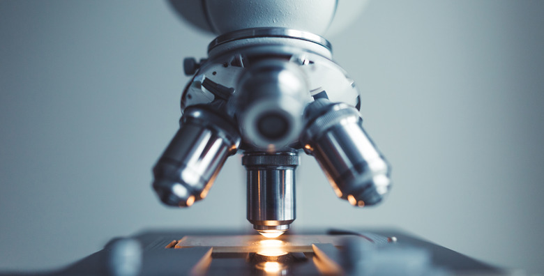
The compound microscope is the most familiar form of optical microscope. A compound microscope utilizes multiple lenses to provide magnification. A typical compound microscope will include a viewing lens that magnifies an object 10 times, and four secondary lenses that magnify an object 10, 40, or 100 times. Light is placed below the sample and travels through one of the secondary lenses and the viewing lens, and is thus magnified twice. For instance, if you use the 40 magnification lens with the 10 magnification viewing lens, the object you're viewing will be magnified 10 times 40, or 400 times. While a compound microscope can provide large amounts of magnification, the image produced by visual light are usually of a lower resolution than those produced by other microscopes.
Purpose of microscopein microbiology
The mental image you probably have of an ordinary microscope is that of an optical microscope. These microscopes use lenses and visual light. You look through the eyepiece of the microscope at a sample in real time. In contrast, imaging microscopes use a beam of radiation or particles. This beam bounces off or passes through the sample and is measured and interpreted by a computer that creates and saves an image of the sample for later viewing.

Imaging microscopes are significantly higher in resolution and magnification than optical microscopes, but are also much more expensive. Different types of imaging microscopes utilize beams of different types of radiation or particles to provide an image of a sample. Confocal microscopes use laser light, scanning acoustic microscopes use sound waves, and X-ray microscopes, predictably, use X-rays. Electron microscopes use electrons and can magnify a sample by up to 2 million times. The transmission electron microscope creates a two-dimensional image, while the scanning electron microscope creates a three-dimensional image.
The microscope gets its name from the Greek words micro, meaning small, and skopion, meaning to see or look, and it literally is a machine for looking at small things. A microscope may be used to look at the anatomy of small organisms such as insects, the fine structure of rocks and crystals, or individual cells. Depending on the type of microscope, the magnified image may be two-dimensional or three-dimensional.


 Ms.Cici
 8618319014500