
Optical Energy Meters – pyroelectric, photodiodes, spectral ... - laser energy measurement
Laserbeam expander
For minimum losses of optical power, the lenses are usually equipped with anti-reflection coatings. These, however, work only within a limited wavelength range.
An optical coherence tomography (OCT) scan is an imaging test that uses low coherence light beams analyse different sections of the human body, obtaining highly precise and reliable pictures of several levels of tissue. This scan is primarily used in ophthalmology (to study the cornea and retina) and cardiology (to assess the state of the blood vessels where there may be a potential narrowing, which could lead to more severe complications).
Beam ExpanderThorlabs
An OCT is similar to an ultrasound, however, light waves are used instead of ultrasound waves. For ophthalmology purposes, this procedure is minimally-invasive, and it uses the infra-red laser rays to analyse the retina and cornea, generating precise and high definition pictures. Thanks to the possibility of analysing each tissue section, this exam can identify several conditions, especially those of the macula and of the optic disk. The test takes just a few minutes, during which the specialist will use certain reference points on the optic disk to receive and store the laser beams. The result is an image of all the strata of the retina, allowing the specialist to determine if there are any abnormalities, from which they can formulate a diagnosis.
There are variable beam expanders (zoom expanders), i.e., devices where the magnification can be adjusted in a certain range (e.g. from 2× to 5× or from 5× to 10×). Those contain at least three lenses and some fine mechanics to adjust the position of at least one of them.
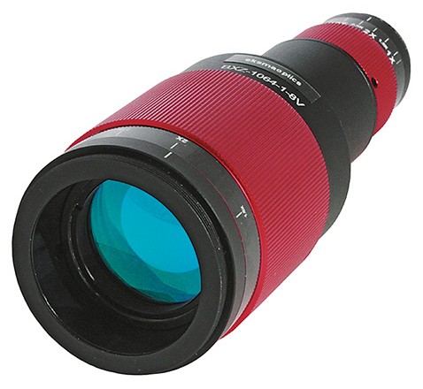
Motorizedbeam expander
Available in singlet, doublet (positive and negative focal lengths) and triplet varieties, Knight Optical can supply customised beam expanders for a range of laser-based, cutting-edge projects. Whether your application works in the ultraviolet (UV), visible, near-infrared (NIR), mid-infrared (MIR) or far-infrared (FIR) spectrum, opting for our custom-made components allows you to specify suitable substrates for your required wavelength.
Beam expanders are optical devices which increases the diameter of the input beam to produce a larger output. Used with collimated light, they have important applications in remote sensing, interferometry, and laser scanning.
In ophthalmology, OCT is used to diagnose and assess the advancement and stage of retinal conditions such as maculopathies, glaucoma, hereditary retinal dystrophies, vitreoretinal diseases and macular oedemas.
Photonic Devices' precision Galilean beam expanders have been sold globally for over 20 years and include an extensive number of standard models. These multi-element beam expanders have been computer-designed to provide excellent wavefront quality with minimum beam deformation. All lens elements are air spaced and the majority of models have variable air spacing for collimation adjustment. All lens surfaces have multilayer antireflection coatings, with power handling capabilities over 1 kW (> 200 W for beam expanders operating at 10.6 microns).

Yes, beam expansion necessarily reduces the scanning angles – just as it also reduces the beam divergence angle. This cannot be avoided simply with additional (fixed) optics.
When doing an eye OCT there is no need for any kind of preparations. You won’t need to use eye drops as the machine won’t physically touch your eyes. However, you won’t be allowed to wear contact lenses during the exam. On the other hand, when doing a heart OCT, you will be under local anaesthetic (the catheter will need to reach the area to be analysed). Therefore, you should not take anticoagulants, to avoid potential complications during or after the procedure.
Laserbeam expanderdesign
Variable beam expanderprice
It is not painful during an OCT scan. During an eye examination, you’ll have to stay still and look at the light from the equipment, whereas during a heart scan you will be under local anaesthetics which will avoid any pain.
For cardiology purposes, this procedure is more invasive, as the light waves are sent from a catheter to the area of interest. That means that the catheter has to physically reach the test area by making an incision. Once the light waves emitted by the transducer hit the clot and bounce back, they produce photon fluctuations which are sent back and received by an interferometer, allowing for an assessment of the problem.
In most cases, a beam expander is realized as an optical telescope consisting of two lenses (or in some cases of two curved mirrors). Two different configurations are common:
Using cylindrical lenses, one can realize beam expanders which work in one transverse direction only. For that purpose, one may also use anamorphic prism pairs.
By submitting the information, you give your consent to the potential publication of your inputs on our website according to our rules. (If you later retract your consent, we will delete those inputs.) As your inputs are first reviewed by the author, they may be published with some delay.
A good familiarity with Gaussian beams is a good basis for understanding the operation of beam expanders and similar devices.
If we expand the scanning beam, will the scanning angle decrease? Is there any way to expand the beam after the scanning optics?
Edmund Optics has a variety of beam expanders, including devices for different wavelength regions (e.g. Nd:YAG and CO2), rotating or non-rotating optics for divergence adjustment and research-grade devices.
Optogama designs and manufactures laser beam expanders, beam reducers and divergence compensators which are used to increase or decrease laser beam diameter and control beam divergence:
For very high laser powers, purely reflective beam expanders (with mirrors instead of lenses) are used. This is because thermal effects such as thermal lensing are weaker on mirrors. Also, that way one can avoid any parasitic reflections. A disadvantage, however, is that some amount of astigmatism is generally introduced by the mirrors.
In cardiology, OCT is used to determine whether there are thromboses, plaque, and calcium build-up or if a stent was placed incorrectly (especially when trying to prevent atherosclerosis).
Here you can submit questions and comments. As far as they get accepted by the author, they will appear above this paragraph together with the author’s answer. The author will decide on acceptance based on certain criteria. Essentially, the issue must be of sufficiently broad interest.
Variable beam expanderdesign
For achieving a given magnifying power (expansion ratio, ratio of beam radii), one may use different values of focal length. Most compact solutions are possible with small focal lengths, but there are limitations. In particular, one may then require lenses with very high numerical aperture, if at the same time a large output beam radius is required. Therefore, beam expanders for operation with large beams are tentatively longer.
Newportbeam expander
Standard off-the-shelf Galilean type zoom beam expanders and compact beam expanders. Our engineers can design a custom beam expander solution to fit your application.
Using our advertising package, you can display your logo, further below your product description, and these will been seen by many photonics professionals.
When doing an eye OCT there is no need for any kind of preparations. You won’t need to use eye drops as the machine won’t physically touch your eyes. However, you won’t be allowed to wear contact lenses during the exam. On the other hand, when doing a heart OCT, you will be under local anaesthetic (the catheter will need to reach the area to be analysed). Therefore, you should not take anticoagulants, to avoid potential complications during or after the procedure.
Please do not enter personal data here. (See also our privacy declaration.) If you wish to receive personal feedback or consultancy from the author, please contact him, e.g. via e-mail.
An OCT is similar to an ultrasound, however, light waves are used instead of ultrasound waves. For ophthalmology purposes, this procedure is minimally-invasive, and it uses the infra-red laser rays to analyse the retina and cornea, generating precise and high definition pictures. Thanks to the possibility of analysing each tissue section, this exam can identify several conditions, especially those of the macula and of the optic disk. The test takes just a few minutes, during which the specialist will use certain reference points on the optic disk to receive and store the laser beams. The result is an image of all the strata of the retina, allowing the specialist to determine if there are any abnormalities, from which they can formulate a diagnosis.
Having glaucoma means undergoing regular eye tests to ensure that vision quality is preserved. There are several tests for monitoring glaucoma and the frequency of these will depend on the individual patient. Mr Kin Sheng Lim explains what glaucoma patients can expect and explains if surgery is necessary for all glaucoma patients. See more
For application with pulsed lasers, the used lens coatings should also have a sufficiently high optical damage threshold. Further, one should avoid operation with misaligned high-power beams, which could lead to overheating of some parts.
In cardiology, OCT is used to determine whether there are thromboses, plaque, and calcium build-up or if a stent was placed incorrectly (especially when trying to prevent atherosclerosis).
Note: this box searches only for keywords in the titles of articles, and for acronyms. For full-text searches on the whole website, use our search page.
We will process your data to send you our newsletter. You can unsubscribe at any time using the unsubscribe links or by notifying us via email at [email protected]. You can also exercise your rights of access, rectification, deletion, objection, limitation of processing, and data portability by writing to the indicated email address. For more information, see Privacy Policy.
In ophthalmology, OCT is used to diagnose and assess the advancement and stage of retinal conditions such as maculopathies, glaucoma, hereditary retinal dystrophies, vitreoretinal diseases and macular oedemas.
For cardiology purposes, this procedure is more invasive, as the light waves are sent from a catheter to the area of interest. That means that the catheter has to physically reach the test area by making an incision. Once the light waves emitted by the transducer hit the clot and bounce back, they produce photon fluctuations which are sent back and received by an interferometer, allowing for an assessment of the problem.
Reflectivebeam expander
In laser technology and general optics, one often works with collimated beams, by definition having a roughly constant beam radius over some length. Sometimes, it is necessary to substantially modify a beam radius, for example in order to achieve a reduced beam divergence for transmitting the beam over a larger distance. For that purpose, beam expanders can be built and are also available as fixed optical components.
Shanghai Optics Inc's custom beam expanders are used in many applications such as laser ranging, laser illumination, interferometry, etc. In high-power laser systems, beam expanders are used increase the beam area without significantly affecting the total laser energy. This results in a reduction of the laser power density which reduce the risk of damaging the coatings and optical materials of optical components. In a laser ranging system, a beam expander is used to minimize the laser divergence, resulting in a smaller collimated beam at a long distance.
It is not painful during an OCT scan. During an eye examination, you’ll have to stay still and look at the light from the equipment, whereas during a heart scan you will be under local anaesthetics which will avoid any pain.
An optical coherence tomography (OCT) scan is an imaging test that uses low coherence light beams analyse different sections of the human body, obtaining highly precise and reliable pictures of several levels of tissue. This scan is primarily used in ophthalmology (to study the cornea and retina) and cardiology (to assess the state of the blood vessels where there may be a potential narrowing, which could lead to more severe complications).
Beam expanders are generally not designed for use with divergent beams, but only for collimated beams, and only within a certain range of beam radii. Otherwise, one may obtain clipping effects and/or not get a collimated beam out. Obviously, a beam can be collimated over a certain length only if its beam waist is large enough. As an example, Figure 3 shows the evolution of beam radius in the same beam expander is considered in Figure 1, but with a five times smaller initial beam radius. Here, the beams can no longer be considered as collimated beams.
Note: the article keyword search field and some other of the site's functionality would require Javascript, which however is turned off in your browser.
When modifying the beam radius, one also modifies the strength of beam pointing deviations. For example, doubling the beam radius implies that angular changes of the output beam are only half as strong as those of the input beam.


 Ms.Cici
 8618319014500